Early retinal and choroidal changes in the macula region associated with axial length in myopic children and adolescents based on swept-source OCTA
Yuan Cao, Qiao Gao, Jun Zhu, Fang Chen

TL;DR
This study shows that longer eye length in myopic children and adolescents is linked to early changes in retinal and choroidal blood flow and structure, detectable with advanced imaging.
Contribution
The study identifies specific early microvascular and structural changes in the macula associated with axial length in myopic youth using swept-source OCTA.
Findings
Increased axial length correlates with reduced deep retinal vessel density and choroidal vessel/stromal volumes in most macular regions.
Regional analysis shows the perifoveal nasal area has the greatest choroidal volumetric loss, while the perifoveal inferior area has the least deep retinal vessel reduction.
Retinal and choroidal thickness decreases with longer axial length, except in the foveal region.
Abstract
The study aimed to investigate the association between axial length (AL) and early retinal/choroidal structural and hemodynamic changes in the macula of myopic children and adolescents using swept-source optical coherence tomography angiography (ssOCTA). This cross-sectional study included 136 eyes from 136 myopic participants aged 4–17 years. The participants underwent a comprehensive ophthalmic examination, including AL measurement and 6×6 mm macular ssOCTA scans. Hemodynamic parameters (vessel density and choroidal vessel/stromal volumes) and thickness metrics across retinal and choroidal layers and Early Treatment of Diabetic Retinopathy Study (ETDRS) subfields were analyzed. The participants were grouped based on AL (<26 mm vs. ≥26 mm). Multivariate linear regression (MLR; adjusted for age and sex) was used to assess correlations between AL and OCTA parameters. Increased AL was…
Genes, proteins, chemicals, diseases, species, mutations and cell lines named across the full text — each resolved to its canonical identifier and authoritative record.
Click any figure to enlarge with its caption.
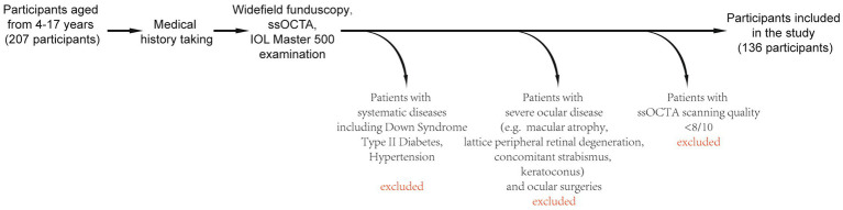 Figure 1
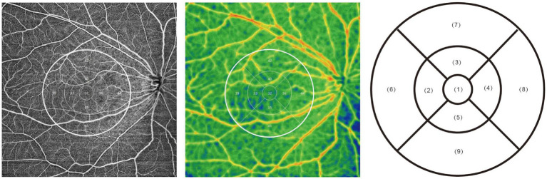 Figure 2
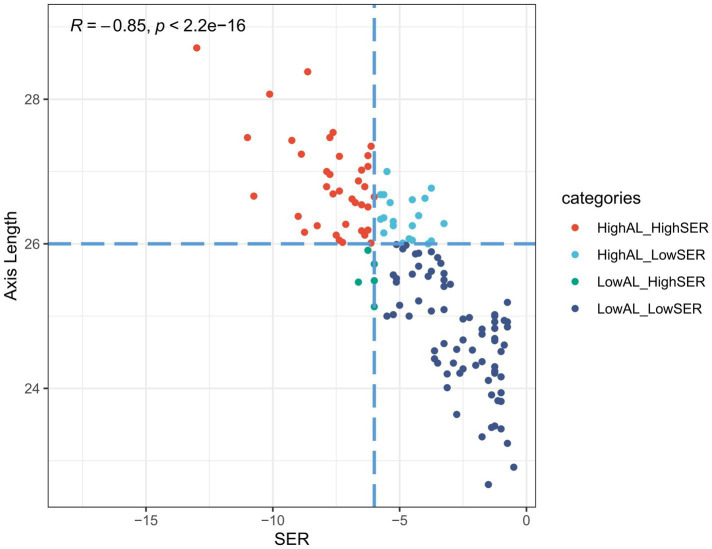 Figure 3
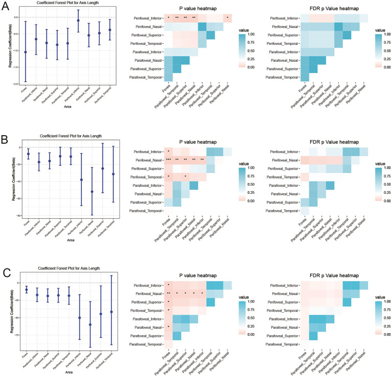 Figure 4
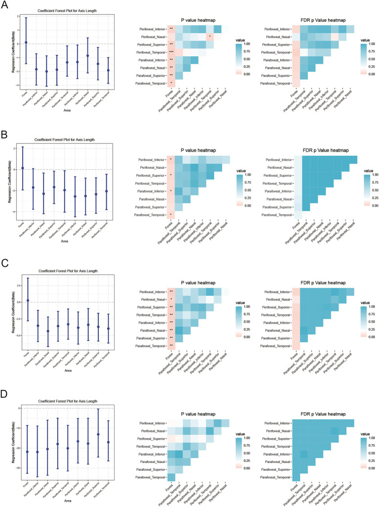 Figure 5
Peer Reviews
No public reviews on file for this paper yet. If you reviewed it on a platform where reviews are public (OpenReview, ICLR, NeurIPS, ICML), you can paste yours below so the community can read it here.
Videos
No videos yet. Explain this paper in a talk, walkthrough, or lecture? Add one.
Taxonomy
TopicsRetinal Diseases and Treatments · Retinal Imaging and Analysis · Glaucoma and retinal disorders