Making deep immunophenotyping accessible: the successful application of a guided 23-parameter mouse immunophenotyping panel package provided through a shared resource
Madison G. Isbell, Alex Wendling, Xinyan Pei, Amit Kumar, Padmanabhan Mannangatti, Bradley A. Krisanits, Stanley Cheatham, Marie Michenkova, Kirill Shumilov, Rachel G. Mendoza, Matthew E. Fernandez, Allyn Bryan, Thuy-An Nguyen, Lauren May, Swadesh K. Das, Victoria J. Findlay

TL;DR
This paper describes a 23-color mouse immunophenotyping panel that makes high-parameter flow cytometry accessible and cost-effective for researchers.
Contribution
A reproducible, modifiable, and cost-effective 23-parameter immunophenotyping panel is provided as a shared resource for mouse studies.
Findings
The panel is broadly applicable to multiple tissue types and streamlines workflow with optimized reagents and protocols.
The method reduces costs and lowers barriers for high-parameter flow cytometry use in research.
The panel serves as a complete resource for other institutions to adopt and adapt.
Abstract
This 23-color mouse immunophenotyping panel was designed and developed by the Virginia Commonwealth University’s (VCU) flow cytometry shared resource (FCSR) to easily bring new use to our high-parameter spectral cytometers. Our method is broadly applicable to multiple tissue types, is modifiable, and provides a reproducible, cost-effective option for utilizing high-parameter flow cytometry. To facilitate the mouse immunophenotyping panel, researchers can be provided with optimized reagents, a step-by-step staining protocol, instrument training, pre-run single-color controls, and acquisition and analysis templates to streamline the workflow. Data analysis is generally done with a traditional manual gating strategy, but t-stochastic neighbor embedding (tSNE) and uniform manifold approximation projection (uMAP) generation can be performed, as desired. In an FCSR, this panel requires only…
Genes, proteins, chemicals, diseases, species, mutations and cell lines named across the full text — each resolved to its canonical identifier and authoritative record.
Click any figure to enlarge with its caption.
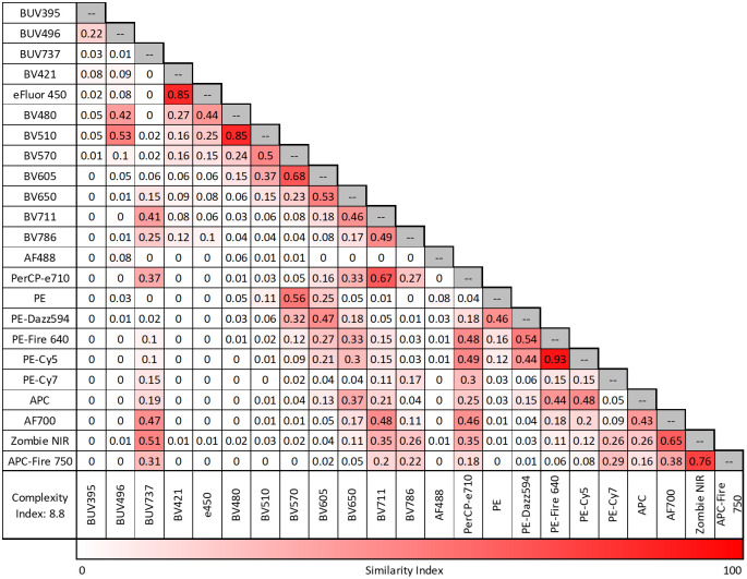 Figure 1
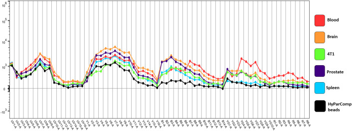 Figure 2
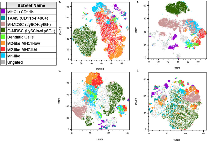 Figure 3
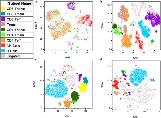 Figure 4
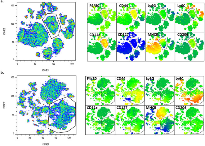 Figure 5
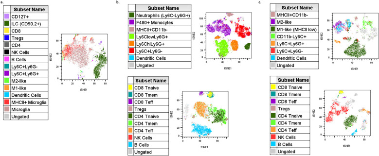 Figure 6
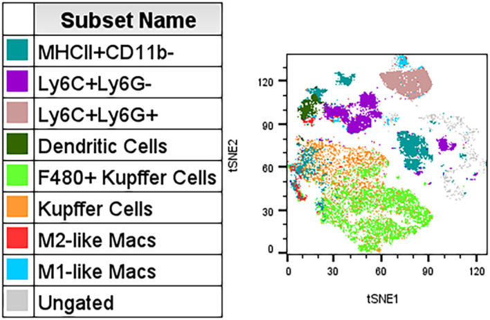 Figure 7
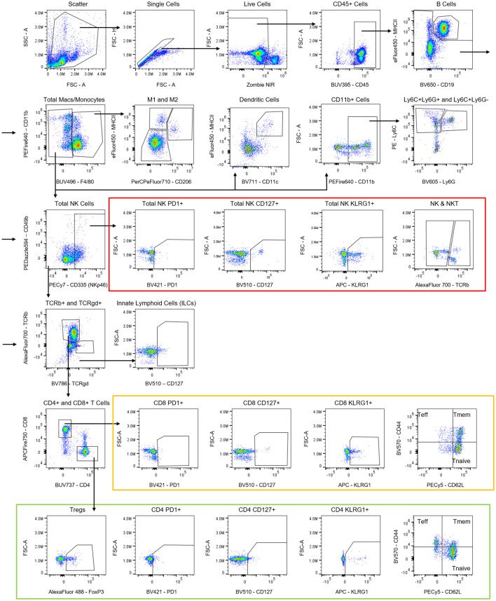 Figure 8
Peer Reviews
No public reviews on file for this paper yet. If you reviewed it on a platform where reviews are public (OpenReview, ICLR, NeurIPS, ICML), you can paste yours below so the community can read it here.
Videos
No videos yet. Explain this paper in a talk, walkthrough, or lecture? Add one.
Taxonomy
TopicsSingle-cell and spatial transcriptomics · Cell Image Analysis Techniques · Zebrafish Biomedical Research Applications