A novel multimodal imaging approach for working diagnosis of acute myocardial infarction with non-obstructive coronary arteries: a promising diagnostic strategy
Giovanni Taverna, Lisa Canton, Lorenza Zilio, Vincenzo Calabrese, Annagrazia Cecere, Maria Teresa Savo, Marco Previtero, Giulia Mattesi, Valeria Pergola, Stefano Da Pozzo, Simone Corradin, Angela Susana, Antonella Cecchetto, Anna Baritussio, Alberto Cipriani, Raffaella Motta

TL;DR
A new imaging method combining CT and MRI helps diagnose heart issues in patients with chest pain and clear arteries.
Contribution
A novel multimodal imaging protocol using CCTA and CMR improves acute diagnosis of MINOCA.
Findings
CCTA-CMR protocol distinguishes ischemic from non-ischemic myocardial injury in MINOCA patients.
Multimodal imaging reveals occult high-risk coronary plaques and enhances diagnostic accuracy.
Late iodine enhancement matches LGE patterns in MINOCA and myocarditis cases.
Abstract
Myocardial infarction with non-obstructive coronary arteries (MINOCA) demands prompt mechanistic clarification. Early integration of coronary CT angiography (CCTA) and cardiovascular magnetic resonance (CMR) can refine diagnosis during the acute phase. Twenty-one consecutive patients (41 ± 10 years; 71% men) presenting with troponin-positive chest pain and unobstructed coronaries underwent CCTA, delayed iodine-enhanced CT for late iodine enhancement (LIE), and CMR imaging within 14 days, with a mean interval of 5 days [interquartile range (IQR) 2–9] between both imaging modalities. CCTA assessed luminal stenosis and high-risk plaque; LIE mapped iodine retention; CMR evaluated myocardial edema and late gadolinium enhancement (LGE). Clinical, electrocardiographic, and laboratory data were collected. Eight patients were classified as MINOCA and 13 as acute myocarditis. Chest pain was…
Genes, proteins, chemicals, diseases, species, mutations and cell lines named across the full text — each resolved to its canonical identifier and authoritative record.
Click any figure to enlarge with its caption.
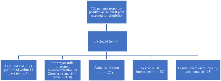 Figure 1
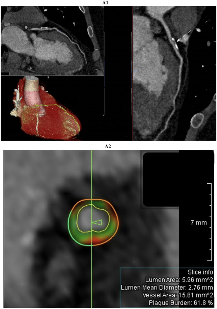 Figure 2
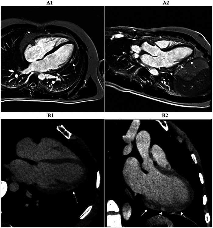 Figure 3
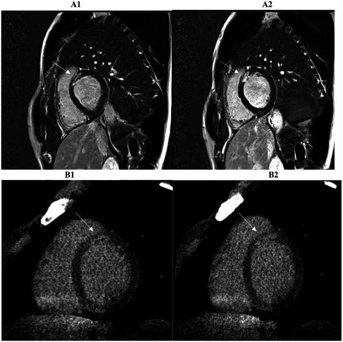 Figure 4
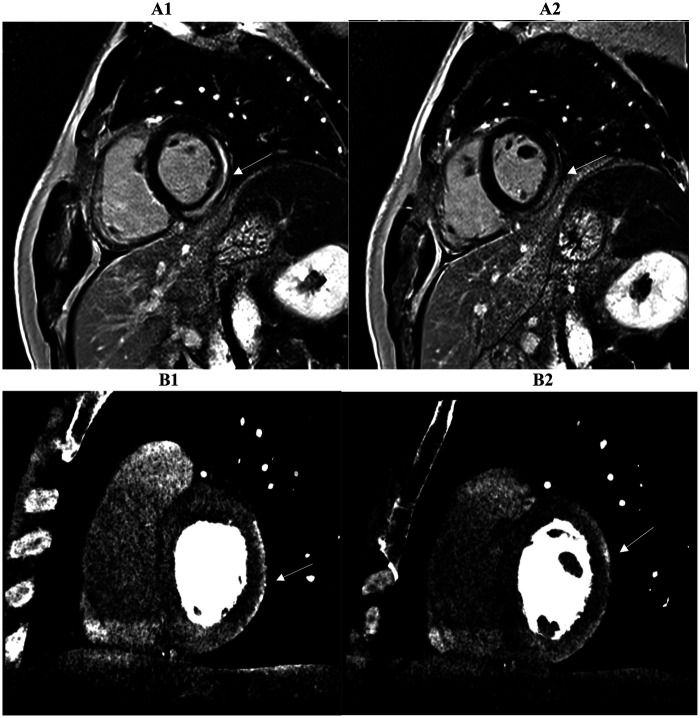 Figure 5
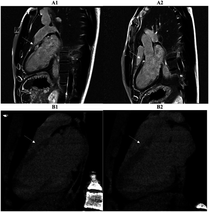 Figure 6
Peer Reviews
No public reviews on file for this paper yet. If you reviewed it on a platform where reviews are public (OpenReview, ICLR, NeurIPS, ICML), you can paste yours below so the community can read it here.
Videos
No videos yet. Explain this paper in a talk, walkthrough, or lecture? Add one.
Taxonomy
TopicsCardiac Imaging and Diagnostics · Acute Myocardial Infarction Research · Coronary Artery Anomalies