Novel use of cholangioscopy-assisted enteroscopy for foreign body removal from a narrow-ostium ileal diverticulum
Jing Guo, Yanbo Yu, Rui Ji, Jun Liu, Xiu-Li Zuo

Abstract
Genes, proteins, chemicals, diseases, species, mutations and cell lines named across the full text — each resolved to its canonical identifier and authoritative record.
Click any figure to enlarge with its caption.
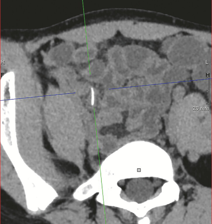 Fig. 1
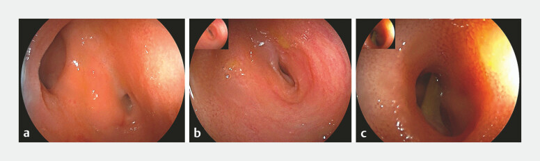 Fig. 2
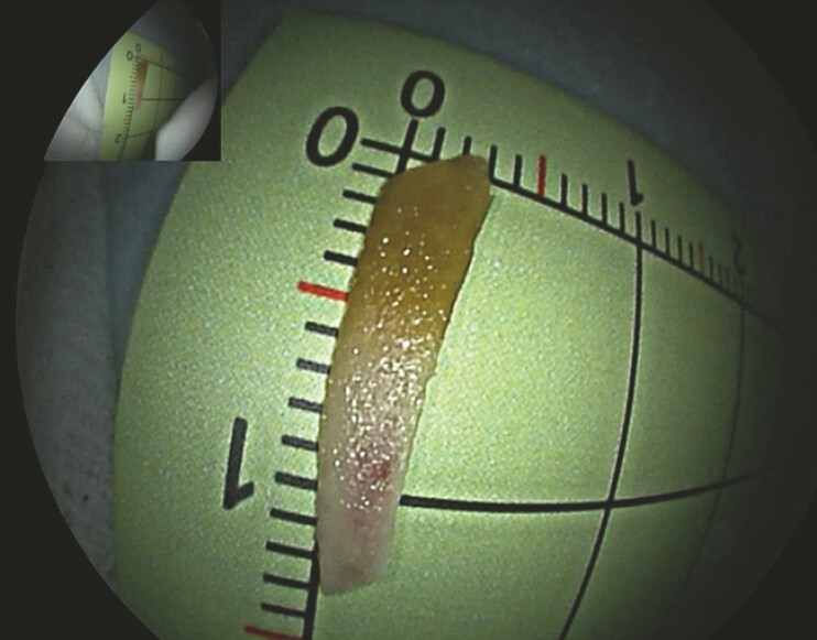 Fig. 3
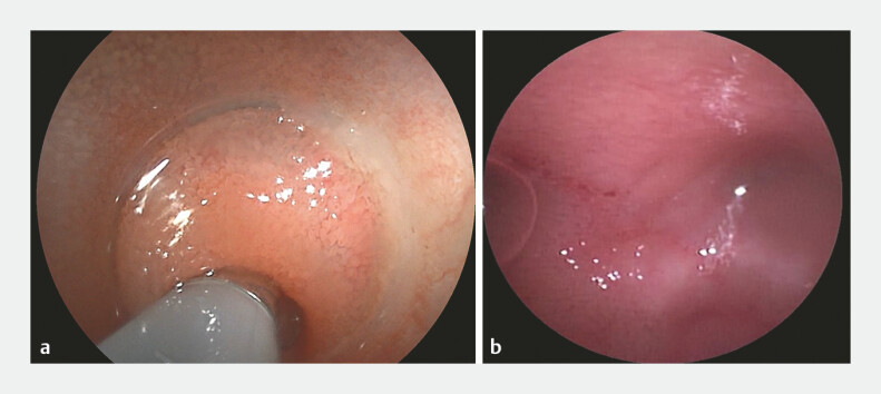 Fig. 4
Peer Reviews
No public reviews on file for this paper yet. If you reviewed it on a platform where reviews are public (OpenReview, ICLR, NeurIPS, ICML), you can paste yours below so the community can read it here.
Videos
No videos yet. Explain this paper in a talk, walkthrough, or lecture? Add one.
Taxonomy
TopicsForeign Body Medical Cases · Gallbladder and Bile Duct Disorders · Biliary and Gastrointestinal Fistulas
A 13-year-old girl was presented with a 20-day history of right-sided abdominal pain. She denied consuming any special foods. Computed tomography (CT) revealed a high-density focus in the right lower abdomen, suggestive of an intestinal foreign body ( Fig. 1 ). Given the high surgical risk and uncertain prognosis, double-balloon enteroscopy was performed via a retrograde approach. At the site 100 cm proximal to an ileocecal valve, a “double lumen” sign was observed ( Fig. 2 a ), comprising the intestinal lumen and a narrow-opening diverticulum ( Fig. 2 b ). Endoscopic visualization identified a rod-like foreign body embedded within the diverticulum ( Fig. 2 c ). A conical transparent cap was attached to the enteroscopy tip to facilitate retrieval. With the cap stabilized at the orifice, the foreign body was successfully retrieved using biopsy forceps, which confirmed a 1.4 cm chicken bone ( Fig. 3 ). Post-retrieval bleeding was noted; however, the narrow opening limited further visualization. For detailed assessment, a digital single-operator cholangioscope was inserted into the diverticulum cavity ( Fig. 4 a ), providing clear visualization of a short cavity with mucosal edema and minor blood clots. After irrigation, no residual foreign body, active bleeding, or perforation was confirmed. ( Fig. 4 b , Video 1 ).
A computed tomographic image showing a high-density shadow in the right lower quadrant of abdomen, indicating an intestinal foreign body.
An image from double-balloon enteroscopy: a the “double lumen” sign at the ileum about 100 cm beyond the ileocecal valve; b the narrow opening of the ileal diverticulum; c a bone foreign body in the diverticulum, and the inner wall of diverticulum cannot be observed.
Photograph of the extracted foreign object, which was found to be a 1.4-cm chicken bone.
Image of the digital single-operator cholangioscope: a Intubation of the diverticulum with the cholangioscope. b Observing the condition of the inner wall of the diverticulum.
Removal of a foreign body from an ileal narrow-opening diverticulum via enteroscopy assisted with a digital single-operator cholangioscope.Video 1
The extraction of sharp foreign bodies from the small intestine is technically challenging with enteroscopic techniques 1 . This difficulty is further exacerbated when the foreign body is lodged within a diverticulum featuring a narrow ostium, which precludes direct visualization. The cholangioscopy 2 can be navigated through these confined openings to effectively address this scenario. To the best of our knowledge, this is the first reported case in which a foreign body was successfully retrieved from an ileal diverticulum with a narrow ostium. Additionally, it marks the novel application of cholangioscopy in conjunction with double-balloon enteroscopy. This integrated approach provided a minimally invasive alternative, avoiding the need for surgery.
Endoscopy_UCTN_Code_TTT_1AP_2AD
The reference list from the paper itself. Each links out to its DOI / PubMed record.
- 1Pennazio M Rondonotti E Despott EJ Small-bowel capsule endoscopy and device-assisted enteroscopy for diagnosis and treatment of small-bowel disorders: European Society of Gastrointestinal Endoscopy (ESGE) Guideline-Update 2022 Endoscopy 202355589510.1055/a-1973-379636423618 · doi ↗ · pubmed ↗
- 2Dezheng Lin Mingli Su Yuping Su Digital single-operator cholangioscope-assisted endoscopic retrograde appendicitis therapy in the management of Crohnʼs disease with acute appendicitis Endoscopy 20245601 E 791E 79210.1055/a-2410-370239299285 PMC 11412757 · doi ↗ · pubmed ↗