3D Quantification of Viral Transduction Efficiency in Living Human Retinal Organoids
Teresa S. Rogler, Katja A. Salbaum, Achim T. Brinkop, Selina M. Sonntag, Rebecca James, Elijah R. Shelton, Alina Thielen, Roland Rose, Sabrina Babutzka, Thomas Klopstock, Stylianos Michalakis, Friedhelm Serwane

TL;DR
This paper introduces a new method to measure how well viruses deliver genes into living human retinal tissues using 3D imaging and machine learning.
Contribution
The novel contribution is a spatiotemporal quantification method for viral transduction in living retinal organoids using confocal imaging and deep learning.
Findings
A pipeline combining live imaging and deep learning enables 3D quantification of transduction efficiency in retinal organoids.
The method preserves spatial and temporal information, allowing time-dependent studies of gene delivery.
This approach can be extended to evaluate drug delivery in various biological systems.
Abstract
The development of therapeutics builds on testing their efficiency in vitro. To optimize gene therapies, for example, fluorescent reporters expressed by treated cells are typically utilized as readouts. Traditionally, their global fluorescence signal has been used as an estimate of transduction efficiency. However, analysis in individual cells within a living 3D tissue remains a challenge. Readout on a single‐cell level can be realized via fluorescence‐based flow cytometry at the cost of tissue dissociation and loss of spatial information. Complementary, spatial information is accessible via immunofluorescence of fixed samples. Both approaches impede time‐dependent studies on the delivery of the vector to the cells. Here, quantitative 3D characterization of viral transduction efficiencies in living retinal organoids is introduced. The approach combines quantification of gene delivery…
Genes, proteins, chemicals, diseases, species, mutations and cell lines named across the full text — each resolved to its canonical identifier and authoritative record.
Click any figure to enlarge with its caption.
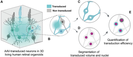 Figure 1
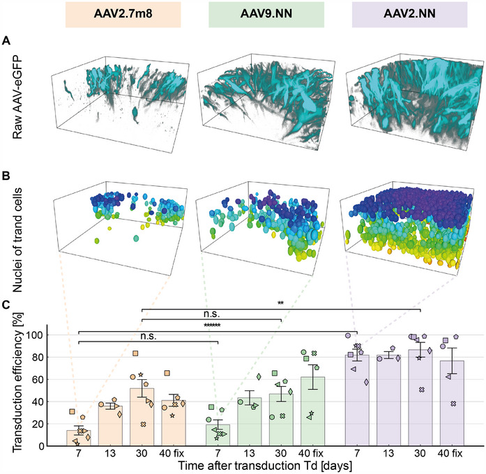 Figure 2
 Figure 3
 Figure 4
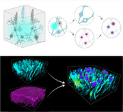 Figure 5
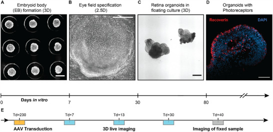 Figure 6
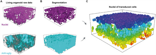 Figure 7
Peer Reviews
No public reviews on file for this paper yet. If you reviewed it on a platform where reviews are public (OpenReview, ICLR, NeurIPS, ICML), you can paste yours below so the community can read it here.
Videos
No videos yet. Explain this paper in a talk, walkthrough, or lecture? Add one.
Taxonomy
TopicsRetinal Development and Disorders · Virus-based gene therapy research · Advanced Fluorescence Microscopy Techniques