Enhanced preoperative planning in congenital polydactyly: superior assessment of MCP/MTP joint angular deformity with 3D-FS-FSPGR MRI compared to conventional radiography
Jie Li, Quan Yun, Yingyu Jia, Jiangtao Long, Qianqian Wang, Yuankai Yang, Shuming Xu

TL;DR
This study shows that 3D-FS-FSPGR MRI provides better visualization of joint deformities in children with polydactyly than x-rays, leading to more accurate surgical planning.
Contribution
The study demonstrates the clinical feasibility and superiority of 3D-FS-FSPGR MRI over conventional radiography for preoperative angular deformity assessment in congenital polydactyly.
Findings
3D-FS-FSPGR MRI offers superior visualization of cartilage and soft tissue compared to x-ray.
Significant discrepancies in joint angle measurements were observed between MRI and x-ray.
MRI enables more accurate and reliable quantification of angular deformities in polydactyly.
Abstract
Precise quantification of angular deformity at the metacarpophalangeal (MCP) or metatarsophalangeal (MTP) joint is paramount in congenital polydactyly surgery. It dictates the surgical center point and informs the necessity for corrective osteotomy. Conventional radiography (x-ray), while standard, suffers from inherent limitations in visualizing cartilage and soft tissue, compromising surgical planning. This study evaluates the clinical feasibility and superiority of a three-dimensional fat suppression rapid phase shifting gradient echo (3D-FS-FSPGR) magnetic resonance imaging (MRI) sequence for overcoming these limitations and achieving accurate preoperative angular assessment. Pediatric patients presenting with congenital polydactyly of the hands or feet underwent preoperative imaging with both standard x-ray and the 3D-FS-FSPGR MRI sequence. Evaluation focused on characterizing…
Genes, proteins, chemicals, diseases, species, mutations and cell lines named across the full text — each resolved to its canonical identifier and authoritative record.
Click any figure to enlarge with its caption.
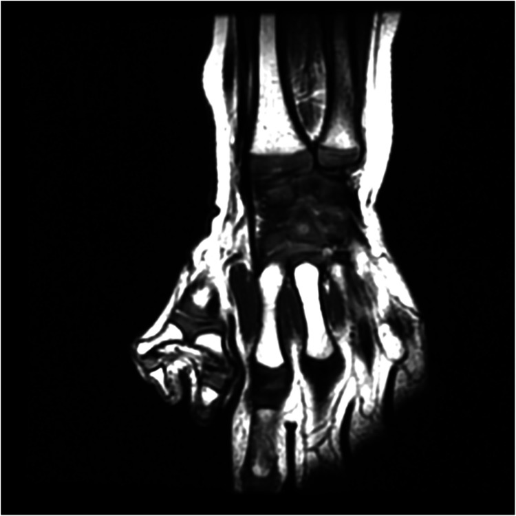 Figure 1
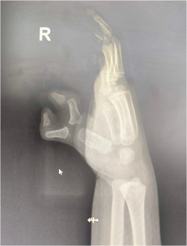 Figure 2
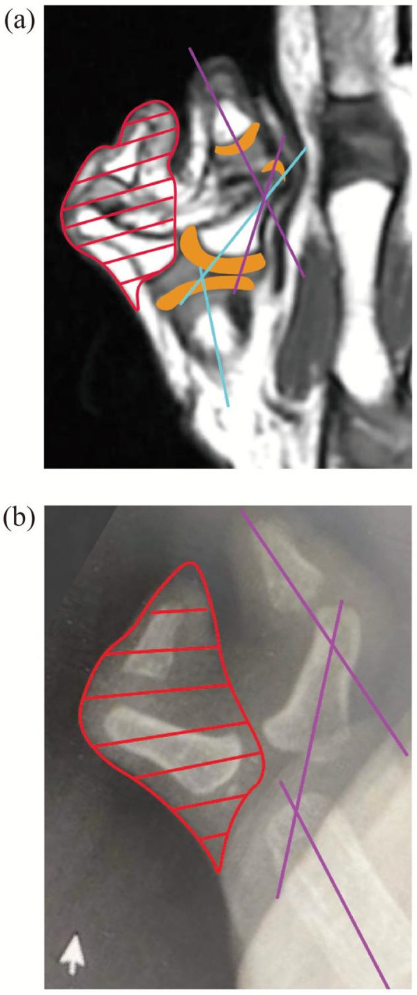 Figure 3
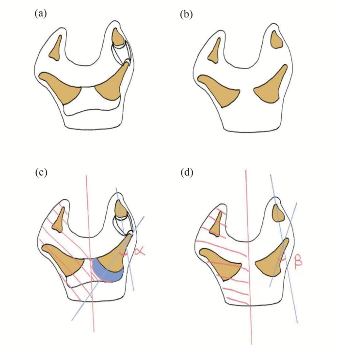 Figure 4
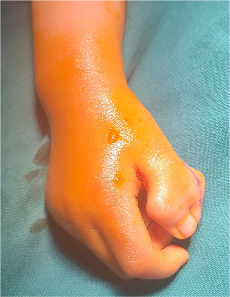 Figure 5
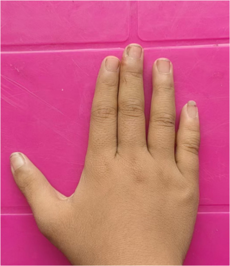 Figure 6
Peer Reviews
No public reviews on file for this paper yet. If you reviewed it on a platform where reviews are public (OpenReview, ICLR, NeurIPS, ICML), you can paste yours below so the community can read it here.
Videos
No videos yet. Explain this paper in a talk, walkthrough, or lecture? Add one.
Taxonomy
TopicsCongenital limb and hand anomalies · Dupuytren's Contracture and Treatments · Foot and Ankle Surgery