An 8-Year-Old Female with Giardiasis-Associated Henoch–Schönlein Purpura: A Case Report and Literature Review
Konstantinos Miliordos, Dimitrios Kapnisis, Christodoulos Chatzigrigoriadis, Emmanouil Koufopoulos, Sokratis Tsantiris, Aris Bertzouanis, Eirini Kostopoulou, Despoina Gkentzi

TL;DR
An 8-year-old girl with giardiasis developed Henoch–Schönlein purpura (HSP), a rare case highlighting the link between parasitic infections and this childhood vasculitis.
Contribution
This is the first reported case in Greece of HSP associated with giardiasis in a pediatric patient.
Findings
Giardiasis can trigger HSP in children, as shown by a confirmed case with stool antigen testing and elevated IgA levels.
The patient's symptoms improved with metronidazole, corticosteroids, and supportive care.
Pediatricians should consider parasitic infections like giardiasis as potential causes of HSP, especially with gastrointestinal symptoms.
Abstract
Background and Clinical Significance: Henoch–Schönlein purpura (HSP), also known as Immunoglobulin A (IgA) vasculitis (IgAV), is a common systemic vasculitis in children characterized by palpable purpura, abdominal pain, and joint and kidney involvement. While respiratory tract viral or bacterial infections are the most common causes of HSP, parasitic infections, such as giardiasis, are occasionally reported. Giardia lamblia is the most common parasite infecting humans and a major cause of infectious diarrhea, which can lead to post-infection complications. To our knowledge, this is the first report in Greece describing a pediatric patient with HSP secondary to giardiasis. A review of pediatric HSP cases caused by parasitic infections is also included. Case presentation: An 8-year-old girl presented with a purpuric rash, joint tenderness, severe abdominal pain, and bloody diarrhea,…
Genes, proteins, chemicals, diseases, species, mutations and cell lines named across the full text — each resolved to its canonical identifier and authoritative record.
Click any figure to enlarge with its caption.
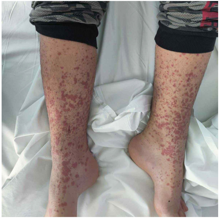 Figure 1
Peer Reviews
No public reviews on file for this paper yet. If you reviewed it on a platform where reviews are public (OpenReview, ICLR, NeurIPS, ICML), you can paste yours below so the community can read it here.
Videos
No videos yet. Explain this paper in a talk, walkthrough, or lecture? Add one.
Taxonomy
TopicsVasculitis and related conditions · Infective Endocarditis Diagnosis and Management · Whipple's Disease and Interleukins
1. Introduction and Clinical Significance
Henoch–Schönlein purpura (HSP), also known as IgA vasculitis (IgAV), is an IgA-mediated inflammatory disease affecting the small blood vessels, especially in the skin, joints, gastrointestinal tract, and kidneys [1,2,3,4,5,6,7]. HSP typically affects children, with an estimated incidence of 3 to 26.7 patients per 100,000 children, but this varies among nationalities [1,3,4,5,6,7,8,9]. HSP is typically preceded by an acute infectious illness, most commonly a respiratory tract infection, followed by infectious diarrhea, skin infections, and urinary tract infections; however, the exact mechanism remains unclear [1,4,5,6,9]. The clinical presentation typically manifests as a combination of palpable purpura, abdominal pain, arthralgia/arthritis, and glomerulonephritis [1,2,3,5,6,7,9,10]. HSP is usually a self-limiting condition requiring supportive care with fluids, analgesics, and management of the underlying cause [1,3,5,7]. Medical management with corticosteroids or other immunosuppressants, as well as surgical management, should only be considered for severe cases [1,3,5,7].
Giardia lamblia (also referred to as G. duodenalis or G. intestinalis) is a microscopic parasite that primarily affects the gastrointestinal tract and is transmitted through the fecal–oral route [11,12,13,14]. The clinical presentation of giardiasis is subclinical or appears as acute or chronic gastroenteritis; post-infectious sequelae, either in the gastrointestinal tract or other organs, may occur [12,13,14,15]. This research paper provides novel insights into the association between HSP and parasites. The case report focuses on an unusual case, which is the first case in Greece, of an 8-year-old female with HSP secondary to Giardia lamblia infection. It highlights that strong clinical suspicion, like in this case, will lead to rapid diagnosis and effective treatment. The literature review focuses on this rare and underreported post-infectious complication, giving robust evidence on the clinical features, diagnosis, and treatment of HSP secondary to parasitic infections [9,16,17,18,19,20,21,22,23,24,25].
2. Case Presentation
An 8-year-old female patient from the Roma ethnic group with an unremarkable medical history presented with a purpuric rash initially on the lower limbs and buttocks, which later spread to the abdomen and upper limbs (Figure 1). The knees, wrists, and distal malleolar regions were tender. She experienced severe, intermittent, diffuse abdominal pain unresponsive to acetaminophen for 2 days, followed by bloody diarrhea. There was no recent history of infections.
The patient’s medical and perinatal history was unremarkable. She was born full-term via cesarean section after an uneventful pregnancy. There was no history of chronic illness, hospitalizations, surgeries, or allergies. She had normal development.
The vital signs were within the normal range. Physical examination revealed a symmetrical purpuric and petechial rash on the buttocks and extremities. Joint tenderness was also present in the lower limbs during passive and active joint movements, as well as non-pitting edema in the knees and in the distal radial and malleolar regions. Abdominal examination revealed mild tenderness without rebound or guarding, normal bowel sounds, and a lack of organomegaly. The head and neck, chest, and neurological examination were unremarkable.
Cell blood count and comprehensive metabolic panel were within the normal range for her age. Serological tests for common viruses and rheumatic diseases were normal, except for an elevated IgA level [Table 1]. Urine analysis was normal, with no evidence of hematuria or proteinuria. Stool analysis was positive for Giardia lamblia antigen on two consecutive samples. Stool and upper respiratory cultures were negative. Abdominal ultrasound was unremarkable.
Upon admission, the patient received intravenous fluids, prednisolone (2 mg/kg/day), and omeprazole due to severe abdominal pain and bloody stools. After the positive antigen testing for Giardia lamblia, a 7-day regimen of metronidazole was administered. A gradual resolution of the patient’s symptoms was observed during the inpatient course. She was discharged after 5 days with a 2-week taper of prednisolone. Follow-up after 1 month revealed complete recovery and negative stool test results.
3. Discussion
3.1. Literature Review
Our literature search of PubMed and Scopus (June 2025) used the search term (IgA vasculitis OR HSP OR Henoch–Schönlein purpura) combined with each parasite name listed on the official website of the United States Centers for Disease Control and Prevention (CDC). We included English-language articles describing cases of HSP secondary to parasitic infections in pediatric patients. The exclusion criteria were non-English literature, patients older than 18 years, and HSP caused by non-parasitic agents. A total of eleven articles met the inclusion criteria, reporting 44 cases [Table 2]. Among these, 15 cases involved Giardia lamblia, 6 involved Trichomonas hominis, 5 involved Entamoeba histolytica, 2 involved Strongyloides stercoralis, 2 involved Ascaris lumbricoides, 1 involved Diploscapter coronata, 1 involved Sarcoptes scabiei, 1 involved Toxoplasma gondii, 1 involved Plasmodium falciparum, and 1 involved Toxocara canis. Of the 44 patients, 30 were males and 14 were females. Reported ages ranged from 2 to 17 years.
3.2. Correlation with Literature
Giardiasis is the most common parasitic disease and a major cause of infectious diarrhea in humans [11,12,13,14]. Waterborne (most common), foodborne, human, and animal transmissions are involved [11,12,13,14]. After ingestion, cysts convert into trophozoites in the small intestine, where they attach to the mucosa, causing immune-mediated disruption rather than invasion [11,12,13]. The trophozoites then detach and transform into cysts, which are excreted in the stool [11,12,13]. The involvement of the intestinal mucosa can be acute or chronic, leading to gastroenteritis, malabsorption, growth and cognitive impairments, and hypokalemic myopathy [11,12,13,14,15]. Persistent inflammation may cause post-infectious functional disorders like dyspepsia and irritable bowel syndrome [11,13,15]. Extra-intestinal complications, such as reactive arthritis, hypersensitivity reactions, chronic fatigue syndrome, and ocular disease, may result from systemic immune activation, even though Giardia lamblia cannot spread through the bloodstream [11,12,13,14,15]. Preventing similar cases of giardiasis is essential. It is of the utmost importance to use soap and water or antiseptics, especially during urination, defecation, and food preparation [12,14]. Purifying the water, preparing the food, avoiding overcrowding, and limiting animal contact are also important [12,13,14,15]. The application of these measures in day-care centers or swimming activities could limit the transmission of giardiasis in children and decrease the incidence of post-infectious complications [12,14]. HSP is a rarely reported immunological complication of Giardia lamblia infection in children, as shown in this review [16,17].
The standard diagnostic test is stool microscopy, while antigen or molecular testing are newer methods [14]. Repeating stool testing is often necessary [12]. Occasionally, diagnosis is established through a fluid aspirate or biopsy from the duodenum [14]. Oral use of an antimicrobial agent and a probiotic, e.g., Lactobacillus spp., along with rehydration and nutritional support, is essential [11,14,26,27]. First-line treatments include nitroimidazoles, such as metronidazole and tinidazole [11,12,14]. Alternative options include benzimidazoles (albendazole and mebendazole), nitazoxamide, paromomycin, acridine, furazolidone, and chloroquine [12,14]. Critical illness or immunosuppression are contraindications for treatment with probiotics [14,26]. In this case, antigen testing confirmed the diagnosis, followed by metronidazole therapy. Probiotics were avoided due to gastrointestinal bleeding and glucocorticoid treatment.
HSP is multi-organ vasculitis resulting from the deposition of IgA immune complexes (ICs) in the microcirculation and the most common vasculitis in pediatric patients [1,3,4,5,7,9]. Risk factors include an age of less than 10 years, male sex, white/Asian race, family history of HSP, and a past medical history of familial Mediterranean fever [1,4,5,6,7,8,16]. Infections are considered the most common trigger of HSP; this fact explains the decrease in incidence during summer [1,4,5,6,7,9,16]. Drugs, vaccines, insect bites, dietary allergens, and neoplasms have been reported as causes of HSP [4,6,10,16,23,24]. The most relevant pathogens are viral and bacterial pathogens [1,4,9,16]. Common examples include coronavirus, parvovirus B19, varicella, rubella, measles, mumps, coxsackie, adenovirus, parainfluenza, respiratory syncytial virus, hepatitis A, hepatitis B, human immunodeficiency virus, Streptococcus, Staphylococcus, Salmonella, Bartonella henselae, Mycoplasma pneumoniae, Helicobacter pylori, Salmonella, Clostridium, and tuberculosis [1,9,18,28]. Parasites, such as Giardia lamblia in our case, are responsible for a minority of pediatric HSP cases, as shown by the literature review [16,17,27,29].
The pathogenesis of HSP is incompletely understood; genetic background and environmental factors are considered essential, as in IgA nephropathy [1,4,5,7,10,16,24]. A plausible mechanism suggests that activated Th2 cells secrete interleukin-6 (IL-6), which induces the production of galactose-deficient IgA1 (Gd-IgA1) [4,5,24]. This leads to tissue deposition of ICs, activation of complement, and inflammatory damage [4,5,6,16,24]. Another theory suggests that molecular mimicry triggers the formation of IgA1 anti-endothelial cell antibodies (AECAs) [4,6]. AECAs target the β2 glycoprotein I receptor on endothelial cells; thus, IL-8 is released, leading to neutrophil infiltration of the microcirculation [4,6].
Possible mechanisms of extraintestinal complications in giardiasis include impairment of the intestinal barrier, dissemination of parasitic toxins or food allergens in the systemic circulation, and tissue deposition of antigens from enteric pathogens [12,13,15]. Notably, humoral immunity is activated after innate immunity to eliminate the parasite [11]. Although the pathogenesis of HSP in the context of giardiasis remains unclear, mucosal infections may trigger the differentiation of B-cells into IgA-producing plasma cells, which is the initial step in this post-infection sequela [27]. It could also be hypothesized that antigens leak into systemic circulation due to intestinal disruption, bind IgA molecules, and promote the formation of ICs [6,12,13,15]. Co-infections may be more likely to cause HSP than isolated giardiasis [27]. Establishing a direct causal relationship between giardiasis and HSP remains challenging due to the multifactorial etiology of IgA vasculitis. Genetic and environmental factors, including multiple pathogens, are all known to contribute to the disease [1,4,5,6,9,16]. Hence, the role of each pathogen is often unclear [1,4,5,6,9,16]. In addition, infectious diseases commonly precede HSP by days to weeks and may be asymptomatic [1,11,12,13,14,15]. Moreover, HSP affects the gastrointestinal tract, predisposing it to superimposed infections [18,20,21]. Thus, the presence of parasites may be either a precipitating factor or a coincidental finding. Further studies in basic research (in vitro experiments and animal models) and clinical research (cohort and case-control studies) could clarify the immunopathogenesis and the causal relationship between parasitic infections and HSP.
The clinical presentation involves multiple organs, but skin involvement is universal [1,2,4,5,7,9,16,18,27]. Rash is defined as palpable purpura and petechiae, mostly in the lower limbs and buttocks, while angioedema may be present [1,2,5,9,16,27]. Atypical cases of rash, such as those with plantar distribution, have been reported in the literature [2,16]. Non-deforming arthritis or arthralgia with knee and ankle predominance is present in most cases [1,2,5,9,16]. Diffuse and colicky abdominal pain with nausea, vomiting, and diarrhea is common [1,2,5,9,16]. Rare but severe complications include gastrointestinal bleeding, intussusception, appendicitis, and bowel perforation [1,2,5,9,16]. Renal involvement presents as hematuria, proteinuria, and hypertension; nephritic syndrome, nephrotic syndrome, and end-stage renal disease represent major complications of HSP [1,2,3,4,7,9,16]. Coagulopathy, testicular, respiratory, neurological, cardiac, and liver involvement are unusual [1,3,5,7,16]. The timing of the symptoms and signs of HSP is variable, and their complete development might occur after a few days or weeks [1,18]. In this case, the diagnosis was straightforward given the combination of rash, arthralgia, and abdominal pain.
The diagnosis of HSP is primarily clinical, although laboratory tests detect potential complications and exclude similar diseases [1,3,5,16,30]. A minimum diagnostic work-up includes cell blood count, coagulation profile, basic metabolic panel, urinalysis, fecal occult blood test, albumin, and blood/urine cultures [1,3,5,16,30]. Additional tests for the investigation of post-streptococcal complications and rheumatic diseases include antistreptolysin O (ASTO), antideoxyribonuclease B (anti-DNAse B), antinuclear antibodies (ANA), anti-double-strand deoxyribonucleic acid (anti-dsDNA), cellular antineutrophil cytoplasmic antibodies (c-ANCA), perinuclear antineutrophil cytoplasmic antibodies (p-ANCA), immunoglobulins, complement, and angiography [7,16]. Imaging (testicular ultrasound, abdominal ultrasound, neuroimaging) and endoscopy of the gastrointestinal or respiratory tract may be necessary for investigating organ-related complications [1,3,7,16,31]. Skin biopsy is indicated in equivocal cases and reveals the deposition of IgA and leukocytoclastic vasculitis [1,3,5,7,28]. Renal biopsy is necessary for the stratification of severe kidney involvement; it reveals findings similar to IgA nephropathy, such as mesangial proliferation and IgA deposition [1,3,7,16,28]. In this case, appropriate laboratory and imaging testing excluded similar diseases, and a skin biopsy was unnecessary given the classic clinical findings.
The differential diagnosis in a pediatric patient with rash and systemic symptoms requires careful consideration of similar diseases [30]. A normal CBC and the lack of lymphadenopathy rule out thrombocytopenic purpura and leukemic infiltration [5,16,30]. The absence of signs of sepsis, such as fever, hypotension, and coagulopathy, rules out meningococcemia [16,30,32,33,34]. Viral and rickettsial exanthematous diseases are important infectious causes of febrile rash that should be considered [30,33,35,36]. The lack of fever, valvular involvement, septic emboli, Janeway lesions, and Osler nodes is inconsistent with infective endocarditis [16]. The absence of recent streptococcal infection, carditis, migratory arthritis, chorea, and erythema marginatum excludes the diagnosis of acute rheumatic fever [16,35,36,37]. Inflammatory bowel disease is more likely to present during adolescence or young adulthood with chronic diarrhea, weight loss, axial spondylarthritis, erythema nodosum, or pyoderma gangrenosum [30]. Serum sickness and serum sickness-like reaction are associated with recent drug exposure and present with urticarial, erythema multiforme-like, or maculopapular rash [35,36,37,38]. However, gastrointestinal involvement is less prominent [36]. Juvenile idiopathic arthritis typically presents with a salmon-like rash, fever, and uveitis, requiring a minimum of six weeks for diagnosis [30,36,38]. The absence of fever, cervical lymphadenopathy, mucositis, palmoplantar rash, and cardiac involvement rules out Kawasaki disease [30,33,36,38]. Polyarteritis nodosa typically presents as a chronic illness in middle-aged male patients [5,16,30]. The lack of antineutrophil cytoplasmic antibodies (ANCA), respiratory, and renal involvement excludes the diagnosis of granulomatosis with polyangiitis [5,30].
HSP is considered self-limiting, with a favorable prognosis [1,3,5,7,9]. Treatment is primarily symptomatic with fluids and analgesics, such as acetaminophen or non-steroidal anti-inflammatory agents (NSAIDs) [1,3,5,7,9]. However, renal damage or gastrointestinal bleeding is a contraindication for the administration of NSAIDs [1,3,5,7,9]. Corticosteroids and non-steroidal immunosuppressants should be administered under specific circumstances for the management of severe complications [1,5,7,9]. It should be noted that immunosuppressants, such as corticosteroids, should be used cautiously because they can worsen the severity of an underlying parasitic infection [18,22,24]. Thus, the management of the underlying cause, such as infections, is essential [7,9,29]. However, the use of antibiotics should be justified, given their potential association with an exacerbation of HSP [21]. In this case, intravenous fluids and corticosteroids were preferred over NSAIDs due to gastrointestinal bleeding, while metronidazole successfully treated giardiasis.
4. Conclusions
Pediatricians should be aware of the rare association between Giardiasis and HSP, as presented in this research paper. Given the overlapping gastrointestinal symptoms, a strong clinical suspicion is essential to initiating the appropriate investigation. Early diagnosis and treatment of the underlying Giardiasis might accelerate the resolution of post-infectious sequelae.
The reference list from the paper itself. Each links out to its DOI / PubMed record.
- 1Reamy B.V. Servey J.T. Williams P.M. Henoch-Schönlein Purpura (Ig A Vasculitis): Rapid Evidence Review Am. Fam. Physician 202010222923332803924 · pubmed ↗
- 2Ozen S. Pistorio A. Iusan S.M. Bakkaloglu A. Herlin T. Brik R. Buoncompagni A. Lazar C. Bilge I. Uziel Y. EULAR/PRINTO/PRES criteria for Henoch–Schönlein purpura, childhood polyarteritis nodosa, childhood Wegener granulomatosis and childhood Takayasu arteritis: Ankara Part II: Final classification criteria Ann. Rheum. Dis.20106979880610.1136/ard.2009.11665720413568 · doi ↗ · pubmed ↗
- 3Ozen S. Marks S.D. Brogan P. Groot N. de Graeff N. Avcin T. Bader-Meunier B. Dolezalova P. Feldman B.M. Kone-Paut I. European consensus-based recommendations for diagnosis and treatment of immunoglobulin A vasculitis—The SHARE initiative Rheumatology 2019581607161610.1093/rheumatology/kez 04130879080 · doi ↗ · pubmed ↗
- 4Xu L. Li Y. Wu X. Ig A vasculitis update: Epidemiology, pathogenesis, and biomarkers Front. Immunol.20221392186410.3389/fimmu.2022.92186436263029 PMC 9574357 · doi ↗ · pubmed ↗
- 5Castañeda S. Quiroga-Colina P. Floranes P. Uriarte-Ecenarro M. Valero-Martínez C. Vicente-Rabaneda E.F. González-Gay M.A. Ig A Vasculitis (Henoch-Schönlein Purpura): An Update on Treatment J. Clin. Med.202413662110.3390/jcm 1321662139518760 PMC 11546386 · doi ↗ · pubmed ↗
- 6Song Y. Huang X. Yu G. Qiao J. Cheng J. Wu J. Chen J. Pathogenesis of Ig A Vasculitis: An Up-To-Date Review Front. Immunol.20211277161910.3389/fimmu.2021.77161934858429 PMC 8630619 · doi ↗ · pubmed ↗
- 7Abu-Zaid M.H. Salah S. Lotfy H.M. El Gaafary M. Abdulhady H. Tabra S.A.A. Salah H. Farag Y. Eissa M. Maher S.E. Consensus evidence-based recommendations for treat-to-target management of im-munoglobulin A vasculitis Ther. Adv. Musculoskelet. Dis.2021131759720 X 21105961010.1177/1759720 X 211059610 PMC 866987434917176 · doi ↗ · pubmed ↗
- 8Gardner-Medwin J.M. Dolezalova P. Cummins C. Southwood T.R. Incidence of Henoch-Schonlein purpura, Kawasaki disease, and rare vasculitides in children of different ethnic origins Lancet 20023601197120210.1016/S 0140-6736(02)11279-712401245 · doi ↗ · pubmed ↗