Use of Artificial Intelligence in Burn Assessment: A Scoping Review with a Large Language Model-Generated Decision Tree
Sebastian Holm, Fredrik Huss, Bahaman Nayyer, Johann Zdolsek

TL;DR
This paper reviews how AI, specifically CNNs and LLMs, can help assess burns by analyzing images and creating a decision tree, but highlights the need for better clinical validation.
Contribution
The novel use of a large language model to synthesize burn assessment literature into a decision tree is a new approach for organizing AI-based findings.
Findings
CNNs show high performance in burn TBSA and depth assessment, but results vary due to differing study designs and datasets.
A single study reported high accuracy for graft prediction, but this cannot be generalized due to data expansion.
LLM-generated decision trees can organize findings but are not intended as clinical decision tools.
Abstract
Background: Burns cause about 180,000 deaths annually and lead to substantial morbidity, especially in low- and middle-income countries. Clinical assessment of burn depth and TBSA relies on visual and bedside examination and remains subjective. Convolutional neural networks (CNNs) have been proposed to improve objectivity in image-based burn assessment, but clinical generalizability and acceptance remain uncertain. Aims: To map current evidence on CNN performance for burn TBSA, burn depth and treatment-related tasks and to explore whether a large language model (LLM) can organize extracted findings into a transparent, literature-derived orientation decision tree. Methods: We performed a scoping review following PRISMA-ScR. PubMed, Web of Science and Cochrane were searched on 5 April 2025. Eligible studies reported CNN analysis of 2D burn images and quantitative performance metrics. We…
Genes, proteins, chemicals, diseases, species, mutations and cell lines named across the full text — each resolved to its canonical identifier and authoritative record.
Click any figure to enlarge with its caption.
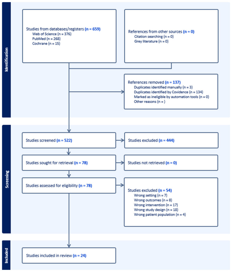 Figure 1
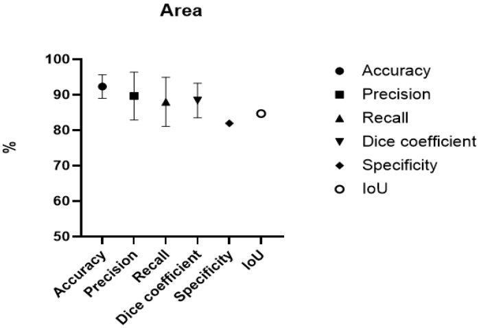 Figure 2
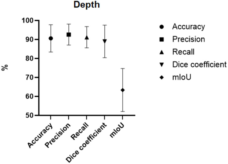 Figure 3
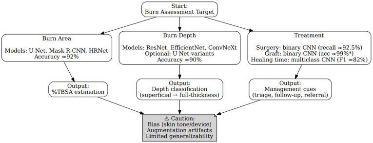 Figure 4
Peer Reviews
No public reviews on file for this paper yet. If you reviewed it on a platform where reviews are public (OpenReview, ICLR, NeurIPS, ICML), you can paste yours below so the community can read it here.
Videos
No videos yet. Explain this paper in a talk, walkthrough, or lecture? Add one.
Taxonomy
TopicsBurn Injury Management and Outcomes · Wound Healing and Treatments · Trauma and Emergency Care Studies
1. Introduction
Burns are a global health problem, causing about 180,000 deaths annually [1]. Most burn injuries occur in low- and middle-income countries (LMICs) [2]. Global overviews indicate that nearly two-thirds of burn injuries arise in the African and South-East Asia regions, and burns are among the leading causes of disability-adjusted life years (DALYs) lost in LMICs [1].
Clinical burn depth assessment relies on inspection and examination. Clinicians assess colour, capillary refill, pinprick or sensation and injury history or mechanism. This remains subjective. Compared with histopathology, specialist burn depth assessment is correct in about 70 to 80% of cases [3]. Accuracy is lower among less experienced clinicians [3]. In contrast, laypeople aided by assessment tools have been reported to estimate TBSA more accurately and consistently than burn professionals using the Rule of Palm and the Lund–Browder chart [4].
Recent advancements in artificial intelligence have led to systems designed to support burn care. Deep learning includes artificial neural networks used for image analysis, including convolutional neural networks (CNNs) [5]. In burn image analysis, CNNs are widely used. They learn patterns through layered processing of image data. They can identify features such as texture, shape and colour across large image sets. Different CNNs share common building blocks, but architectures differ in how layers are arranged and connected (e.g., depth, kernel sizes, skip connections, encoder–decoder designs).
Several CNN architectures have achieved strong performance in medical imaging, including AlexNet, VGG, U-Net, ResNet and DenseNet, as described in recent reviews of deep learning in medical imaging [6]. Performance depends heavily on data quality and training approach. Attention modules, such as Bidirectional Associative Memory (BAM), can be integrated into these backbones [7,8]. These architectures can be trained by datasets from scratch or by the use of pretrained models that use transfer learning, which is then further trained with a smaller training set [5]. One method to increase the medical images in the training set is by using Generative Adversarial Networks (GANs) that attempt to produce photorealistic images of burn wounds [9].
To evaluate CNN performance for TBSA and burn depth tasks, studies report different metrics. These include diagnostic accuracy, recall, precision, Dice coefficient (DC), specificity, Intersection over Union (IoU) and many more. Diagnostic accuracy refers to the ability of a CNN to correctly classify burn depth or delineate injured skin for TBSA estimation [10]. Recall and precision analyze the same thing but from different perspectives; recall reflects how many true positives are identified. Precision reflects how many predicted positives are true positives. Although recall and precision are quite similar, they are both important for the information they give. To optimize recall and precision, they are combined in the Dice coefficient, defined as 2 x precision x recall divided by precision plus recall (2PR/(P + R)). Specificity reflects the number of predicted negative cases that are true negatives. It thus describes the test’s ability to recognize healthy skin [10]. The Intersection over Union analyzes the test’s segmentation capabilities. Here, it is a measure of how well the CNN determines %TBSA. It is similar to the Dice coefficient in that both quantify overlap between predicted and ground-truth segmentation masks. In fact, for binary segmentation the Dice coefficient = 2 × IoU/(1 + IoU) [11].
The aim of this study was to evaluate the current evidence for artificial intelligence, specifically CNNs, in the assessment of burn TBSA, burn depth and treatment-related prediction tasks (e.g., surgery vs. non-surgery, graft vs. non-graft and healing-time category prediction) rather than treatment efficiency. In addition, to evaluate one large language model’s (LLM) (ChatGPT, Version 5.0) ability to generate clinical decision trees based on this scoping review.
2. Methods
This scoping review followed the PRISMA extension for scoping reviews (PRISMA-ScR) checklist to ensure transparent reporting and methodological rigor [12]. No protocol was pre-registered as PROSPERO does not currently accept scoping reviews [13]. In accordance with PRISMA-ScR guidance, we did not perform a formal risk-of-bias assessment using a single appraisal tool due to heterogeneity in designs, datasets and outcome metrics.
The following inclusion and exclusion criteria were used to gather studies.
Inclusion criteria:
- Written in English.
- Reported use of CNN for analysis of two-dimensional burn images.
- Reported quantitative model performance metrics.
- Exclusion criteria:
- Not focused on burn assessment tasks (TBSA, burn depth or treatment-related tasks).
- Did not use CNN-based methods.
- Did not report quantitative performance metrics.
- Conference abstracts, editorial, letters, protocols or non-peer-reviewed records.
A comprehensive search of three databases: PubMed, Cochrane Library (Wiley) and Web of Science (Clarivate) was conducted on 5 April 2025. Search strategies were designed for each database using Boolean operators and keywords related to “burns”, “artificial intelligence” and “convolutional neural networks”. Full search strategies are provided in Supplement S1. All retrieved records were imported into Covidence systematic review software (Veritas Health Innovation, Melbourne, Australia). Duplicates were removed automatically. Blinded title and abstract screening were performed in duplicate using Covidence by two reviewers. Disagreements were resolved through discussion with a third reviewer. The same procedure was used for the full text screening.
Data extraction was performed using a standardized template. Extracted information included author, paper, year, country, study design, artificial intelligence (AI) model, dataset, training set, outcome and statistical performance metrics. All quantitative summaries in this scoping review are descriptive aggregations of reported study-level values. They are not pooled estimates and should not be interpreted as meta-analytic performance.
LLM-Derived Decision Model
After data extraction, we compiled a structured, study-level summary containing task domain (TBSA/area, depth, treatment), imaging modality (RGB smartphone/clinical photography, LDPI or other), model family or architecture as reported by the authors and the published performance metrics already extracted into our tables. We provided this structured summary to ChatGPT version 5.0 (OpenAI, San Francisco, CA, USA) via the ChatGPT web application and instructed it to organize the extracted information into a one-page orientation decision tree. The prompt required the model to reproduce only the extracted models and extracted numeric values and not to generate new estimates, new statistics or new claims beyond the extracted dataset.
The LLM output was used to draft the layout and wording. Two consultant burn surgeons (J.Z., F.H.) independently reviewed the figure for clarity and plausibility and suggested wording changes. No patient data were used, and no clinical decisions were made. This figure is therefore a literature-synthesis visualization and not a trained or validated clinical tool. The full prompt and the consultant evaluation form are provided in Supplements S2 and S4. The model was queried using default system parameters in the ChatGPT web interface, without iterative prompt refinement or temperature adjustment, to minimize prompting bias.
The results were divided into three categories: burn area, burn depth and treatment, which included the AI’s capability to recommend appropriate treatment based on the assessments. We extracted performance metrics: accuracy, precision, recall, Dice coefficient, specificity and Intersection over Union (IoU) as outcome measures. For burn depth tasks, specificity was not used because the included studies did not report a meaningful negative class. The focus was on classifying depth within injured skin, not distinguishing burns from non-burns. When studies reported class-wise mean IoU (mIoU) instead of IoU, we recorded mIoU and summarized it separately rather than pooling it with IoU. For terminology, “Generated images” are synthetic samples created with generative models (e.g., GANs). “Expanded images” are augmented versions of real images (e.g., rotations, flips, crops, colour).
3. Results
In total, the database search yielded 659 studies. After removal of 137 duplicates, 522 studies remained. The title and abstract screening were followed by full-text screening. Non-eligible studies were discarded leaving 24 studies eligible for data extraction [11,14,15,16,17,18,19,20,21,22,23,24,25,26,27,28,29,30,31,32,33,34,35]. The screening process was documented and is presented in Figure 1.
3.1. Summary of Included Studies
Of the 24 eligible and thus included studies, 10 included information about burn area evaluation, 17 about burn depth assessment and 4 about treatment. Most studies originated from Asia (n = 15, 62.5%), followed by Europe (n = 7, 29.1%) and North America (n = 2, 8.3%). The distribution between comparative and experimental studies was even among the included studies. All studies, but one (2019), were published after the year 2020. The number of studies in “reported outcome” is greater than the total number of studies, as some studies included information on more than one outcome. Details of each study, including results, are found under the corresponding subheading depending on which of the three categories they focused on, see Table 1.
3.2. Burn Area
In total, 11,768 images were collected and analysed using CNNs across nine different studies to help assess the burn area [11,14,15,16,17,18,19,20,21]. Of these images, 184 were Laser Doppler Imaging (LDPI) images and 1200 images were acquired through expansion. LDPI captures physiologic perfusion-related signals and is not directly comparable with RGB photography. Reported performance values across LDPI and RGB studies should therefore not be interpreted as comparative. All ten studies used %TBSA to determine the area of the burn. Different metrics were reported: accuracy, precision, recall, Dice coefficient, specificity and IoU. Reported descriptive means and ranges in this review span multiple imaging modalities and study designs, and they are presented for overview purposes only. Across burn-area studies, the descriptive mean accuracy was 92.3% (SD 3.33) and the descriptive mean recall was 88.0% (SD 6.93). The descriptive mean precision was 89.6% (SD 6.72) and the descriptive mean Dice coefficient was 88.4% (SD 4.88). IoU was reported in a single study (84.6%). See Table 2 and Figure 2. (For a collection of results, see Supplement S3).
3.3. Burn Depth
A total of 15,781 images across 17 studies were collected and processed to help diagnose the burn depth [11,19,20,21,22,23,24,25,26,27,28,29,30,31,32,33,34]. Of these images, 184 were LDPI images and 9323 images were obtained through expansion of the existing dataset. LDPI captures physiologic perfusion-related signals and is not directly comparable with RGB photography. Reported performance values across LDPI and RGB studies should therefore not be interpreted as comparative. Similarly to burn area tasks, studies reported multiple metrics. Reported descriptive means and ranges in this review span multiple imaging modalities and study designs, and they are presented for overview purposes only. Accuracy was the most reported metric. The mean accuracy attained was 90.24 with a standard deviation of 7.61. Table 3 and Figure 3. (For a collection of results, see Supplement S3)
3.4. Treatment
The studies that focused on outcomes beyond TBSA and depth were grouped as treatment-related prediction tasks, including surgery vs. non-surgery, graft vs. non-graft and healing-time category prediction. In this category, a new column is introduced because of the different outcomes that can be incorporated in the broader meaning of treatment. Outcomes included in the treatment category are surgical vs. non-surgical, graft vs. non-graft and healing time with focus on depth and colour tracking (Table 4).
3.5. Large Language Model Evaluation
3.5.1. ChatGPT
Using the extracted evidence from the scoping review, ChatGPT produced a concise decision tree that begins with the user’s burn assessment target and branches to the model that was most used and their typical outputs (Figure 4). For burn area, segmentation backbones such as U-Net, Mask R-CNN and HRNet were linked to automated %TBSA estimation, with a descriptive mean accuracy ≈ 92% across the included studies. For burn depth, classifiers such as ResNet, EfficientNet and ConvNeXt (with optional U-Net variants for pixel-level maps) were linked to depth classification (superficial to full-thickness burn) with descriptive mean accuracy ≈ 90%. For treatment-oriented tasks, binary CNNs were linked to surgery vs. no surgery (recall ≈ 92.5% in one study) and graft vs. no graft (accuracy ≈ 99% in one study, largely augmentation-dependent), while multiclass CNNs were linked to healing-time categories (F1 ≈ 82%). The tree ends in a caution box summarizing cross-cutting limitations (dataset bias by skin tone/device, augmentation artefacts, limited external validation). These values and models reproduce our tabulated findings and do not constitute clinical recommendations.
Starting from the user’s burn assessment target, tree branches lead to three domains that show which model is most commonly used in our review and the typical outputs they produce. Burn area: representative segmentation backbones (U-Net, Mask R-CNN, HRNet) with outputs %TBSA estimation, descriptive mean accuracy ≈ 92% in our dataset. Burn depth: representative classifiers (ResNet, EfficientNet, ConvNeXt, optional U-Net variants for pixel-level depth maps), with output depth classification (superficial to full thickness) and descriptive mean accuracy ≈ 90%. Treatment: task-specific models with outputs, surgery vs. no surgery (binary CNN, recall ≈ 92.5%), graft vs. no graft (binary CNN, accuracy ≈ 99%*) and healing-time category (multiclass CNN, F1 ≈ 82%). The bottom caution box summarizes cross-cutting limitations observed across studies: dataset bias (e.g., skin tone/device), augmentation artefacts and limited external validation/generalizability. All models and performance values shown are taken from the included studies and our descriptive summaries; no new training or estimates were produced by ChatGPT, which only organized our extracted results.
3.5.2. Evaluation of the LLM Decision Tree
Two consultant burn surgeons (J.Z., F.H.) reviewed the decision tree using the three-item form in Supplement S2. Both reviewers answered yes to all items. This yields perfect agreement on clarity, relevance and perceived usefulness. It suggests the figure communicates the intended structure and outputs without ambiguity. It also indicates that outputs match clinical expectations for area, depth and treatment. Both surgeons also evaluated the tree—it could potentially guide model choice at the level of an orientation aid.
4. Discussion
This scoping review mapped how convolutional neural networks (CNNs) are used for burn assessment. We focused on TBSA, burn depth and treatment-related tasks. We summarized reported performance, common model types and limitations for clinical translation. Across the included studies, reported performance for area and depth tasks was often high and treatment-related prediction tasks showed early promise. High reported performance does not necessarily imply clinical robustness or real-world accuracy, since many studies used small datasets, heavy augmentation or synthetic image generation and internal-only validation without external testing. We refined these findings by entering them into an LLM-derived orientation tree to aid readers in matching targets to typical model approaches. However, since the studies used different datasets, labels, and validation methods, the results may not generalize well. This variability likely also explains why overlap metrics like mIoU look weaker than others. Several factors likely inflate reported performance and limit transfer to routine clinical use. Many studies relied on small single-centre datasets, heavy augmentation or synthetic image generation and internal test-sets drawn from the same distribution as the training data. These design choices increase the risk of overfitting and optimistic test performance. External validation across centres, devices and skin tones was uncommon. This limitation is especially important for treatment-oriented tasks where errors affect operative decisions. Single-study results with heavily expanded datasets, such as graft versus non-graft prediction, should therefore be interpreted cautiously.
Cirillo et al. 2019 [24] compared multiple CNN architectures for burn depth assessment and reported accuracy ranging from 77.79% to 81.66%, with ResNet-101 performing highest in their study. A year later, Khan et al. 2020 [31] managed to achieve an accuracy of 79.4% when assessing burn depth. He also stated that these were the best results compared to previous studies and results. More recent studies report higher values, but heterogeneity in datasets, labels and validation limits comparability. Most of the authors state that CNNs are ready to be implemented in healthcare and that, if the performance increase continues to follow the path of the last recent years into the coming years, there is potential for CNNs to act as a valuable tool in burn healthcare. Boissin et al. 2023 [17] compared performance across lighter and darker skin groups using images from Sweden and South Africa. They reported higher recall in darker skin than in lighter skin. This finding highlights two issues. First, model performance can differ across skin tone groups due to contrast, lighting, camera properties and label noise. Second, many datasets remain limited in skin tone diversity, which raises fairness and safety concerns if models are deployed without representative training and external validation. Future studies should report skin tone distribution, device characteristics and subgroup performance to support safer translation.
As described before, the mean Intersection over Union, mIoU, is considerably lower than every other tested metric. Various reasons could cause this, with the first and most prominent being the limited data on the metric. In the data extraction, only one study, Zhang et al. [26], was found to measure the mIoU. This makes the interpretation of the results harder and increases the risk for statistical distortion as mean values and confidence intervals cannot be calculated. The study in question also raised concerns about their limited dataset due to an absence of publicly available datasets and sole reliance on a single internal dataset. Another explanation for this could be the metric itself and its characteristics. Müller et al. [38] explain that the metric tends to be less forgiving. This indicates that a few low values are enough to decrease the mean by a higher amount than other metrics used for the same analysis, like the Dice coefficient.
Regarding the treatment aspect of burns, there are fewer studies and each of these addresses a different aspect of burn management. As for the ability to forecast whether surgery is needed or not, Boissin et al. [17] shows a high recall of 92.5%. Yadav et al. [33] focused on graft vs. non-graft reaching a 99.67% accuracy. Both studies’ outcomes may be explained by the usage of a simpler binary classification that allows for less error. Wang et al. [35] assessed the healing time by examining if the burn was shallow/superficial (0–10 days to healing), moderate (11–20 days), deep (more than 21 days) or needed skin graft, showing a recall of 82.34%. Another study addressing healing time, Ethier et al. [22], did so by assessing the colours of the burn where red was granulation/inflammation, yellow was slough, black was necrotic tissue and white was scabbing or epithelialization. Binary models work well for triage as a quick, accurate first-pass filter; multiclass models can support more detailed assessments in complex clinical situations. CNNs seem to have the potential to support the future of burn care; this is shown by their achieved results across multiple studies. Despite the positive results, limitations exist and improvements can be made. Large datasets can create new relevant ethical considerations, such as ensuring data protection with increasing digitalization. Still, studies need to increase and diversify the training of the models, so they are weighted and properly generalized in order not to suffer when applied to unseen data. This is especially important as many studies include the integration of mobile platforms. Analyzing and drawing conclusions is currently difficult due to methodological differences between the studies. To combat this hurdle, future studies should strive for more standardized methods and outcome parameters to make comparisons and larger-scale meta-analyses possible.
Limitations
This scoping review includes studies with substantial heterogeneity in datasets, skin tone distribution, imaging modality, augmentation strategy, model type and reported metrics. Many studies used different outcome definitions and validation approaches. For this reason, the descriptive means and SDs reported here do not represent pooled performance and do not support direct cross-study comparison. The values should be read as an overview of reported results rather than as benchmarks for model selection or clinical deployment. Some included studies used LDPI-based imaging or other physiologic modalities, while others used RGB photography from smartphones or clinical cameras. These modalities capture different signals and are not directly comparable. Mixing modality-specific results can bias the interpretation of performance values and clinical transfer. We therefore emphasize modality as a key limitation when interpreting reported metrics and generalizability.
This review has several limitations. Many of the included studies relied on small datasets, often expanded through augmentation or GAN-generated images, which may introduce unrealistic features and reduce external validity. The reference standards used for labelling, such as LDPI and clinical assessment, are themselves imperfect, which could reduce the reliability of the models. Conflicts of interest were inconsistently reported, reducing transparency. Finally, the restriction to only three databases for the search may have limited coverage. The decision tree generated by the LLM has several limitations. It inherits all biases and errors present in the underlying studies (e.g., small, single-centre datasets, variable labels, heterogeneous validation) and adds additional constraints. One of them is comparability; the performance numbers come from different tasks’ datasets, splits and metrics and are not directly comparable. Abstraction is another constraint, as the figure simplifies diverse architectures and training schemes into a decision tree. LLMs can misread and/or over-generalise summaries. The tree has also not been validated prospectively and must not guide patient care. The LLM only used values extracted in this review and obtained an independent review by two consultant burn surgeons; nonetheless, any recommendation by this tree should be interpreted cautiously and verified in prospective studies. In accordance with PRISMA-ScR guidance, a formal risk-of-bias assessment was not performed.
5. Conclusions
Current studies have proven the inclusion of AI and CNNs to be promising, achieving high results in diagnostic metrics for burn assessment. With more standardized methods and outcome parameters in the future to minimize their limitations, CNNs can function as a solution to many of the limitations of current methods of assessment. The clinical value of CNNs in burn care remains to be established through standardized datasets and external validation. The LLM-derived decision tree could potentially be used as an orientation aid, not as a clinical tool in burn assessment.
The reference list from the paper itself. Each links out to its DOI / PubMed record.
- 1World Health Organization (WHO) Burns Available online: https://www.who.int/news-room/fact-sheets/detail/burns(accessed on 25 September 2025)
- 2Rybarczyk M.M. Schafer J.M. Elm C.M. Sarvepalli S. Vaswani P.A. Balhara K.S. Carlson L.C. Jacquet G.A. A systematic review of burn injuries in low- and middle-income countries: Epidemiology in the WHO-defined African Region Afr. J. Emerg. Med. Rev. Afr. Med. D’urgence 20177303710.1016/j.afjem.2017.01.00630456103 PMC 6234151 · doi ↗ · pubmed ↗
- 3Phelan H.A. Holmes Iv J.H. Hickerson W.L. Cockerell C.J. Shupp J.W. Carter J.E. Use of 816 Consecutive Burn Wound Biopsies to Inform a Histologic Algorithm for Burn Depth Categorization J. Burn Care Res. Off. Publ. Am. Burn Assoc.2021421162116710.1093/jbcr/irab 15834387313 · doi ↗ · pubmed ↗
- 4Tocco-Tussardi I. Presman B. Huss F. Want Correct Percentage of TBSA Burned? Let a Layman Do the Assessment J. Burn Care Res. Off. Publ. Am. Burn Assoc.20183929530110.1097/BCR.000000000000061328877135 · doi ↗ · pubmed ↗
- 5Chen M.Y. Progress in the application of artificial intelligence in skin wound assessment and prediction of healing time Am. J. Transl. Res.2024162765277610.62347/MYHE 348839114681 PMC 11301465 · doi ↗ · pubmed ↗
- 6Litjens G. Kooi T. Bejnordi B.E. Setio A.A.A. Ciompi F. Ghafoorian M. van der Laak J.A.W.M. van Ginneken B. Sánchez C.I. A survey on deep learning in medical image analysis Med. Image Anal.201742608810.1016/j.media.2017.07.00528778026 · doi ↗ · pubmed ↗
- 7Sharma N. Ray A.K. Sharma S. Shukla K.K. Pradhan S. Aggarwal L.M. Segmentation and classification of medical images using texture-primitive features: Application of BAM-type artificial neural network J. Med. Phys.20083311912610.4103/0971-6203.4276319893702 PMC 2772042 · doi ↗ · pubmed ↗
- 8Mienye I.D. Swart T.G. Obaido G. Jordan M. Ilono P. Deep Convolutional Neural Networks in Medical Image Analysis: A Review Information 20251619510.3390/info 16030195 · doi ↗