Correction: The C-terminal of CASY-1/Calsyntenin regulates GABAergic synaptic transmission at the Caenorhabditis elegans neuromuscular junction
Shruti Thapliyal, Amruta Vasudevan, Yongming Dong, Jihong Bai, Sandhya P. Koushika, Kavita Babu

Abstract
Genes, proteins, chemicals, diseases, species, mutations and cell lines named across the full text — each resolved to its canonical identifier and authoritative record.
Click any figure to enlarge with its caption.
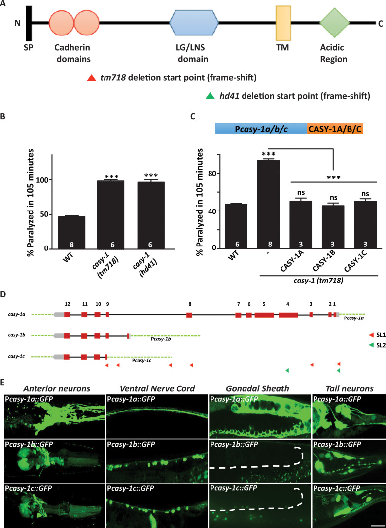 Figure 1
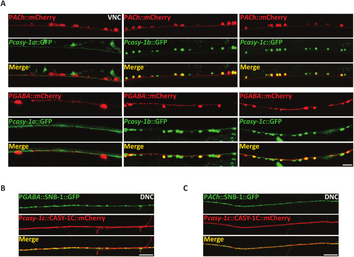 Figure 2
Peer Reviews
No public reviews on file for this paper yet. If you reviewed it on a platform where reviews are public (OpenReview, ICLR, NeurIPS, ICML), you can paste yours below so the community can read it here.
Videos
No videos yet. Explain this paper in a talk, walkthrough, or lecture? Add one.
Taxonomy
TopicsGenetics, Aging, and Longevity in Model Organisms · Cellular transport and secretion · Genetic Neurodegenerative Diseases
After publication of this article [1], errors were identified in Figs 1, 5, and S7.
In the originally published Figs 1E and 5A:
With this Correction, the first author provides revised Figs 1 and 5 including the correct image for the Pcasy-1c::GFP panel of Fig 1E from the original experiments.
Regarding the PGABA::mCherry panel in the first column of Fig 5A, the first author stated that at approximately z-stack position #7, the worm underwent a slight movement, creating the impression of two cell bodies in the maximum intensity projection (MIP) of all stacks. With this Correction, the PGABA::mCherry, Pcasy-1a::GFP and Merge panels are replaced with a reduced number of images of the same image stack from after the movement occurred, to remove this artifact present in the merged image used in the published panel. Based on the explanation and underlying image data provided, which is supportive of the revised figure, PLOS considers this concern resolved.
The first author confirms that the same images used to represent the Pcasy-1b::GFP and Pcasy-1c::GFP panels in the Ventral Nerve Cord column of Fig 1E are also used in the lower part of Fig 5A (now correctly aligned in the revised Fig 5) and that the images correctly represent the labeled conditions in each revised figure. This is also reflected in the updated figure legend for Fig 1.
In both S7A and S7C Figs, one additional WT sample was incorrectly included during the preparation of the bar graphs, resulting in incorrect raw WT data and subsequently also casy-1 data, which are normalized to WT data. The correct number of WT samples in S7A Fig is 17, and in S7C Fig is 13. The corresponding author confirmed that the changes in S7A and S7C Figs do not change the significance of the results presented. With this Correction, they provide the corrected bar graphs in a revised S7 Fig.
The first and corresponding authors have shared the original images underlying all panels in Figs 1, 2, 5, S2, and S7 in S1-5 Files and the individual-level quantitative data underlying the updated Figs 1 and S7 are provided in S6–7 Files. The first author stated that all other underlying data are available and can be provided upon request.
With this Correction the PLOS Genetics Editors inform readers that there was a potential competing interest between the authors and one or more people involved in peer review. After reviewing this matter PLOS concluded that the article’s publication is supported based on expert input that was not affected by the concern. We regret that the issues were not addressed prior to the article’s publication.
Supporting information
S7 Fig(A) Representative image for Mitochondrial marker (Punc-25::MITO::GFP) in GABAergic motor neurons in WT and casy-1 mutants.(B) Representative image for early endosomal marker [juIs198 (Punc-25:: YFP::RAB-5)] in GABAergic motor neurons in WT and casy-1 mutants. (C) Representative image for Lysosomal marker (Punc-25::CTNS-1::GFP) in GABAergic motor neurons of WT and casy-1 mutants. Scale bar, 10μm. The fluorescence intensity for mitochondrial and early endosomal marker are largely normal in casy-1 mutants, while lysosomal marker showed a subtle but significant decrease in fluorescent intensity when compared to WT animals. Quantification of fluorescent intensity is normalized to WT values. The number of animals analyzed for each genotype is indicated at the base of the bar graph. Quantified data are displayed as mean ± S.E.M. (*p < 0.05 using two-tailed Student’s t-test, “ns” indicates not significant in all Figures).(TIF)
S1 FileFig 1E underlying image data.This file includes the original images underlying Fig 1E. Each image file included in S1 File is generated from image stacks taken in the original experiments.(ZIP)
S2 FileFig 2A underlying image data.This file includes the original images underlying Fig 2A. Each image file included in S2 File is generated from image stacks taken in the original experiments.(ZIP)
S3 FileFig 5 underlying image data.This file includes the original images underlying Fig 5. Each image file included in S3 File is generated from image stacks taken in the original experiments.(ZIP)
S4 FileFig S2A underlying image data.This file includes the original images underlying S2A Fig. Each image file included in S4 File is generated from image stacks taken in the original experiments.(ZIP)
S5 FileFig S7 underlying image data.This file includes the original images underlying S7A-C Figs. Each image file included in S5 File is generated from image stacks taken in the original experiments.(ZIP)
S6 FileFig 1 individual-level quantitative data.This file includes the underlying individual-level quantitative data underlying Figs 1B and 1C.(XLSX)
S7 FileFig S7 individual-level quantitative data.This file includes the underlying individual-level quantitative data underlying S7A-C Figs, including the raw and normalized data.(XLSX)
The reference list from the paper itself. Each links out to its DOI / PubMed record.
- 1Thapliyal S, Vasudevan A, Dong Y, Bai J, Koushika SP, Babu K. The C-terminal of CASY-1/Calsyntenin regulates GAB Aergic synaptic transmission at the Caenorhabditis elegans neuromuscular junction. P Lo S Genet. 2018;14(3):e 1007263. doi: 10.1371/journal.pgen.1007263 29529030 PMC 5864096 · doi ↗ · pubmed ↗