Complex case of congenital pulmonary sequestration with successful “EXIT” procedure
Rūta Bernatavičienė, Gabija Pikturnaitė, Gilvydas Verkauskas, Arūnas Strumila

TL;DR
A rare case of fetal lung anomaly was successfully managed using a special delivery procedure to ensure the baby's survival.
Contribution
This paper presents a rare case of congenital pulmonary sequestration successfully managed with an EXIT procedure and thoracoscopy.
Findings
The EXIT procedure preserved fetal oxygenation during delivery, enabling neonatal breathing.
Elective thoracoscopy at six months confirmed pedicle torsion and successfully removed the sequestration.
Early diagnosis and multidisciplinary care improved outcomes for a life-threatening fetal lung condition.
Abstract
The ex-utero intrapartum treatment (EXIT) procedure preserves fetal oxygenation via the umbilical cord in urgent respiratory distress. We report a rare case of congenital extralobar pulmonary sequestration with pedicle torsion. A 22-year-old primigravida at 29 + 4 weeks presented with a fetal supradiaphragmatic mass, hydrothorax, and cardiac displacement; fetal magnetic resonance imaging (MRI) confirmed pulmonary sequestration. Rapid fetal deterioration led to the termination of the pregnancy and the application of ex-utero intrapartum treatment procedure during Cesarean section. Before cord clamping, intubation along with thoracocentesis enabled neonatal breathing. The 2140 g newborn required intensive care for respiratory distress, heart failure, and pulmonary hypertension and was discharged stable after 3 weeks. Elective thoracoscopy at the age of 6 months removed the sequestration,…
Genes, proteins, chemicals, diseases, species, mutations and cell lines named across the full text — each resolved to its canonical identifier and authoritative record.
Click any figure to enlarge with its caption.
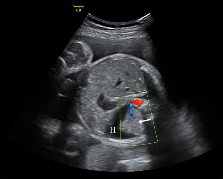 Figure 1
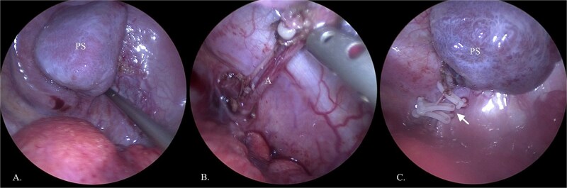 Figure 2
Peer Reviews
No public reviews on file for this paper yet. If you reviewed it on a platform where reviews are public (OpenReview, ICLR, NeurIPS, ICML), you can paste yours below so the community can read it here.
Videos
No videos yet. Explain this paper in a talk, walkthrough, or lecture? Add one.
Taxonomy
TopicsCongenital Diaphragmatic Hernia Studies · Congenital Anomalies and Fetal Surgery · Gastrointestinal disorders and treatments
Introduction
Pulmonary sequestration (PS) is a rare congenital lung defect of non-functional lung tissue with an autonomous arterial blood supply. Its prevalence among congenital lung malformations ranges from 0.15% to 6.40% [1]. The clinical significance largely hinges on size and location of lung sequestration. Minor cases often remain asymptomatic and are discovered incidentally in adulthood while severe forms can cause prenatal complications such as intrauterine growth restriction, preterm birth, or miscarriage, and postnatal issues including respiratory distress, heart failure, pulmonary hemorrhage, hydrops, and feeding difficulties [2]. When lung development rapidly deteriorates, the ex-utero intrapartum treatment (EXIT) procedure may sustain breathing before full delivery [3]. During EXIT, tracheostomy, mass excision, extracorporeal membrane oxygenation (ECMO) cannulation, thoracentesis, and others may be performed [4]. Although this procedure demands a considerable level of expertise, the overall survival rate for fetuses stands at 90% [5]. This report highlights the importance of the EXIT procedure in a rare case of congenital extralobar PS with pedicle torsion.
Case report
A 22-year-old primigravida was referred at 29 + 4 weeks due to abnormal fetal sonography showing a left supradiaphragmatic mass, hydrothorax, and rightward heart displacement. Magnetic resonance imaging (MRI) revealed a 2.6 × 1.9 × 1.6 cm supradiaphragmatic structure with an aortic arterial supply, confirming PS.
At 31 + 5 weeks, worsening hydrothorax prompted initiation of pulmonary maturation (Fig. 1). One week later, ultrasound showed bilateral hydrothorax compressing both lungs, dextrocardia, and hydrocele, indicating impending hydrops. A multidisciplinary team decided on EXIT to alleviate lung compression and facilitate breathing before cord clamping.
Fetal ultrasound with doppler. The arrow points to PS having a separate feeding branch from the aorta. Around sequestration the hydrothorax is present (H).
Under general anesthesia, a Cesarean section was performed. Before cord clamping, the neonate appeared hypotonic without reflexes or spontaneous activity. Immediate intubation, resuscitation and thoracentesis were done simultaneously. A total of 190 ml of fluid was drained from the left pleural cavity, and the umbilical cord was clamped after 3 min 30 s. Maternal recovery was uneventful.
The 2140 g neonate (Apgar 4/5) was shifted to intensive care for respiratory distress, heart failure, and pulmonary hypertension. Patient was discharged after 3 weeks being stable. At 6 months of age, PS was electively removed by thoracoscopy, confirming its torsion (Fig. 2). Recovery was uncomplicated, and the child remains healthy at 2 years of age. Genetic evaluation revealed isolated PS without associated anomalies.
Thoracoscopic removal of PS. (A) Visualized PS in the thoracic cavity. (B) Displayed feeding artery (A) of lung sequestration. (C) Feeding artery is clamped with clips (arrow).
Discussion
PS is classified as intralobar (within normal pleura) or extralobar (separate pleura and venous drainage). Extralobar lesions, as in this case, more often impair lung maturation and present prenatally [1, 2]. Additional lung structure having its autonomous arterial blood supply branching from the aorta is the pathognomonic sign for PS [6]. Antenatally, Doppler sonography and MRI can detect abnormal feeding vessel with 72% and 80% accuracy, respectively. If PS is undiagnosed prenatally, postnatal computed tomography is the primary diagnostic tool with 90% accuracy [7]. In this case, ultrasound revealed indirect signs—additional lung mass, hydrothorax, heart shift; however, the feeding artery was not visualized, prompting MRI for diagnostic clarification.
Perinatal management of intra or extralobar sequestration demands complex management in a well-equipped hospital capable of aggressive resuscitation and treatment for pulmonary hypoplasia due to high mortality risk from hypoxia. For instance, thoracoamniotic shunting is indicated up to 30 weeks of fetal age in those with hydrops [1, 8]. Other possible interventions to improve perinatal outcomes may be thoracocentesis, laser coagulation of the feeding vessel, sclerotherapy or combination of several treatment methods [1, 9]. Postnatal care may require high-frequency oscillatory ventilation or ECMO [10]. In our presented case, the lung development started to deteriorate rapidly at 31 gestation weeks allowing the initiation of pulmonary maturation followed with complete pregnancy termination and C-section 1 week later. This decision was made by a multidisciplinary team after agreeing to adopt lifesaving EXIT strategy to drainage neonate’s pleura before cutting off maternal oxygen support through umbilical cord.
Originally, the EXIT procedure was developed for removing tracheal clip in fetuses after intrapartum tracheal occlusion due to severe congenital diaphragmatic hernia [11]. Over time, EXIT indications expanded to manage various airway obstructions, most often due to congenital high airway obstruction syndrome or giant neck masses [12]. Although no strict guidelines exist, EXIT is recommended when airway obstruction or mass may cause immediate respiratory failure after birth. Hence, EXIT is used in cases requiring surgical airway access, ECMO cannulation, separation of conjoined twins, resection of large thoracic masses such as congenital pulmonary airway malformation or mediastinal/pericardial teratoma [5, 13, 14]. EXIT demands precise planning due to potential risks including maternal hemorrhage and hypotension, fetal bradyarrhythmias, inadequate uterine relaxation leading to reduced oxygen delivery [14, 15]. Risk mitigation requires a multidisciplinary team involving anesthesiologists, fetal medicine specialists, neonatologists, pediatric surgeons, and experienced operating room staff [14, 16]. All team members should participate in detailed preoperative planning for maternal and fetal anesthesia, uterine relaxation to maintain placental circulation; discuss anticipated interventions and potential challenges and complications. Accurate implementation ensures EXIT safety and favorable perinatal outcomes [17]. Our patient benefited from coordinated team management, resulting in a successful EXIT and complete recovery for both mother and infant.
Conclusion
PS can cause severe prenatal complications such as lung hypoplasia and bilateral hydrothorax. When pregnancy continuation is unfeasible, the EXIT procedure provides temporary placental oxygenation while securing neonatal airway and cardiopulmonary function. This case demonstrates that with multidisciplinary cooperation, meticulous planning, and advanced perinatal techniques, favorable outcomes are achievable even in complex congenital pulmonary anomalies.
The reference list from the paper itself. Each links out to its DOI / PubMed record.
- 1Chakraborty RK, Modi P, Sharma S. Pulmonary Sequestration. Treasure Island: Stat Pearls Publishing, 2023. http://www.ncbi.nlm.nih.gov/books/NBK 532314/ (14 August 2023, date last accessed).30335347 · pubmed ↗
- 2Pedata R, Palermo M, Maiello M, et al. Fetal lung lesions diagnosis: the crucial role of ultrasonography. J Prenat Med 2009;3:49–52.22439045 PMC 3279113 · pubmed ↗
- 3Sangaletti M, Garzon S, Raffaelli R, et al. The ex-utero intrapartum treatment (EXIT) procedure: case report of a multidisciplinary team approach. Acta Biomed 2021;92:e 2021142.33944827 10.23750/abm.v 92i S 1.9964 PMC 8142784 · doi ↗ · pubmed ↗
- 4Bence CM, Wagner AJ. Ex utero intrapartum treatment (EXIT) procedures. Semin Pediatr Surg 2019;28:150820.31451172 10.1053/j.sempedsurg.2019.07.003 · doi ↗ · pubmed ↗
- 5Spiers A, Legendre G, Biquard F, et al. Ex utero intrapartum technique (EXIT): indications, procedure methods and materno-fetal complications—a literature review. J Gynecol Obstet Hum Reprod 2022;51:102252.34638008 10.1016/j.jogoh.2021.102252 · doi ↗ · pubmed ↗
- 6Gabelloni M, Faggioni L, Accogli S, et al. Pulmonary sequestration: what the radiologist should know. Clin Imaging 2021;73:61–72.33310586 10.1016/j.clinimag.2020.11.040 · doi ↗ · pubmed ↗
- 7Mon RA, Johnson KN, Ladino-Torres M, et al. Diagnostic accuracy of imaging studies in congenital lung malformations. Arch Dis Child Fetal Neonatal Ed 2019;104:F 372–7.30049725 10.1136/archdischild-2018-314979 · doi ↗ · pubmed ↗
- 8Stoiber B, Moehrlen U, Kurmanavicius J, et al. Congenital lung lesion: prenatal course, therapy and predictors of perinatal outcome. Ultraschall Med 2017;38:158–65.26126151 10.1055/s-0035-1553261 · doi ↗ · pubmed ↗