Molecular identification of hookworm infection in humans, dogs and cats in Lao People’s Democratic Republic
Somphou Sayasone, Chomseng Norkhankham, Sysouphanh Many, Anousin Homsana, Tawin Inpankaew, Peter Odermatt

TL;DR
This study identifies hookworm species in humans, dogs, and cats in Laos, showing high infection rates and the need for combined human and animal control strategies.
Contribution
The study provides the first molecular identification of hookworm species in humans and animals in Laos, revealing interspecies transmission risks.
Findings
Hookworm infection prevalence was 24.1% in humans and 76.1% in dogs and cats combined, with higher rates in the southern region.
Necator americanus was the most common species in humans, while Ancylostoma caninum and N. americanus were prevalent in dogs and cats.
The study highlights the need for integrated One Health strategies to control zoonotic hookworm infections.
Abstract
Hookworm infection remains a major public health concern in the Lao People’s Democratic Republic (Lao PDR), where it is endemic nationwide and particularly prevalent in remote communities. However, information regarding species-specific identification is limited. This study aimed to determine the prevalence of hookworm infection and identify the infecting species among humans, dogs, and cats in both northern and southern regions of the Lao PDR. A cross-sectional study was conducted between May and July 2023 in Luangprabang Province (northern Lao PDR) and Champasak Province (southern Lao PDR). All residents aged 18 years and older who were present during the survey period and met the inclusion criteria were enrolled. In addition, dogs and cats owned by enrolled participants and/or their household members were included. Hookworm infection was detected by identifying eggs in preserved…
Genes, proteins, chemicals, diseases, species, mutations and cell lines named across the full text — each resolved to its canonical identifier and authoritative record.
Click any figure to enlarge with its caption.
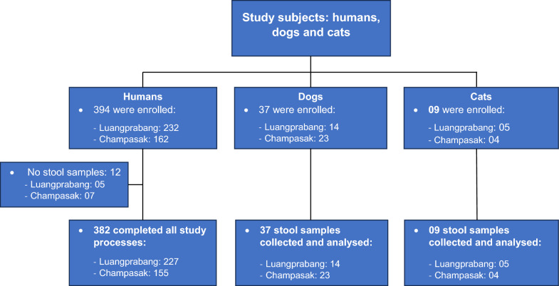 Figure 1
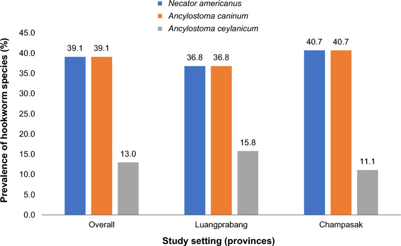 Figure 2
- —This study was funded by the Southeast Asia-Europe Joint Funding Scheme for Research and Innovation and the Swiss National Science Foundation
Peer Reviews
No public reviews on file for this paper yet. If you reviewed it on a platform where reviews are public (OpenReview, ICLR, NeurIPS, ICML), you can paste yours below so the community can read it here.
Videos
No videos yet. Explain this paper in a talk, walkthrough, or lecture? Add one.
Taxonomy
TopicsParasites and Host Interactions · Parasitic Diseases Research and Treatment · Dermatological diseases and infestations
Background
Hookworm, a nematode intestinal parasite, is a common soil-transmitted helminth infection that poses a significant health problem worldwide, particularly in rural areas of tropical and subtropical developing countries [1]. Infection with hookworm primarily occurs through skin penetration upon contact with the infective stage of the larvae in contaminated soil [2, 3]. It is one of the most prevalent chronic infections, affecting people in remote settings with poor hygiene, inadequate sanitation, and limited access to safe water [4]. Although hookworm infection rarely results in death, it can lead to intestinal blood loss, poor iron status, and potentially cause iron deficiency anemia [1]. Importantly, the infection may impair growth and cognitive development, particularly in children and women of reproductive age [1, 5].
Necator americanus and Ancylostoma duodenale are the predominant human hookworm species globally [2, 3]. Zoonotic hookworms, including Ancylostoma ceylanicum and Ancylostoma caninum, predominantly infect dogs and cats but have occasionally been reported in humans [6, 7]. The presence of these zoonotic hookworms complicates diagnosis. While parasitological methods widely employed in surveillance and control programs in developing countries can detect hookworm eggs in stool samples, species identification requires advanced molecular techniques, such as polymerase chain reaction (PCR) and sequencing [7, 8]. Consequently, species identification of hookworms is rarely performed in most resource-constrained areas.
In Lao People’s Democratic Republic (Lao PDR), hookworm infection remains a significant public health problem. The nationwide prevalence in adults is approximately 21.6%, with particularly high rates in remote areas where most inhabitants engage in subsistence agricultural farming with high contact with soil during farming activities and limited access to improved sanitation and safe water [9]. Although this study showed highly prevalent of hookworm infection, but its species distribution and cross-species infection were unidentified. Decades ago, few studies suggested that N. americanus is a predominant hookworm infection and A. ceylanicum is the zoonotic species identified among study participants [6]. However, there is a lack of recent assessments on species distribution. This study aims to identify hookworm species circulating in Luangprabang and Champasak provinces of the Lao PDR, where our previous national survey had documented a high prevalence of hookworm infection [9]. The results generate from this study would provide evidence to improve the national control programme.
Methods
Study area and population
We conducted a cross-sectional study between May and July 2023 to identify hookworm species circulating in Luangprabang and Champasak provinces of the Lao PDR. These provinces were selected based on findings from our previous national survey, which had documented a high prevalence of hookworm infection [9]. Luangprabang is the largest province in northern Lao PDR. It comprises 12 districts and has a population of 431,889 [10]. Our study was conducted in the Nambak district, approximately 100 km north of Luangprabang city (geocoordinates: 20.62° N, 102.47° E). Champasak province is the largest province in the southern part of Lao PDR. It encompasses 10 districts and has a total population of 694,023 [10]. Our fieldwork was conducted in the Batiengchaleunsouk district, which is located approximately 10 km from Pakse city (geocoordinates: 15.25° N, 105.95° E).
Sampling procedure
From each district, two villages known to have a high prevalence of hookworm (two villages in Nambak District, Luangprabang Province, and two villages in Batiengchaleunsouk District, Champasak Province), were selected as study villages [9]. We enrolled all adults aged 18 and older who were present in the selected villages on the survey day to our study. Only participants who agreed to sign an informed consent form were enrolled. Enrolled participants provided two stool samples for parasitological examination. Additionally, all dogs and cats owned by the study participants or their household members were included in the study for parasitological assessment.
Sample size calculation
The sample size calculation for this study followed the formula: \documentclass[12pt]{minimal} \usepackage{amsmath} \usepackage{wasysym} \usepackage{amsfonts} \usepackage{amssymb} \usepackage{amsbsy} \usepackage{mathrsfs} \usepackage{upgreek} \setlength{\oddsidemargin}{-69pt} \begin{document}$$\mathrm{N}=\frac{P\left(P-1\right){Z}^{2}}{{E}^{2}}$$\end{document} , with the components: (i) P (prevalence), the prevalence was 21%, as determined from our previous study [9], (ii) Z (level of confidence) set at 95% and E (margin of error) was 0.05. Based on these parameters, the initial sample size required was 255 individuals. To account for potential dropouts or non-compliance during the study, we added 20%. Consequently, a total sample size of 306 participants was targeted.
Field and laboratory procedures
The field team selected a temporary research station as a working place in each village; usually a temple, village office, or school. A meeting was organized with village authorities and villagers to explain the objective and plan the field activities. The villagers who met the study criteria and were willing to participate in the study were invited to register. Later, the research team assigned a unique identity number to each registered villager and arranged an appointment with her/him on the following day for the written informed consent and enrolment process. Each study participant received a pre-labelled container (30 ml) with a unique ID. After signing an informed consent form, a laboratory technician provided clear instructions on how to collect the stool sample, where to return it, and when. For each stool sample, we aliquoted 2 g into a 15 ml tube containing 10 ml of sodium acetate, acetic acid, and formalin (SAF) solution for parasitological analysis. Additionally, 1 g of the sample was fixed in 70% ethanol for polymerase chain reaction (PCR) analysis.
In parallel with human sample collection, each dog and cat owned by a study participant or a member of their household was confined in an individual cage, clearly labelled with a unique identification number. The dog and/or cat was given a gel Unison enema (Sodium chloride 15%) and left it in the cage until it defecated. A sample of 2 g was collected and fixed in SAF for parasitological analysis, and 1 g was fixed in 70% ethanol for PCR analysis.
The samples fixed in SAF solution were transported to the laboratory at the Lao Tropical and Public Health Institute (Lao TPHI) for parasitological analysis using the formalin-ethyl-acetate concentration technique (FECT) [11, 12]. All eggs of parasites detected under the light microscope were identified and recorded separately.
Samples fixed in 70% ethanol were sent to the Department of Parasitology at the Faculty of Veterinary Medicine, Kasetsart University, Bangkok, Thailand, for identification of the hookworm species using PCR.
DNA extraction
The Genomic DNA from the human and animal stool samples was extracted directly from 200 mg stool samples and Glass Beads X using a commercial extraction kit, E.Z.N.A® Stool DNA kit (Omega Bio-tek, Inc., Norcross, Georgia, USA), following the manufacturer's instructions. The final eluted volume of 100 µl was stored at − 20 °C until it was analysed by conventional polymerase chain reaction.
Molecular characterization
In humans
All 92 samples confirmed as hookworm-positive by microscopic analysis using FECT were subjected to molecular analysis. A subset of 35 microscopically negative samples (10% per province) was included alongside the confirmed hookworm-positive samples to assess potential false-negative results by microscopy, particularly in cases of low-intensity infections. Genomic DNA was extracted from all samples and screened for Necator americanus and Ancylostoma spp. infections using polymerase chain reaction. This method amplified a fragment of 380–485 bp from the internal transcribed spacer (ITS)-1, 5.8S, and ITS-2 regions, as described by Traub et al., 2008 (Table 1) [8]. The suspected N. americanus (485 bp) (Figure S1) and Ancylostoma spp. (380 bp) (Figure S2) band were cut and purified using the E.Z.N.A® Gel Extraction kit (Omega Bio-tek, Inc., Norcross, Georgia, USA), following the manufacturer’s instructions. The purified samples were then sent to Macrogen® in Gangnam-gu, Seoul, Republic of Korea, for sequencing. The sequences were analysed for similarity using the Basic Local Alignment Search Tool (BLAST) (https://blast.ncbi.nlm.nih.gov/Blast.cgi).Table 1. Primer used for PCRs for the characterization of hookworms in humans and domestic animals (dogs and cats)Tested speciesPrimersParasiteProduct (bp)ReferencesHumansRTHW1FRTHW1RNecator americanus**Ancylostoma spp.485380Traub et al. [13]Dogs/CatsRTGHF1RTABCR1Ancylostoma caninum**Ancylostoma ceylanicum545Traub et al. [8], Palmer et al. [14]; Inpankaew et al. [7]Dogs/CatsRTGHF1 RTAYR1Ancylostoma braziliense673Traub et al. [8], Palmer et al. [14]PCR polymerase chain reaction
In dogs and cats
All 46 stool samples collected from dogs and cats were molecularly analysed using diagnostic PCR-restriction fragment length polymorphism (RFLP) to characterize hookworm species, following previously described protocols [7]. Primers sets listed in Table 1 were used to amplify a region of the internal transcribed spacer (ITS)-1, 5.8S, and ITS-2. The amplified PCR products of RTGHF1–RTABCR1 were digested with RsaI to differentiate Ancylostoma tubaeforme from A. ceylanicum and A. caninum. HinF1 was used to distinguish A. caninum from A. ceylanicum. RFLP profiles obtained from each sample were compared with established reference patterns for each hookworm species [13, 14].
Representative sequences from both humans and animal samples were deposited in GenBank under accession numbers PX497935–PX497937 (A. ceylanicum), and PX497932-PX497934 (A. caninum).
Data management and analysis
Data was digitally collected using tablets. The questionnaire was developed in the CommCare server (www.commcarehq.org) to capture demographic information from study participants, including age, sex, gender, ethnicity, educational level, occupation, availability of a household latrine and ownership of pets (dogs or cats). The Commcare ODK application was installed on the tablet for daily field data collection. Collected data were synchronized to the CommCare server and downloaded into an Excel spreadsheet for consistency and completeness checks. The double-checked data was transferred to STATA, version 14 (Stata Corporation, College Station, TX, USA) for analysis. Frequencies described hookworm prevalence in humans, dogs, and cats. The mean summarized the intensity of hookworm infection, presented as eggs per gram of stool (EPG). Associations between infection and demographic data were analysed using Chi-squared tests. Wilcoxon rank-sum and Kruskal–Wallis tests were used to compare means where appropriate. P-value of less than 0.05 was considered statistically significant.
Results
Study subjects
In total, 394 villagers, 37 dogs and 9 cats were initial enrolled in the study. Of these, 12 villagers failed to submit stool samples for parasitological analysis (5 from Luangprabang and 7 from Champasak). Consequently, 382 villagers (Luangprabang: 227; Champasak: 162), 37 dogs (Luangprabang: 14; Champasak: 23), and 9 cats (Luangprabang: 5; Champasak: 4) successfully completed all study procedures and were included in the final data analysis (Fig. 1). Among the 382 villagers, gender distribution was comparable between Luangprabang and Champasak (P = 0.597). The median age was slightly higher in Luangprabang (55 years) than in Champasak (51 years). The age group analysis showed a statistically significant difference (P = 0.049), with younger participant profile in Champasak. Ethnic composition differed significantly between the two provinces (P < 0.001): Lao-Tai participants predominated in Luangprabang (46.3%), whereas ethnic minorities constituted the majority in Champasak (88.4%). Access to sanitation facilities also differed significantly (P = 0.006), with nearly universal latrine coverage in Luangprabang (99.6%) compared to lower access in Champasak (95.5%) (Table 2).Fig. 1. Study subjects (humans, dogs and cats) enrolled in the study by provinceTable 2Sociodemographic characteristics of 382 study participants, stratified by provinceVariablesOveralln = 382, % (n)Luangprabangn = 227, % (n)Champasakn = 155, % (n) \documentclass[12pt]{minimal} \usepackage{amsmath} \usepackage{wasysym} \usepackage{amsfonts} \usepackage{amssymb} \usepackage{amsbsy} \usepackage{mathrsfs} \usepackage{upgreek} \setlength{\oddsidemargin}{-69pt} \begin{document}$${x}^{2}$$\end{document} P-valueGender Male40.3 (154)41.4 (94)38.7 (60) Female59.7 (228)58.6 (133)61.3 (95)0.280.597Age in year Median age (IQR)54 (40–60)55 (43–60)51 (36–60)Age group (years) 18–3517.3 (66)13.2 (30)23.2 (36) 36–4924.1 (92)23.8 (54)24.5 (38) 50–5920.9 (80)23.8 (54)17.8 (26) ≥ 6037.7 (144)39.2 (89)3.5 (55)7.860.049Ethnicity Lao-Tai31.4 (120)46.3 (101)11.6 (19) Minorities68.6 (262)53.7 (117)88.4 (145)44.42 < 0.001Education level Illiterate16.5 (63)14.2 (31)19.5 (32) Primary school45.3 (173)64.8 (102)43.3 (71) Secondary/high school33.5 (128)14.4 (75)32.3 (53) College or higher4.7 (18)4.6 (10)4.9 (8)1.130.771Occupation Employees17.8 (68)16.1 (25)18.9 (43) Farmers82.2 (314)83.9 (130)81.1 (184)0.490.480Having pets at home No63.9 (244)49.7 (77)73.6 (167) Yes36.1 (138)50.3 (78)26.4 (60)22.78 < 0.001Latrine at home No2.1 (8)0.4 (1)4.5 (7) Yes97.9 (374)99.6 (226)95.5 (148)7.460.006^*^ *P-*value was obtained from Chi-square test; *P-*value < 0.05 was counted as statistical significance
Intestinal parasitic detection in FECT
Among the 382 villagers, analysis of preserved stool samples using the formalin-ether concentration technique (FECT) revealed an overall prevalence of intestinal helminth infections of 36.6%. Hookworm was the most prevalent parasite, detected in 24.1% of samples, followed by Strongyloides stercoralis (10.7%) and Opisthorchis viverrini (6.0%). The prevalence of these intestinal parasites was significantly higher in Campasak compared to Laungprabang: hookworm (36.6% vs 16.5%, P < 0.001), S. stercoralis (16.5% vs 6.4%, P = 0.002) and O. viverrini (9.7% vs 3.2%, P = 0.008), respectively. Other intestinal parasites, including Ascaris lumbricoides, Trichuris trichiura, Taenia spp., and Giardia intestinalis, were identified at prevalences below 5%, with no statistically significant differences (P > 0.05) between province (Table 3).Table 3. Prevalence of intestinal parasitic infections among 382 study participants detected in the stool analysis using formalin-ethyl acetate concentration technique stratified by provinceParasiteOveralln (%), n = 382Luangprabangn (%), n = 227Champasakn (%), n = 155 \documentclass[12pt]{minimal} \usepackage{amsmath} \usepackage{wasysym} \usepackage{amsfonts} \usepackage{amssymb} \usepackage{amsbsy} \usepackage{mathrsfs} \usepackage{upgreek} \setlength{\oddsidemargin}{-69pt} \begin{document}$${x}^{2}$$\end{document} P-valueNematodes Hookworm24.1 (96)16.5 (36)36.6 (60)20.04 < 0.001 Strongyloides stercoralis10.7 (41)6.4 (14)16.5 (27)9.850.002 Trichuris trichiura1.6 (6)2.4 (4)0.9 (2)1.400.236 Ascarid lumbricoides1.0 (4)0.4 (1)1.9 (3)1.690.193 Enterobius vermicularis0.5 (2)02 (1.2)2.670.102Trematodes Opisthorchis viverrini6.0 (23)3.2 (7)9.7 (16)7.090.008 Big trematode egg0.3 (1)0.5 (1)00.750.385Cestodes Taenia spp.2.9 (11)1.8 (4)4.3 (7)1.950.162Protozoal Giardia intestinalis0.5 (2)0.4 (1)0.6 (1)0.230.895^^ Statistical significance if *P-*value < 0.05 based on Chi-square test
Regarding hookworm infection, women harboured a slightly higher prevalence (25.0%) and mean intensity of infection (6.6 EPG vs. 2.1 EPG) compared to men (22.7%; 2.1 EPG), although these differences were not statistically significant (P > 0.05). The highest prevalence was observed among individuals aged 18–35 years (27.3%). Notably, ethnic minority groups demonstrated significantly higher prevalence (27.1% vs. 17.5%, P = 0.042) and intensity of infection (6.4 EPG vs. 1.4 EPG, P = 0.025) compared to the Lao-Tai group (Table 4).Table 4. Prevalence and intensity of hookworm infections among 382 study participants detected in stool analysis using formalin-ethyl acetate concentration techniqueIndicatorsNo. of sample analysisPrevalenceIntensity% (n)P-valueMean EPG (95% CI)P-valueGender Male15422.7 (35)2.1 (1.1–3.2) Female22825.0 (57)0.610^a^6.6 (2.8–10.4)0.449^a^Age group 18–356627.3 (18)4.8 (1.5–8.2) 36–499222.8 (21)5.0 (0.7–9.3) 50–598021.3 (17)1.9 (0.2–3.6) ≥ 6014425.0 (36)0.834^a^6.3 (1.1–11.4)0.657^b^Ethnicity Lao-Tai12017.5 (21)1.4 (0.5–2.3) Minorities26227.1 (71)0.042^a^6.4 (3.0–9.7)0.025^c^EPG Mean egg per gram of stool, CI Confidence intervala) *P-*value obtained from Chi-square testb) *P-*value obtained from Wilcoxon rank-sum testc) *P-*value obtained from Kruskal-Wallis test
For dogs and cats, the analysis of preserved stool samples revealed intestinal parasitic infections in 87.0% (40 out of 46 animals) of the animals. Hookworm was the most prevalent parasite (76.1%), with significantly higher prevalence (P = 0.002) in Champasak (92.6%) compared to Luangprabang (52.6%). Other intestinal parasites, included Trichuris spp. (10.9%), Ascaris spp. (10.9%), Strongyloides stercoralis (8.7%), Enterobius vermicularis (2.2%), Taenia spp., (4.3%), Dipylidium caninum (4.3%), large trematode eggs (2.2%) and Toxoplasma gondii (6.5%), showed no statistically significant differences between provinces (P > 0.05) (Table 5).Table 5. Prevalence of intestinal helminth infections among dogs and cats detected in ormalin-ethyl acetate concentration technique, n = 46Intestinal parasitesOveralln = 46, % (n)Luangprabangn, % (n)Champasak% (n) \documentclass[12pt]{minimal} \usepackage{amsmath} \usepackage{wasysym} \usepackage{amsfonts} \usepackage{amssymb} \usepackage{amsbsy} \usepackage{mathrsfs} \usepackage{upgreek} \setlength{\oddsidemargin}{-69pt} \begin{document}$${x}^{2}$$\end{document} P-valueNematodes Hookworm76.1 (35)52.6 (10)92.6 (25)9.790.002 Trichuris spp.10.9 (5)10.5 (2)11.1 (3) < 0.010.950 Ascaris lumbricoides10.9 (5)15.8 (3)7.4 (2)0.810.368 Strongyloides spp.8.7 (4)15.8 (3)3.7 (1)2.050.152 Enterobius vermicularis2.2 (1)03.7 (1)0.720.396Cestodes *Taenia spp.4.3 (2)10.5 (2)02.970.085 Dipylidium caninum4.3 (2)07.4 (2)1.470.225Trematodes Large trematode eggs2.2 (1)03.7 (1)0.710.396Protozoa Toxoplasma gondii6.5 (3)011.1 (3)2.260.133^^ *P-*value was obtained from Chi-square test
Hookworm species identification
A sub-sample of 127 human stool samples, comprising 92 microscopically confirmed hookworm-positive cases and 35 negatives, was subjected to PCR amplification for species identification. PCR analysis successfully amplified hookworm DNA in 98 samples (77.2%), including 88 from microscopy-confirmed infections and 10 from microscopy-negative samples. The remaining 29 samples (22.8%) failed to yield species-specific sequences, consisting of 4 microscopy-positive and 25 microscopy-negative cases. Table 6 summarizes the molecular characterization of hookworm species among human samples, stratified by province. Sequencing of the 98 PCR-positive samples identified two distinct hookworm species. Necator americanus, a human-specific species, was predominant, detected in 82 of 98 samples (83.7%). Its prevalence was significantly higher in Champasak compared to Luangprabang (90.9% vs 68.8%, P = 0.005). A. ceylanicum was identified in 26 of 98 samples (26.5%), with a higher prevalence in Luangprabang than in Champasak (53.1% vs. 13.6%, P < 0.001). Single-species infections, either N. americanus or A. ceylanicum, accounted for 89.8% (88/98), while double infections were observed in 10.2% (10/98), respectively. No co-infections of triple or more species were identified in these samples.Table 6. Molecular characterization of hookworm species among 98 PCR-positive samples, stratified by provinceHookworm speciesOverall, % (n)Luangprabang, % (n)Champasak, % (n) \documentclass[12pt]{minimal} \usepackage{amsmath} \usepackage{wasysym} \usepackage{amsfonts} \usepackage{amssymb} \usepackage{amsbsy} \usepackage{mathrsfs} \usepackage{upgreek} \setlength{\oddsidemargin}{-69pt} \begin{document}$${x}^{2}$$\end{document} P-valuen = 98n = 32n = 66Necator americanus No16.3 (16)31.2 (10)9.1 (6) Yes83.7 (82)68.8 (22)90.9 (60)7.750.005Ancylostoma ceylanicum No73.5 (72)46.9 (15)86.4 (57) Yes26.5 (26)53.1 (17)13.6 (9)17.24 < 0.001Co-infection Necator americanus73.5 (72)46.9 (15)86.4 (57) Ancylsotoma ceylanicum16.3 (16)31.2 (10)9.1 (6) Double infections10.2 (10)21.9 (7)4.5 (3)17.39 < 0.001PCR Polymerase Chain Reaction^**^ P-*value obtained from Chi-square test; *P-*value < 0.05 was counted as significant
Among the 46 animal stool samples subjected to PCR analysis, 35 were microscopically confirmed as hookworm-positive, while 11 samples tested negative. PCR amplification successfully detected hookworm DNA in 34 samples (77.9%), including 30 of the 35 microscopy-confirmed positives and 4 of the 11 microscopy-negative samples. In five microscopy-positive samples (14.3%), the PCR amplification failed to identify products suitable for species identification. Sequencing of the 34 PCR-positive samples revealed three hookworm species. N. americanus and A. caninum were each detected in 18 samples (39.1%), with the highest prevalence observed in Champasak Province (40.7% each), while A. ceylanicum was detected in 6 of the 46 samples (13.0%), showing its highest prevalence in Luangprabang Province (15.8%) (Fig. 2). A. ceylanicum was exclusively identified in 6 of the 39 canine hosts (15.4%). In contrast, N. americanus and A. caninum were detected in both dogs and cats, with prevalence rates of 41.0% and 28.6%, respectively. Single species infections accounted for 56.5% of the sequenced samples. All feline samples were positive for only a single hookworm species, either N. americanus (28.6%) or A. caninum (28.6%). Double-species infections were observed exclusively in canine samples, involving N. americanus with either A. caninum (15.2%) or A. ceylanicum (2.2%). No triple co-infections were identified in these animal stool samples (Table 7).Fig. 2. Hookworm species characterization among 46 dogs and cats by provincesTable 7Hookworm species characterization among dogs and cats enrolled in the studySpecies characterizationOveralln (%), n = 46Dogsn (%), n = 39Catsn (%), n = 7Necator americanus39.1 (18)41.0 (16)28.6 (2)Ancylostoma caninum39.1 (18)41.0 (16)28.6 (2)Ancylostoma ceylanicum13.0 (6)15.4 (6)0Single infection No infection26.1 (12)23.1 (9)42.8 (3) Only Necator americanus15.2 (7)12.8 (5)28.6 (2) Only Ancylostoma caninum28.3 (13)28.2 (11)28.6 (2)* Only Ancylostoma ceylanicum*13.0 (6)15.4 (6)0Double infections Necator americanus + Ancylostoma caninum15.2 (7)17.9 (7)0 Necator americanus + Ancylostoma ceylanicum2.2 (1)2.6 (1)0Single infection: sequencing analysis identified only A. americanus or A. Ceylanicum or A. caninum in the samplesDouble infections: sequencing analysis identified N. americanus and A. Ceylanicum or N. americanus and A. caninum in the samplesOverall infections: sequencing analysis identified any N. americanus or A. Ceylanicum or A. caninum in the samples
Discussion
Hookworm infection remains a significant public health concern in the Lao PDR, with high endemicity reported nationwide [9]. Despite its considerable burden, species-level data, and information on potential animal reservoirs have been limited, hindering the development of targeted and effective interventions. To address these gaps, the current study employed both parasitological and molecular methods to characterize hookworm infections in humans, dogs, and cats in two endemic provinces.
Analysis of stool samples preserved in SAF and examined using the FECT revealed the high prevalence of hookworm at 24.1% in humans and 76.1% in dogs and cats. These findings align with a previous studies conducted a decade earlier in northern provinces, which also reported the high prevalence rates of 46.3% in humans and 89.5% in dogs based on conventional parasitological techniques [6]. Additional studies in Champasak province reported hookworm prevalence of 62.2% in dogs [6] and 48.8% in humans [15]. However, in these two subsequent studies, species-level identification was not performed. The consistently high prevalence across host populations underscores the enduring public health and veterinary importance of hookworm in Lao PDR and suggests a possible zoonotic interface between human and animal reservoirs. This study observed the geographical variation in prevalence. Champasak province exhibited significantly higher rates of hookworm infection compared to Luangprabang, both in humans (36.6% vs 16.5%, P < 0.001) and in animals (92.6% vs 52.6%, P = 0.002). Several ecological and environmental factors may contribute to this disparity. Champasak, located in the lowland Mekong basin, is characterized by a warmer, more humid climate and extensive agricultural activity, conditions that favour the survival and development of hookworm larvae in soil. In contrast, Luangprabang’s cooler, more mountainous environment may limit larval development and reduce transmission intensity. Although this study did not directly assess risk factors associated with infection, previous studies have identified key determinants, including household socio-economic status, participation in agricultural work, and sanitation practices, as significantly associated with infection [9, 15].
In this study, all human and animal stool samples preserved in 70% ethanol and testing positive by microscopy were subjected to PCR and sequencing analyses to characterize hookworm species. Additionally, a subset of microscopy-negative samples was also analysed to investigate potential false negatives, which can occur when using parasitological methods, particularly in cases of low infection intensity [16]. Sequencing analysis identified three hookworm species in preserved human and animal samples: Necator americanus, a human specific species and two zoonotic species, Ancylostoma ceylanicum and A. caninum. N. americanus was predominant species detected across all three hosts, including humans, dogs and cats. A. ceylanicum was identified in both human and dog samples, while A. caninum was exclusively identified in animal samples (dogs and cats). The presence of N. americanus and A. ceylanicum in both human and animal hosts underscore their public health and veterinary significance in Lao PDR and supports growing evidence of cross-species transmission at the human–animal interface. A. ceylanium was the principal zoonotic hookworm species infected humans in Lao PDR. This finding aligns with a recent review and other studies conducted in Southeast Asia indicating that A. ceylanicum is the predominant zoonotic species responsible for human infections in the region [6, 7, 17, 18].
Interestingly, the molecular analysis of microscopy-negative samples identified hookworm DNA in 10 of the 35 human samples and 4 of the 11 animal samples, indicating false-negative results by conventional parasitological methods. These findings highlight the superior sensitivity of molecular diagnostics in detecting infections and demonstrate their value in providing a more accurate assessment of infection burden compared with traditional parasitological techniques. This observation is consistent with previous studies reporting higher sensitivity of molecular methods for detecting soil-transmitted helminth infections, including hookworm than conventional parasitological techniques [11, 19–21]. However, it is noteworthy that 4 of the 92 human samples and 5 of the 35 animal samples confirmed as hookworm-positive by FECT were molecularly negative. Such discrepancies between parasitological and molecular results have been reported previously and may be attributed to several factors, including DNA degradation in ethanol-preserved samples, the presence of PCR inhibitors in stool, or low worm burden below the detection threshold of the assay [21, 22]. In some cases, microscopy may detect eggs that are no longer viable or contain insufficient DNA for amplification. Conversely, misidentification of hookworm eggs under microscopy cannot be entirely excluded, particularly in regions such as Lao PDR where human Trichostrongylus colubriformis infections have been documented in rural communities [23, 24]. The eggs of Trichostrongylus spp. are morphologically similar to those of hookworms, and this resemblance can lead to diagnostic confusion when relying solely on parasitological methods, which may consequently explain some of the PCR-negative results.
Furthermore, the sequencing analysis revealed double-species hookworm infections in approximately one-tenth of human samples and one-fifth of animal samples. These co-infections have important implications for both epidemiological analysis and diagnostic interpretation [6, 20]. First, co-infections complicate prevalence estimates when species cannot be distinguished by microscopy, leading to potential underestimation of zoonotic species such as A. ceylanicum, which is highly prevalence in both humans and animals in the region. Second, the presence of more than one species within a single specimen may influence amplification efficiency in molecular analyses, occasionally resulting in preferential detection of the dominant species and underrepresentation of the secondary species. Such challenges highlight the necessity of employing molecular methods capable of resolving mixed-species infections to ensure accurate species attribution, which is critical for understanding transmission dynamics and designing effective integrated control strategies.
This study was conducted in purposively selected villages with known high hookworm prevalence [9]. Therefore, the reported prevalence in humans, dogs, and cats should not be generalized to the wider population. The relatively small number of animal samples also limits the reliability of host-specific analyses. In addition, some discrepancies between microscopy and molecular results may reflect sample preservation issues, PCR inhibition, or misidentification of morphologically similar eggs such as Trichostrongylus spp. Future studies with larger, representative animals are needed to clarify host roles in the transmission dynamic, which may be to inform evidence-based policy and the design of sustainable integrated interventions.
Conclusions
This study demonstrates the added value of molecular diagnostics in elucidating the epidemiology of hookworm infections in Lao PDR. PCR not only detected infections missed by microscopy but also enabled species-level identification, revealing the co-circulation of Necator americanus, Ancylostoma ceylanicum, and A. caninum in both humans and animals. The detection of double-species infections in approximately one-tenth of human samples and one-fifth of animal samples highlights the complexity of transmission dynamics and the limitations of relying solely on conventional parasitological methods. Importantly, the identification of zoonotic A. ceylanicum in both humans and dogs underscores the role of animal reservoirs and the necessity of a One Health approach to surveillance and control. Together, these findings emphasize that accurate species attribution is critical for refining prevalence estimates, understanding transmission pathways, and designing effective, integrated intervention strategies.
Supplementary Information
Supplementary Material 1.Supplementary Material 2.