Cephalometric characteristics of individuals with impacted maxillary canines regarding their impaction sector: A comparative study
Hugo Rodrigo Mendoza-Trujillo, Yalil Augusto Rodríguez-Cárdenas, Gustavo Armando Ruíz-Mora, Pedro Luis Tinedo-López, Luis Ernesto Arriola-Guillén

TL;DR
This study compares cephalometric features of individuals with impacted maxillary canines in different sectors, finding distinct skeletal and soft tissue patterns.
Contribution
The study identifies specific cephalometric differences based on the impaction sector of maxillary canines.
Findings
Impacted canines in sectors 4 and 5 show increased SNA angles compared to controls.
The 1-NA distance is reduced in sectors 1, 2, and 3 compared to controls.
A higher nasolabial angle is observed in sectors 4 and 5 compared to controls.
Abstract
Impacted maxillary canines (ICM) can significantly change the shape of the anterior maxilla, particularly when they are situated near the dental midline. This research aimed to evaluate the cephalometric characteristics of individuals with ICM considering their impaction sector. This retrospective cross-sectional study included 135 sets of panoramic and lateral head radiographs divided into three groups. Group A consisted of 45 patients with IMC in sectors 1, 2, and 3. Group B included 45 patients with IMC in sectors 4 and 5, and the control group, matched for sex and age, comprised 45 patients without IMC. Cephalometric measurements were collected using WebCeph software, focusing on variables such as the impaction sector, growth pattern, SNA angle, Wits appraisal, 1/NA angle, 1-NA distance, nasolabial angle, and maxillary length. One-way ANOVA with Tukey's post hoc test and multiple…
Genes, proteins, chemicals, diseases, species, mutations and cell lines named across the full text — each resolved to its canonical identifier and authoritative record.
Click any figure to enlarge with its caption.
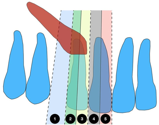 Figure 1
Peer Reviews
No public reviews on file for this paper yet. If you reviewed it on a platform where reviews are public (OpenReview, ICLR, NeurIPS, ICML), you can paste yours below so the community can read it here.
Videos
No videos yet. Explain this paper in a talk, walkthrough, or lecture? Add one.
Taxonomy
Topicsdental development and anomalies · Orthodontics and Dentofacial Orthopedics · Temporomandibular Joint Disorders
Introduction
Impacted maxillary canine (IMC) is a dental developmental anomaly with a prevalence of 0.8% and 3% in the general population. This condition can lead to significant functional and aesthetic issues, including displacement of adjacent teeth, root resorption of incisors, and disturbances in maxillofacial development. Therefore, early diagnosis is crucial for facilitating traction, reducing treatment time, and minimizing adverse effects (1 - 5). Thus, the sagittal position of the impacted canine is a vital factor in treatment planning, and it can be assessed using the impaction sector classification proposed by Ericson and Kurol (6 - 9). Maxillary canine impaction may alter the morphology of the anterior maxilla, especially when the impacted canines are positioned near the dental midline (8 , 10). This condition can also affect the positioning of the central incisors in the premaxilla. However, these differences should be demonstrated through cephalometric analysis comparing groups with various impaction sectors or a control group (10 - 12). Although prior studies have explored skeletal characteristics in patients with impacted canines, (12 - 14) none have specifically compared these characteristics based on their impaction sector. Additionally, most existing research has focused on populations in Europe and North America, highlighting the need for more studies on diverse populations to compare findings (13 - 20). In this way, this study aimed to compare the cephalometric characteristics of individuals with IMC considering their impaction sector. The research aims to improve our understanding of the dentoalveolar, and skeletal factors involved in canine impaction and contribute to developing early diagnostic strategies that enhance orthodontic and surgical planning.
Material and Methods
- Study characteristics and ethical approval This observational, cross-sectional, retrospective study received approval from the Científica del Sur University Ethics Committee in Lima, Peru (protocol number: POS-53-2025-00308). The study adhered to the STROBE (Strengthening the Reporting of Observational Studies in Epidemiology) guidelines and followed the principles of the Declaration of Helsinki. No interventions were performed on patients, nor was any new clinical or personal data collected. All radiographic images were anonymized to ensure confidentiality and compliance with ethical standards. - Sample size and population The study sample included 135 panoramic and lateral head radiographs from young adult patients who visited a radiology center in Lima, Peru. The participants were divided into three groups. Group A comprised 45 patients with IMC in sectors 1, 2, and 3. Group B included 45 patients with IMC in sectors 4 and 5. The control group, matched for sex and age, consisted of 45 patients without IMC. The sample size was calculated using Stata/SE 18.0 (StataCorp LLC, College Station, TX, USA) with a power of 90% and a significance level of 5%, based on the comparative means reported in the study by Cernochova et al. (4). The minimum required sample size for each group was 40 participants. - Inclusion criteria The inclusion criteria consisted of high-quality digital lateral cephalometric and panoramic radiographs of young adult patients aged 13 to 40 years who had IMC. We excluded radiographs from patients who had undergone orthodontic treatment, presented with supernumerary teeth, odontomas, cysts, or had any syndromes, cleft lip/palate, a history of maxillary trauma, or missing maxillary incisors. - Calibration and observer agreement Two trained and calibrated observers conducted all measurements. The training was supervised by an orthodontist with over 10 years of experience. Additionally, inter-observer agreement for qualitative variables was evaluated using Cohen's Kappa test, while intra-observer reliability was assessed using the Intraclass Correlation Coefficient (ICC). - Cephalometric Measurements Cephalometric analyses were conducted using WebCeph software (AsyRaM Inc., 2025). Each patient was registered along with their lateral cephalometric and panoramic radiographs. The investigator reviewed the software's AI-assisted cephalometric tracing for accuracy. The radiographic scale marker located in the upper right corner of the lateral cephalogram was used for calibration (Table 1, Fig. 1).
Figure 1. Maxillary canine impaction sector by Ericson and Kurol classification.1 from the mesial aspect of the first premolar to the distal aspect of the lateral incisor. (sector 1). From the distal aspect of the lateral incisor to the long axis of the lateral incisor (sector 2). From the long axis of the lateral incisor to the distal aspect of the central incisor (sector 3). From the distal aspect of the central incisor to the long axis of the central incisor (sector 4). From the long axis of the central incisor to the interincisor median line (sector 5).
- Statistical analyses The biostatistical analysis was conducted using Stata/SE 18.0. The mean and standard deviation (SD) were calculated for each variable. The Shapiro-Wilk test was employed to assess normality for the bivariate analysis. If normality was confirmed, ANOVA and Tukey post hoc tests were performed to compare outcome variables comparing three groups, with a significance level at p < 0.05.
Results
- Reliability The Cohen's Kappa test values were 1 for intra and inter-observer reliability. The ICC values ranged from 0.90-1.00 in the intra-observer reliability for all evaluations. The error method was less than 1 mm. - Main results Table 2 shows the initial characteristics of the sample, highlighting the similarity among the three groups in terms of the Wits appraisal (p=0.876) and skeletal relationship (p=0.195).
However, the control group was older (p<0.001), and significant differences were observed in the distribution of impaction sectors across the groups (p<0.001). Table 3 presents the cephalometric characteristics of the anterior maxilla, comparing the three groups with and without canine impaction.
Significant differences were found in the SNA angle between the control group (81.19°) and the group with impaction in sectors 4 and 5 (82.96°). The position of the upper central incisor (1-NA) in the control group was 4.20mm, while in the impaction groups it was lower, showing statistical significance only in sectors 1, 2, and 3 (2.96mm). The nasolabial angle was smaller in the control group (93.70°) compared to both impaction groups, with the highest value observed in sectors 4 and 5 (102.22°). Table 4 presents three multiple linear regression models evaluating the influence of predictive variables (age, sex, impaction group, and skeletal relationship) on incisor position and inclination, as well as on the nasolabial angle, but no significant associations were found (p>0.05).
Discussion
Maxillary canine impaction is an anomaly that affects dental occlusion, maxillary development, and dental esthetics; for this reason, it is studied in orthodontics to improve early diagnosis and long-term prognosis. Beyond this, the clinical and radiographic characteristics of individuals with impacted maxillary canines (IMC), compared to a control group, have been scarcely reported in the literature and even less so when considering the sector of impaction, which may alter certain cephalometric landmarks, particularly in the anterior region. In this context, the present study compared the cephalometric characteristics from lateral head radiographs of patients with IMC in different sectors of impaction, with the aim of identifying whether differences exist in the dentoskeletal patterns of patients presenting this anomaly across different sectors of impaction, along with a control group. The results of this study showed a statistically significant difference in SNA angle measurements (p< 0.05). An increase in this angle was found in the IMC groups compared to the control group. Radiographs with IMC in sectors 4 or 5 showed significantly greater SNA values than those with impaction in sectors 1, 2, or 3. This finding indicates that individuals with IMC exhibit a protruded premaxilla, which is more pronounced in cases of impaction near the midline. Similar results were reported by Ciavarella et al. (20), who found an increase in the SNA angle in patients with IMC when comparing craniofacial morphology to a control group. Although that study did not consider the sector of impaction, its findings align with the present analysis. Likewise, Cernochová et al. (4) also reported greater SNA values in patients with IMC. Furthermore, Athanasiou et al. (18) using three-dimensional morphometric analysis demonstrated greater sagittal projection of the premaxilla in IMC cases, which is consistent with an increased SNA angle within a conventional cephalometric framework. The findings of the present study confirm an increased SNA angle in patients with IMC and suggest greater severity in those with impaction in sectors 4 and 5, probably because most of these midline-closer impactions are buccal and, therefore, exert direct influence on point A. The results from the comparison of the 1-NA distance also showed significant differences. Contrary to the increase observed in the SNA angle with the presence of IMC, the 1-NA distance was reduced in the group with impaction in sectors 1, 2, and 3, indicating greater retrusion of the upper incisors. Cernochová et al. (4) reported a reduced 1-NA distance in the IMC group with palatal projection. More retroclined incisors were found compared to the control group. These findings help define a cephalometric pattern regarding the position of the upper incisors in patients with this anomaly. Regarding the nasolabial angle, a significant increase was observed in the IMC groups. This finding may be directly related to the position of the upper incisor, as dentoalveolar retrusion can displace the upper lip posteriorly and, consequently, increase the nasolabial angle. Although this association between the nasolabial angle and IMC has not been described in the literature, an orthodontic relationship between lip projection and the position and inclination of the incisors does exist. This finding represents a new contribution to literature by proposing the nasolabial angle as a useful cephalometric parameter in the assessment of IMC cases. The findings of this study enhance our understanding of the cephalometric patterns in patients with internal maxillary constriction (IMC). We observed an increased SNA angle, with greater values noted in sectors 4 and 5. Additionally, there was a reduction in the 1-NA distance, suggesting greater upper incisor retrusion in impactions located in sectors 1, 2, and 3. These results may indicate a dentoalveolar compensation related to the site of impaction. Additionally, we observed an increase in the nasolabial angle, a finding not previously documented in the literature regarding IMCs. This increase is a potential factor for aesthetic and cephalometric evaluation in patients with this condition. Limitations This study did not include the complete clinical records of the patients whose radiographs were analyzed. This limitation prevented a broader generalization of the results. It is recommended that future studies collect full clinical records to allow for a deeper analysis of the findings and complement this research with longitudinal studies to observe whether these features resolve following canine traction.
Conclusions
Patients with IMCs exhibit a modified cephalometric pattern, characterized by an increased SNA angle more pronounced when the impaction is located near the midline and incisor retrusion, as evidenced by the reduced 1-NA distance. Additionally, an increased nasolabial angle was observed, likely related to the incisor retrusion described above. These findings suggest that the sector of canine impaction influences both the dentoskeletal pattern and soft tissue profile, which is relevant for the diagnosis and treatment planning of this dental anomaly.
The reference list from the paper itself. Each links out to its DOI / PubMed record.
- 1Ericson S Kurol J Early treatment of palatally erupting maxillary canines by extraction of the primary canines Eur J Orthod 198810428395.320884310.1093/ejo/10.4.283 · doi ↗ · pubmed ↗
- 2Proffit WR Skinner’s 2 Science of Dental Materials 1991 Philadelphia PA Sounders 1646
- 3Ericson S Kurol PJ Resorption of incisors after ectopic eruption of maxillary canines: a CT study Angle Orthod 200070641523.1113864410.1043/0003-3219(2000)070<0415:ROIAEE>2.0.CO;2 · doi ↗ · pubmed ↗
- 4Cernochova P Izakovicova-Holla L Dentoskeletal characteristics in patients with palatally and buccally displaced maxillary permanent canines Eur J Orthod 201234675461.2175024610.1093/ejo/cjr 069 · doi ↗ · pubmed ↗
- 5Arriola-Guillén LE Ruíz-Mora GA Rodríguez-Cárdenas YA Aliaga-Del Castillo A Boessio-Vizzotto M Dias-Da Silveira HL Influence of impacted maxillary canine orthodontic traction complexity on root resorption of incisors: A retrospective longitudinal study Am J Orthod Dentofacial Orthop 201915512839.3059116010.1016/j.ajodo.2018.02.011 · doi ↗ · pubmed ↗
- 6Arriola-Guillén LE Ruíz-Mora GA Rodríguez-Cárdenas YA Aliaga-Del Castillo A Dias-Da Silveira HL Root resorption of maxillary incisors after traction of unilateral vs bilateral impacted canines with reinforced anchorage Am J Orthod Dentofacial Orthop 20181545645656.3038493510.1016/j.ajodo.2018.01.015 · doi ↗ · pubmed ↗
- 7Cicek O Gurel T Demir Cicek B Investigation of the Relationship of Impacted Maxillary Canines with Orthodontic Malocclusion: A Retrospective Study Children (Basel)2023106950.3737118210.3390/children 10060950 PMC 10296910 · doi ↗ · pubmed ↗
- 8Arriola-Guillén LE Aliaga-Del Castillo A Ruíz-Mora GA Rodríguez-Cárdenas YA Dias-Da Silveira HL Influence of maxillary canine impaction characteristics and factors associated with orthodontic treatment on the duration of active orthodontic traction Am J Orthod Dentofacial Orthop 20191563391400.3147426910.1016/j.ajodo.2018.10.018 · doi ↗ · pubmed ↗