Research of imaging in left Atrium: A Bibliometric Analysis
Can Cui, Jiang-Hua Zhu, Ya-Hong Tao, Zhen-Yi Zhao, Yun Peng, Minjing Zuo

TL;DR
This paper analyzes 20 years of research on left atrial imaging using bibliometric methods to identify trends and future directions.
Contribution
The study provides a comprehensive bibliometric analysis of left atrial imaging research from 2004 to 2023, highlighting growth and emerging topics.
Findings
The number of published articles on left atrial imaging has increased year by year.
Research on left atrial appendage occlusion and LA strain has become a notable focus in recent years.
Abstract
The evaluation of the left atrial (LA) by imaging is becoming increasingly essential due to its significant role in numerous diseases. This study aimed to analyze and summarize research on LA imaging in the past 20 years through bibliometric analysis and offer insights into future research prospects. The Web of Science (WOS) core collection database was retrieved for literature in LA imaging research from 2004 to 2023. Subsequently, the literature was processed and visualized by the VOSviewer and CiteSpace. VOSviewer was used to create cooperation networks for countries/regions and institutions. CiteSpace was used to analyze burst keywords in citation analysis. A total of 3664 articles published in this field between January 2004 and December 2023 were analyzed. The number of published articles is increasing year by year. The USA contributed the most articles (1072). Hugh Calkins (44)…
Genes, proteins, chemicals, diseases, species, mutations and cell lines named across the full text — each resolved to its canonical identifier and authoritative record.
Click any figure to enlarge with its caption.
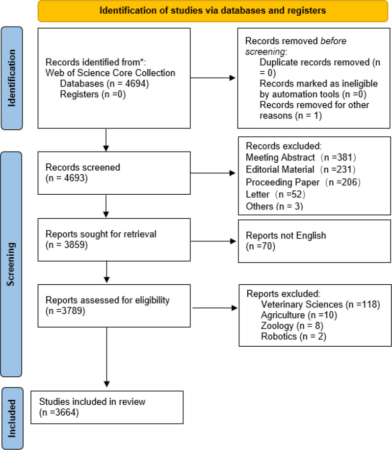 Figure 1
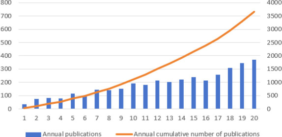 Figure 2
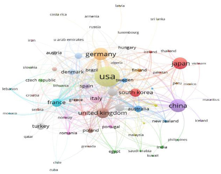 Figure 3
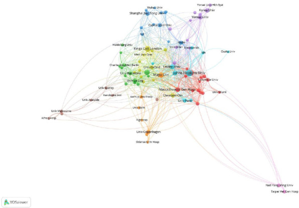 Figure 4
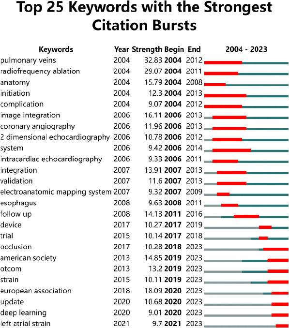 Figure 5
Peer Reviews
No public reviews on file for this paper yet. If you reviewed it on a platform where reviews are public (OpenReview, ICLR, NeurIPS, ICML), you can paste yours below so the community can read it here.
Videos
No videos yet. Explain this paper in a talk, walkthrough, or lecture? Add one.
Taxonomy
TopicsAdvanced X-ray and CT Imaging
INTRODUCTION
1
The LA serves as a crucial link between the left ventricle and the pulmonary veins [1] and plays a vital role in the process of filling the left ventricle and in maintaining overall cardiac function. The evaluation of the LA is essential, particularly in patients with atrial fibrillation (AF) [2]Assessing the structure and function can help stratify risk, guide treatment, and predict prognosis [3]. Clinical evaluation of LA includes biochemical markers like NT-proBNP, ANP, BNP, etc [4, 5]. The alterations of these biochemical markers involve a variety of molecular and pathological mechanisms [6]. Imaging assessment is also an important method of LA evaluation [7], such as ultrasound, Cardiac Computed Tomography (CT), and Cardiac Magnetic Resonance Imaging (MRI). Imaging can offer a thorough evaluation of the LA structure and function [8], thus offering valuable information for the treatment and prognosis. For example, CT can clearly show the structure and anatomical variation of the pulmonary veins [9, 10], which is of great significance for the preoperative evaluation of radiofrequency ablation of AF.
Bibliometrics is a method of statistical analysis [11] that examines the authors, journals, citation frequency, and publication year of literature [12]. It is used to discover patterns within the literature, summarize research hotspots, and identify development trends in the analyzed disciplines. There are many studies on LA, but quantitative analysis is still lacking. This study aimed to analyze and visualize the research on LA imaging in the past 20 years through bibliometric analysis, provide insights into the field’s status, and indicate the research hotspots.
METHODS
2
We conducted a literature search on the WOS Core Collection on 17 July 2024 using the following search formula: (TS= ((“Left Atrium” or “Left atrial”) AND (“CT” or “MRI” or “Magnetic Resonance Imaging” or “PET” or “positron emis-sion tomography” or “computed tomography” or “Coronary angiography” or “radiography” or “ultrasonography”)) AND (FPY=2004-2023). The criteria for literature inclusion were as follows: (I) LA imaging as the topic of study, (II) articles and reviews, and (III) English-language literature. Meanwhile, the exclusion criteria were as follows: (I) meeting abstract, editorial material, proceeding paper, and letter; (II) literature not published between January 2004 and December 2023, and (III)the research field of the literature does not match the topic, such as veterinary sciences, agriculture, and zoology. The process is shown in Fig. (1).
Data Analysis
2.1
CiteSpace (version 6.2) and VOSviewer (version 1.6.2) were applied for bibliometric and visualization analysis of the included literature. VOSviewer was used to create cooperation networks for countries/regions and institutions. Citespace presents the research structure and development trend of a certain discipline in specific fields [13]. which was used to analyze citation burst keywords.
RESULTS
3
Temporal Distribution of the Literature
3.1
Fig. (2) is a graph of the number of publications drawn by Excel, which depicts the trend of the number of publications per year. The number of published articles has been increasing year by year from 2004 to 2023 and reached 370 in 2023.
Contribution of Countries and Regions
3.2
Table 1 shows the top 10 countries with the largest number of publications. The United States, China, and Germany ranked in the top three, with 1072, 445, and 416 articles, respectively. Fig. (3) shows the cooperation map of countries or regions in LA imaging studies. The United States, the United Kingdom, South Korea, Australia, and Lithuania cooperate closely; China has more cooperation with Germany, the United States, the United Kingdom, Australia, and France.
Distribution of Research Institutions
3.3
Fig. (4) shows the institution’s cooperation network diagram by using VOSviewer. It demonstrated that the cooperation between different institutions within China was intense, among which Shanghai Jiao Tong University established close cooperation with Wuhan University and capital Medical University. However, there is less cooperation with institutions abroad.
Author Analysis
3.4
Table 2 lists the top 10 authors with the highest number of publications in the field of LA imaging. The author with the highest number of publications was Hugh Calkins, with a total of 44 publications, followed by Nassir F. Marrouche and Eugene Kholmovski with 39 and 36 publications. We also conducted an h-index analysis of the authors, and the results showed that Yun-Yu Chen had the highest h-index (18), followed by Hugh Calkins.
Journal Distribution
3.5
As shown in Table 3, the Journal of Cardiovascular Electrophysiology is the most productive in the field of LA imaging, with a total of 171 articles. It was followed by Europace (124), the International Journal of Cardiovascular Imaging (110), Heart Rhythm (103), and the International Journal of Cardiology (95).
Analysis of Keyword Burst
3.6
As shown in Fig. (5), a pulmonary vein was the strongest burst word with an intensity of 22.92, followed by radiofrequency ablation with an intensity of 20.03. In 2003-2017, the strongest citation bursts included: pulmonary vein, radiofrequency ablation, initiation, anatomy, complication, etc. Keywords that appeared after 2017 and continued to have the strongest citation bursts include occlusion, LAAO, strain, and clinical outcome.
DISCUSSION
4
The study analyzed the use of imaging in LA through bibliometrics. The number of published articles in this field has increased steadily from 35 in 2004 to 370 in 2023. This upward trend suggests that LA imaging research is garnering significant attention among scholars.
The top three countries with the largest number of publications are the United States, China, and Germany, indicating their strong interest in research in this field. In institutional analysis, Harvard University ranks first. Seven of the top 10 institutions are from the United States, which also shows that the United States is leading in this field. The United States has very close exchanges with other countries, which have also contributed to the development of the field of LA imaging. Analysis of cited journals reveals that Circulation is the most frequently cited journal in the field of LA imaging research, underscoring its popularity and authoritative standing.
Keyword burst analysis can reflect the main research hotspots and their trends in a specific period. According to (Fig. 5), the keyword burst is divided into two phases. The first phase is 2004-2017, emphasis is placed on the anatomy of the LA and pulmonary veins, and some preliminary imaging techniques. AF is a highly prevalent cardiovascular disease that is closely related to LA remodeling [14]. Hugh Calkins and his team demonstrated that radiofrequency ablation for the treatment of AF reports higher efficacy rates than anti-arrhythmic drug therapy and a lower rate of complications [15]. Radiofrequency ablation requires only limited fluoroscopy, is catheter-guided, and is achieved using an electroanatomic mapping system [16]. It is important to note that during radiofrequency ablation, the relative position of the esophagus to the posterior wall of the LA needs to be identified to avoid complications such as esophageal injury during surgery [17]. Michel Haissaguerre et al. have shown that the spontaneous initiation of AF by ectopic beats originating in the pulmonary veins [18]. These ectopic rhythms originate from the muscle bundles that extend from the atria to the pulmonary veins, so pulmonary vein isolation is the cornerstone of radiofrequency ablation. Besides, the changes in the number and location of pulmonary veins correlated with the outcome of AF radiofrequency catheter ablation procedures. Recommended by Kirchhof et al. in the 2016 ESC Guidelines [19] for the management of AF, developed in collaboration with EACTS, routine LA size and anatomy assessment in patients with AF is essential. LA size is usually assessed using standard 2D echocardiography [20] in the past. With the development of imaging technology, intracardiac echocardiography [21]. It is complementary to transesophageal echocardiography and is much better tolerated by patients, helping to meet the growing need for monitoring of patient anatomy, catheter placement, and intraoperative complications such as pericardial effusion or thrombosis.
In the last few years, it has become possible to integrate different imaging modalities to guide catheter ablation procedures for AF [22]. There are now a variety of specialized hybrid imaging systems or software image co-registration techniques [23] that combine anatomical information from fluoroscopy, CT, MRI, or intracardiac echocardiography with information provided by electroanatomic mapping to guide catheter ablation of AF [24]. The development of imaging methods and techniques has led to a better understanding of the LA and pulmonary veins [25].
From 2017 to 2023, the focus shifted towards clinical efficacy and patient prognosis, as well as devices for LAAO. Furthermore, the LA strain emerged as a significant research topic [26]. Left atrial appendage thrombosis stands as the primary cause of thromboembolic stroke in almost 90% of AF patients [27], LAAO represents a swiftly evolving technique for individuals unable to sustain long-term oral anticoagulation [28]. LAAO emerged in 1949 as a surgical procedure, with percutaneous closure first conducted on human patients in 2001. There are many LAAO devices, such as Watchman, PLAATO, LARIAT, WaveCrest, and Amplatzer [29]. Among them, Watchman is the best-studied LAAO device. In 2017, a clinical trial called PREVAIL and PROTECT AF Trials showed that LAAO with Watchman devices prevents stroke in patients with nonvalvular AF to a similar extent to oral anticoagulation with warfarin [30]. In addition, LAAO causes less disability or death than warfarin [31]. Recent studies from American scholars have shown that LAAO surgery has significant advantages over oral anticoagulant therapy in improving long-term cognitive function, regardless of gender and type of AF. Imaging modalities like transesophageal echocardiography (TEE) or LA CT serve as the primary imaging techniques for post-LAAO assessment [32]. Typically conducted 1 to 6 months post-surgery. These modalities can detect peri-device leakage, device-related thrombosis, and device embolism to inform subsequent management decisions.
LA strain has garnered attention as a significant area of research in recent years [33]. Strain denotes the myocardial deformation during the cardiac cycle, representing the percentage change in myocardial length from diastolic relaxation to systolic contraction [34]. Echocardiographic dot tracking technology, LA CT, and cardiac MRI all enable the evaluation of LA strain, providing a comprehensive assessment of LA storage, conduit, and contractile pump functions. The strain rate refers to [35] the speed of myocardial deformation, which can more accurately and sensitively reflect the early functional impairment of the myocardium. There is growing interest in the use of artificial intelligence (AI) in LA imaging, especially deep learning-based AI. Deep learning was more reproducible than imaging measurements [36]. Deep learning can also integrate latent information from imaging that may inform disease risk. DROID, a deep learning echocardiographic interpretation model built by Emily Lau and her team [37], accurately quantifies standard measurements of LA structure and function [38] and can combine information from other heart chambers to generate predictions of LA size, which may provide additional predictive value for disease risk. Medical imaging provides information in a noninvasive, reproducible manner, has grown significantly in importance in clinical decision-making [39].
This bibliometrics article has certain limitations. Firstly, citation data can be influenced by factors such as language preferences and subject selections, potentially impacting the reliability of bibliometric analysis. Secondly, the data were retrieved solely from the WOS, excluding articles from other databases, potentially resulting in the omission of relevant literature. Lastly, the collection and statistical analysis of literature data may vary among individuals and could be subjectively influenced by personal perceptions.
CONCLUSION
The top 3 countries with the largest number of publications are the United States, the United Kingdom, and Germany, which suggests that these countries are interested in this area and have a lot of research results. In recent years, research on LAAO and LA strain has been notably focused and garnered considerable attention. The ongoing trend of utilizing multimodal LA imaging to evaluate the prognosis and LA strain post-LAAO is expected to remain a prominent topic in future research.
The reference list from the paper itself. Each links out to its DOI / PubMed record.
- 1Marino P.N. Left atrial conduit function: A short review.Physiol. Rep.20219191505310.14814/phy 2.1505334605214 PMC 8488566 · doi ↗ · pubmed ↗
- 2Artur E. Marta S. Guillaume J. Multimodality imaging in thoracic aortic diseases: A clinical consensus statement from the european association of cardiovascular imaging and the european society of cardiology working group on aorta and peripheral vascular diseases.Eur. Heart J. Cardiovasc. Imaging 2023245 e 65e 8510.1093/ehjci/jead 02436881779 · doi ↗ · pubmed ↗
- 3Han R. Mei Y.C. Li H.W. Li R-J. He Y-H. Wang Z-F. Wu Y-Q. Evaluation of cardiac function recovery in patients with paroxysmal atrial fibrillation after catheter radiofrequency ablation using two-dimensional speckle tracking imaging and real-time three-dimensional echocardiography.Cardiology 202411210.1159/00054124739342934 PMC 11965856 · doi ↗ · pubmed ↗
- 4Hoeper M.M. Bogaard H.J. Condliffe R. Frantz R. Khanna D. Kurzyna M. Langleben D. Manes A. Satoh T. Torres F. Wilkins M.R. Badesch D.B. Definitions and diagnosis of pulmonary hypertension.J. Am. Coll. Cardiol.20136225 Suppl.D 42D 5010.1016/j.jacc.2013.10.03224355641 · doi ↗ · pubmed ↗
- 5NouguéH. Pezel T. Picard F. Sadoune M. Arrigo M. Beauvais F. Launay J.M. Cohen-Solal A. Vodovar N. Logeart D. Effects of sacubitril/valsartan on neprilysin targets and the metabolism of natriuretic peptides in chronic heart failure: A mechanistic clinical study.Eur. J. Heart Fail.201921559860510.1002/ejhf.134230520545 · doi ↗ · pubmed ↗
- 6Sun M. Wu X. Yu Y. Wang L. Xie D. Zhang Z. Chen L. Lu A. Zhang G. Li F. Disorders of calcium and phosphorus metabolism and the proteomics/metabolomics-based research.Front. Cell Dev. Biol.2020857611010.3389/fcell.2020.57611033015068 PMC 7511772 · doi ↗ · pubmed ↗
- 7Spencer K.T. Flachskampf F.A. Focused cardiac ultrasonography.JACC Cardiovasc. Imaging 20191271243125310.1016/j.jcmg.2018.12.03631272607 · doi ↗ · pubmed ↗
- 8Silva J. Azevedo T. Ginja M. Oliveira P.A. Duarte J.A. Faustino-Rocha A.I. Realistic aspects of cardiac ultrasound in rats: Practical tips for improved examination.J. Imaging 202410921910.3390/jimaging 1009021939330439 PMC 11433567 · doi ↗ · pubmed ↗