Diffusion kurtosis imaging detects cortical microstructural alterations in amyloid-positive MCI patients
Rune B. Nielsen, Peter Parbo, Rola Ismail, Rikke B. Dalby, Anna Tietze, Hans Brændgaard, Hanne Gottrup, David J. Brooks, Leif Østergaard, Simon F. Eskildsen

TL;DR
Diffusion kurtosis imaging reveals early brain changes in people with mild cognitive impairment who have amyloid buildup, potentially aiding early Alzheimer's detection.
Contribution
This study shows that mean kurtosis from DKI can detect cortical microstructural changes linked to amyloid pathology before atrophy occurs.
Findings
Aβ-positive MCI patients had higher cortical mean kurtosis in specific brain regions compared to controls and Aβ-negative MCI.
Mean kurtosis correlated with Aβ burden in parietal and temporal cortices, even without cortical atrophy.
Mean diffusivity showed weaker and less consistent associations with Aβ compared to mean kurtosis.
Abstract
Alzheimer's disease (AD) is characterized by early accumulation of amyloid-β (Aβ) plaques and tau pathology which precede overt neurodegeneration and cognitive decline. Detecting microstructural brain changes associated with Aβ deposition before the onset of atrophy is critical for early diagnosis and intervention. This study investigates whether diffusion kurtosis imaging (DKI) can detect early microstructural alterations in cortical and subcortical gray matter (GM) associated with Aβ pathology in individuals with mild cognitive impairment (MCI). Using DKI-derived metrics—mean kurtosis (MK) and mean diffusivity (MD) – we assessed cortical and subcortical microstructure in 67 participants (23 cognitively normal [CN], 44 MCI, including 29 Aβ-positive). Aβ burden was quantified using 11C-PiB PET imaging. Cortical atrophy, hippocampal volume, and white matter hyperintensities (WMH) were…
Genes, proteins, chemicals, diseases, species, mutations and cell lines named across the full text — each resolved to its canonical identifier and authoritative record.
Click any figure to enlarge with its caption.
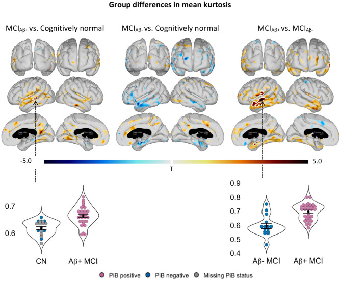 Figure 1
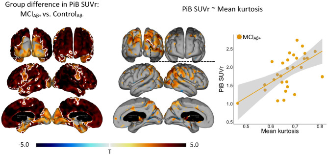 Figure 2
 Figure 3
Peer Reviews
No public reviews on file for this paper yet. If you reviewed it on a platform where reviews are public (OpenReview, ICLR, NeurIPS, ICML), you can paste yours below so the community can read it here.
Videos
No videos yet. Explain this paper in a talk, walkthrough, or lecture? Add one.
Taxonomy
TopicsAdvanced Neuroimaging Techniques and Applications · Dementia and Cognitive Impairment Research · Functional Brain Connectivity Studies