Vomeric Chondrosarcoma: A Case Report of a Rare Entity With CT and MRI Findings
Amine Benfaida, Badr Soudi, El Mehdi Mniai, Samy Ammari, Amal Rami

TL;DR
This case report describes a rare cartilage tumor in the nose area, diagnosed using CT and MRI scans.
Contribution
The report provides a detailed case of vomeric chondrosarcoma with radiologic and histologic confirmation.
Findings
CT and MRI helped define the tumor extent and guide surgery.
The patient presented with progressive right-sided epiphora.
Radiologic and histologic correlation confirmed the diagnosis.
Abstract
Chondrosarcoma of the vomer is an exceptionally rare malignant tumor of cartilaginous origin within the sinonasal region. Due to its rarity, the diagnosis can often be challenging, and imaging features may overlap with other cartilaginous or bone lesions. We describe a case of a patient presenting with progressive right-sided epiphora, in whom radiologic and histologic correlation confirmed the diagnosis of vomeric chondrosarcoma. This report highlights the complementary role of CT and MRI in defining tumor extent and guiding surgical management.
Genes, proteins, chemicals, diseases, species, mutations and cell lines named across the full text — each resolved to its canonical identifier and authoritative record.
Click any figure to enlarge with its caption.
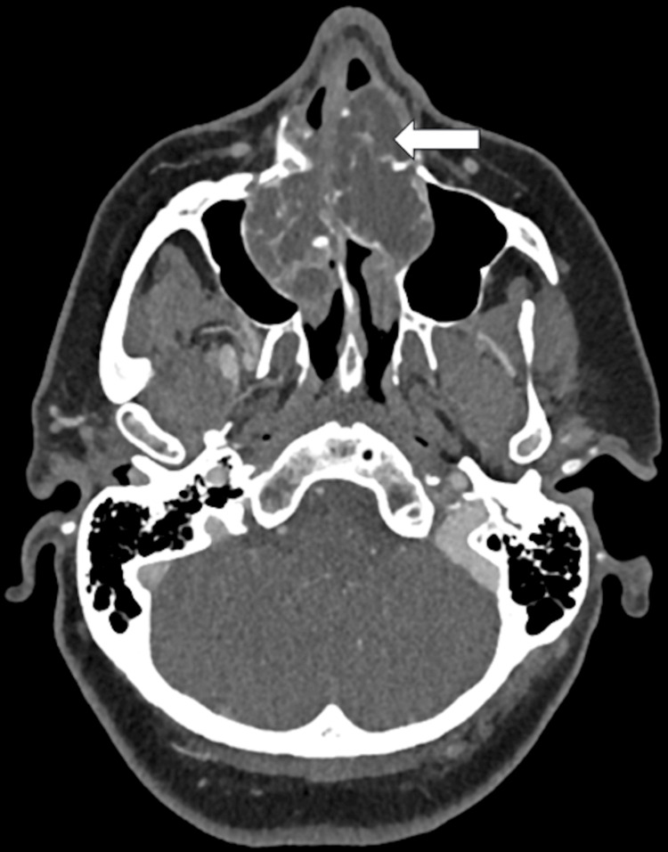 Figure 1
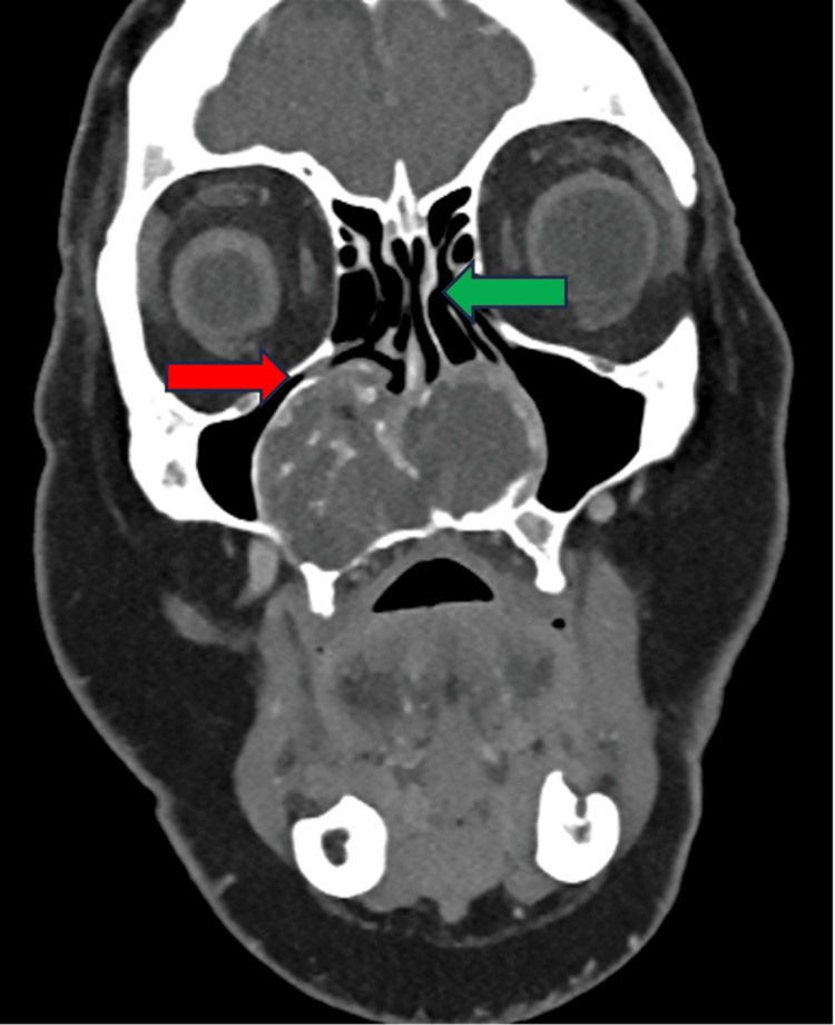 Figure 2
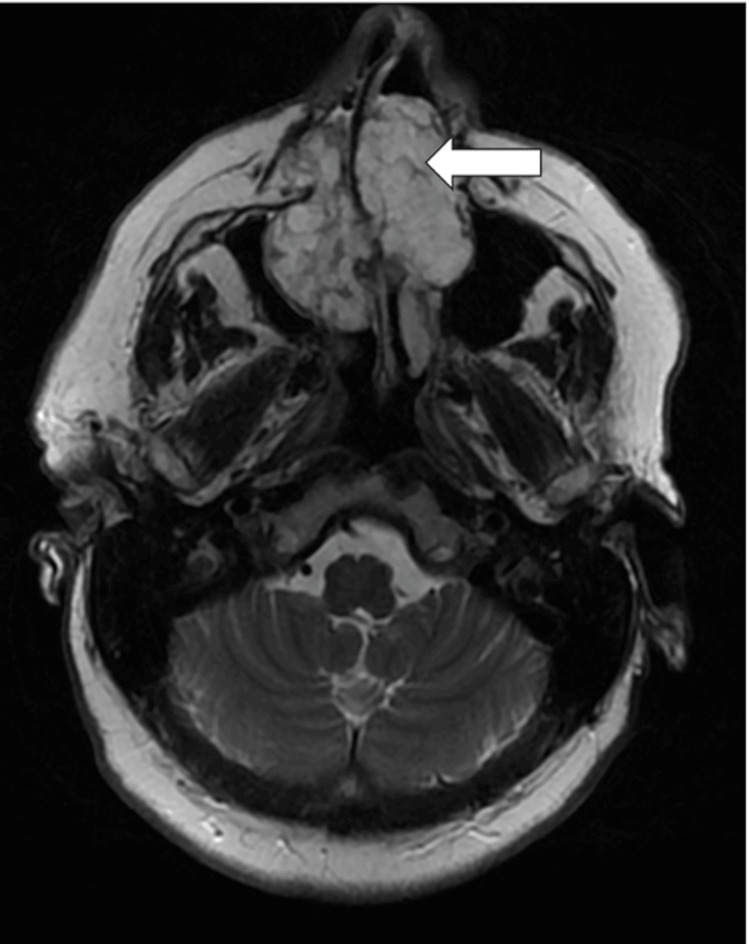 Figure 3
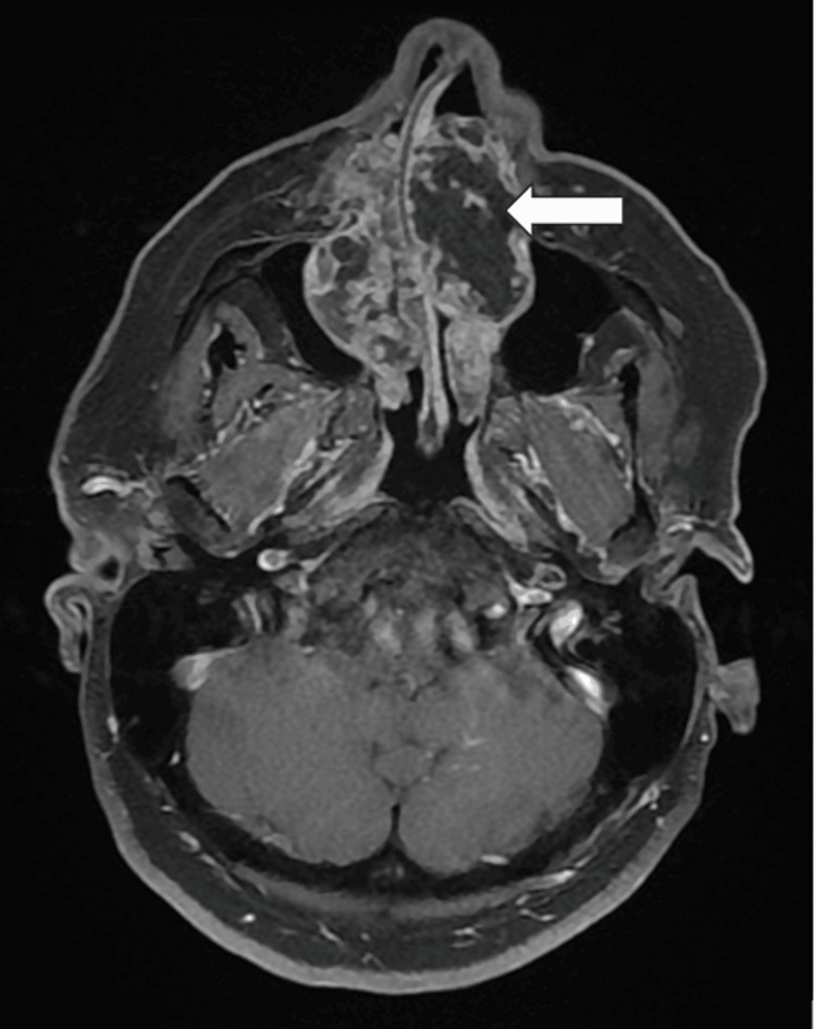 Figure 4
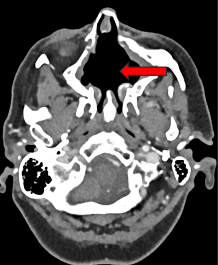 Figure 5
 Figure 6
Peer Reviews
No public reviews on file for this paper yet. If you reviewed it on a platform where reviews are public (OpenReview, ICLR, NeurIPS, ICML), you can paste yours below so the community can read it here.
Videos
No videos yet. Explain this paper in a talk, walkthrough, or lecture? Add one.
Taxonomy
TopicsBone Tumor Diagnosis and Treatments · Head and Neck Surgical Oncology · Teratomas and Epidermoid Cysts