Prevalence of Mandibular Third Molar Impaction, Associated Pathologies, and Correlation With Temporomandibular Joint Morphology in a Hospital‐Based Spanish Cohort: A Panoramic Radiography Study
Hassan Ahmed Assiri, Albert Estrugo-Devesa, Sonia Egido-Moreno, Xavier Roselló Llabrés, Mohammad Shahul Hameed, Abdullah Alqarni, Jose López-López

TL;DR
This study examines the prevalence of impacted lower wisdom teeth and their connection to jaw joint shape in a Spanish hospital cohort using panoramic X-rays.
Contribution
The study provides new insights into the prevalence and characteristics of impacted mandibular third molars and their potential association with TMJ morphology in a specific population.
Findings
Vertical impaction and oval-shaped condyles were the most common findings.
Dental caries and bone loss were frequently observed pathologies.
No significant correlation was found between TMJ pathologies and condyle shape.
Abstract
Mandibular third molar is the most frequent impacted tooth in the oral cavity. Its presence can be associated with complications including the temporomandibular joint (TMJ) symptoms. Therefore, the present study aimed to assess the prevalence of impacted mandibular third molar (IMTM), associated pathologies, and its correlation with TMJ morphology in a hospital‐based Spanish cohort. We retrospectively reviewed existing orthopantomographs (OPGs) records, panoramic images of patients aged ≥18 with at least one IMTM who attended the Dental Hospital of the University of Barcelona (HOUB) between September 2021 and May 2023. The OPGs were assessed and interpreted by an experienced oral and maxillofacial radiologist for the type of impaction according to Winter’s classification system, associated pathologies, and shape of mandibular condyle. Out of 80 OPGs, 60% (95% confidence interval [CI]:…
Genes, proteins, chemicals, diseases, species, mutations and cell lines named across the full text — each resolved to its canonical identifier and authoritative record.
Click any figure to enlarge with its caption.
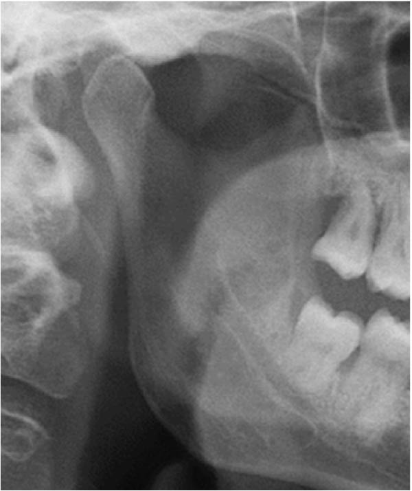 Figure 1
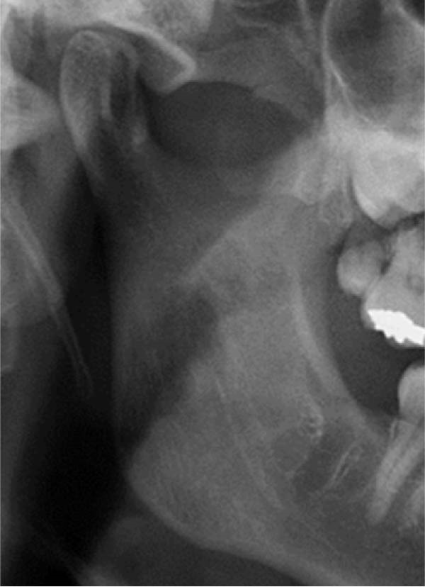 Figure 2
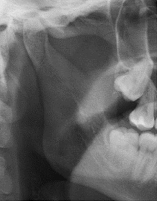 Figure 3
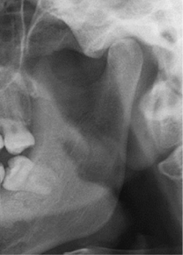 Figure 4
 Figure 5
 Figure 6
 Figure 7
 Figure 8
 Figure 9
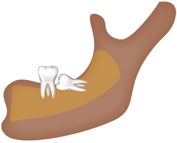 Figure 10
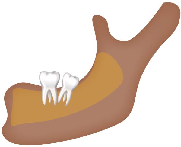 Figure 11
Peer Reviews
No public reviews on file for this paper yet. If you reviewed it on a platform where reviews are public (OpenReview, ICLR, NeurIPS, ICML), you can paste yours below so the community can read it here.
Videos
No videos yet. Explain this paper in a talk, walkthrough, or lecture? Add one.
Taxonomy
TopicsDental Radiography and Imaging · dental development and anomalies · Temporomandibular Joint Disorders