Muscle Imaging in Inclusion Body Myositis: Refinement of MRI Criteria and Insights Into Upper Body Involvement
Eleonora Torchia, Matteo Lucchini, José Verdu‐Diaz, Sara Bortolani, Beatrice Ravera, Vincenzo Carlomagno, Alessandra Cicia, Daniela Bernardo, Mauro Monforte, Robert Rehmann, Rudolf Andre Kley, Mario Sabatelli, Jordi Díaz‐Manera, Enzo Ricci, Massimiliano Mirabella, Giorgio Tasca

TL;DR
This study improves MRI criteria for diagnosing inclusion body myositis and reveals new insights into upper body muscle involvement and disease patterns.
Contribution
The study refines MRI criteria for IBM and identifies distinct radiological phenotypes through cluster analysis.
Findings
Revised MRI criteria achieved 96% sensitivity and showed consistent performance across clinical subgroups.
Whole-body analysis revealed frequent mild wasting in paraspinal, neck, and scapular girdle muscles.
Cluster analysis identified two distinct imaging phenotypes with sex-related differences in muscle involvement.
Abstract
The diagnosis of inclusion body myositis (IBM) can be delayed because of its heterogeneous clinical presentation and the lack of specific biomarkers. Muscle imaging has gained increasing relevance over the past decade and is now included among the supportive criteria in the international diagnostic guidelines. This study aimed to refine MRI criteria for IBM to facilitate clearer pattern recognition, increase their reproducibility and broader clinical applicability. We also aimed to provide a comprehensive evaluation of muscle wasting across the entire body, including less frequently assessed regions such as the neck, scapular girdle and trunk muscles, and to explore the presence of radiological IBM phenotypes through cluster analysis. Sixty‐eight MRI scans and clinical records from patients diagnosed with IBM between 2003 and 2024 (60% males; mean age: 66 years, range: 46–85) were…
Genes, proteins, chemicals, diseases, species, mutations and cell lines named across the full text — each resolved to its canonical identifier and authoritative record.
Click any figure to enlarge with its caption.
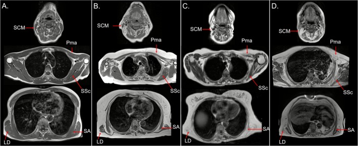 Figure 1
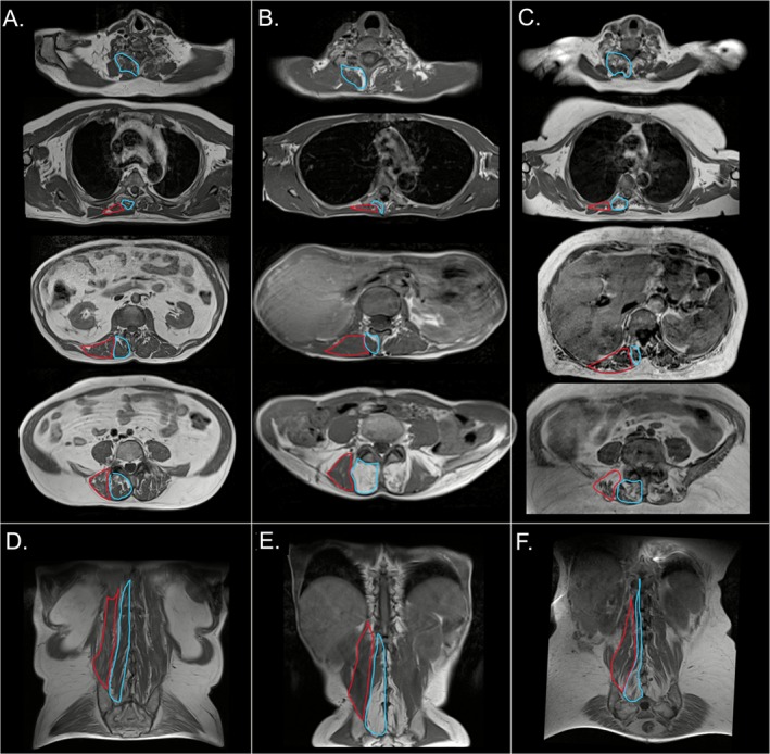 Figure 2
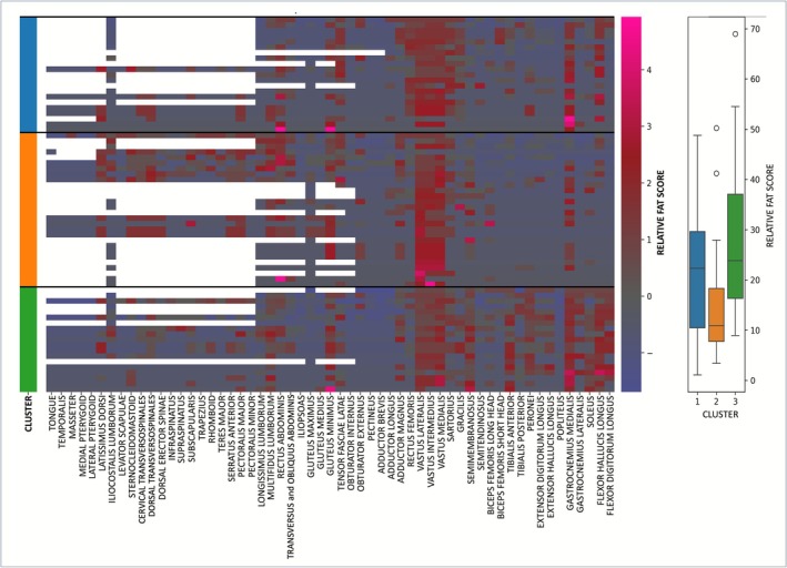 Figure 3
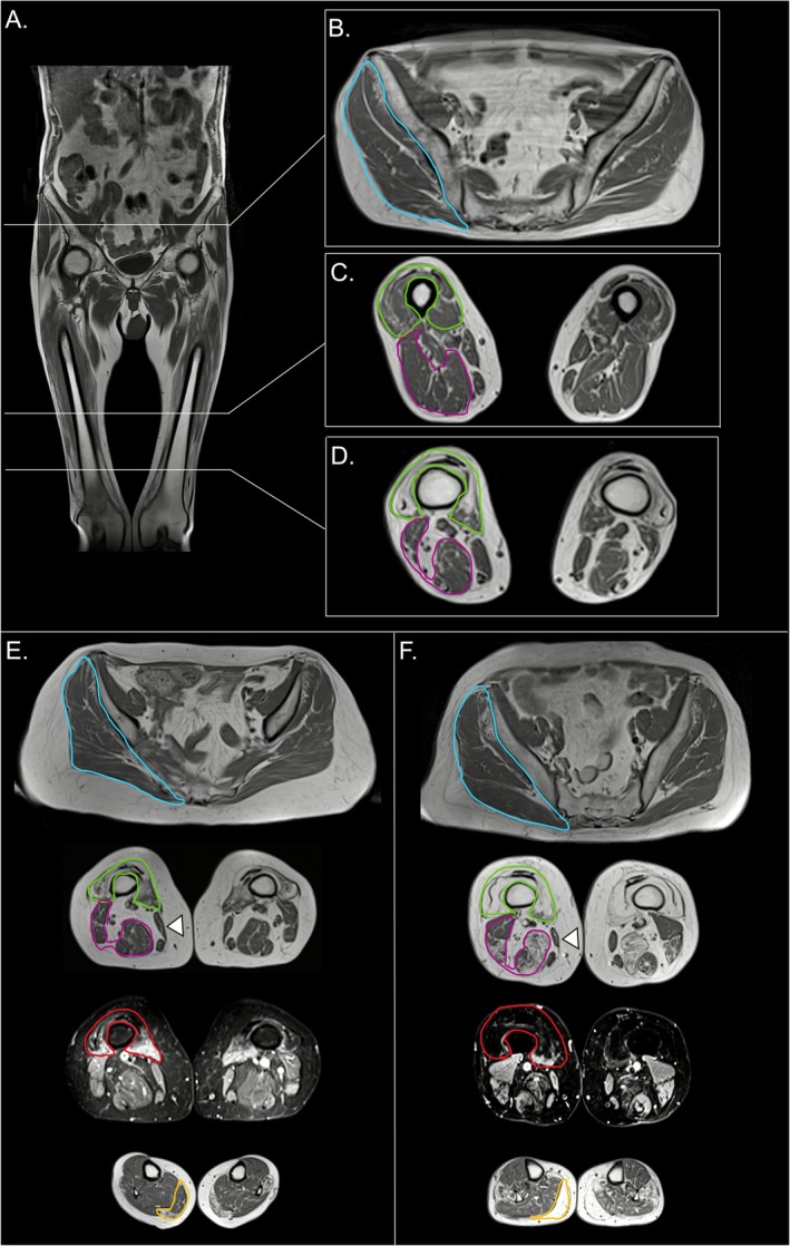 Figure 4
Peer Reviews
No public reviews on file for this paper yet. If you reviewed it on a platform where reviews are public (OpenReview, ICLR, NeurIPS, ICML), you can paste yours below so the community can read it here.
Videos
No videos yet. Explain this paper in a talk, walkthrough, or lecture? Add one.
Taxonomy
TopicsInflammatory Myopathies and Dermatomyositis · Parkinson's Disease and Spinal Disorders · Muscle and Compartmental Disorders