Noninvasive Evaluation of the Rat Adenomyosis Model Constructed by Autologous Endometrial Implantation using Magnetic Resonance Imaging
Qi Zhang, Qianwen Zhu, Linghui Xu, Yujia Shen, Junhai Zhang

TL;DR
This study shows that MRI can noninvasively evaluate a rat model of adenomyosis, a condition where endometrial tissue grows into the uterine muscle.
Contribution
The study introduces a noninvasive MRI method to evaluate a rat adenomyosis model and visualizes the junctional zone for the first time in this context.
Findings
MRI successfully detected adenomyosis lesions in all model group rats, showing myometrial thickening and T2 hypersignal spots.
Histological analysis confirmed MRI findings, showing hyperplasia and compact cell arrangement in the junctional zone.
MRI can distinguish between ectopic endometrial tissue and muscle hyperplasia based on signal intensity patterns.
Abstract
Dynamic changes in adenomyotic lesions in animal models have been difficult to observe and evaluate in vivo on a regular basis. Therefore, this study aims to investigate the feasibility of establishing a rat model of adenomyosis through autologous endometrial implantation and to assess the value of magnetic resonance imaging (MRI) for noninvasive evaluation of the model. Forty rats were randomly divided into two groups (20 rats in the control group, 20 rats in the model group). A rat adenomyosis model was constructed through autologous endometrial implantation. Three months after the modeling surgery, the rats underwent MRI examination, including T2-weighted axial imaging and T1-weighted axial imaging. The thickness of the uterine myometrium and junctional zone was measured. Following the MRI, the rat uterus was sliced for hematoxylin-eosin (HE) staining. In the model group, lesions…
Genes, proteins, chemicals, diseases, species, mutations and cell lines named across the full text — each resolved to its canonical identifier and authoritative record.
Click any figure to enlarge with its caption.
 Figure 1
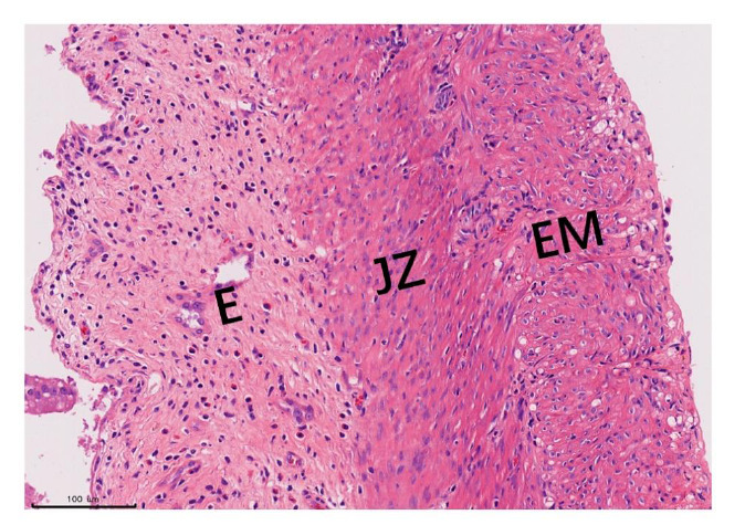 Figure 2
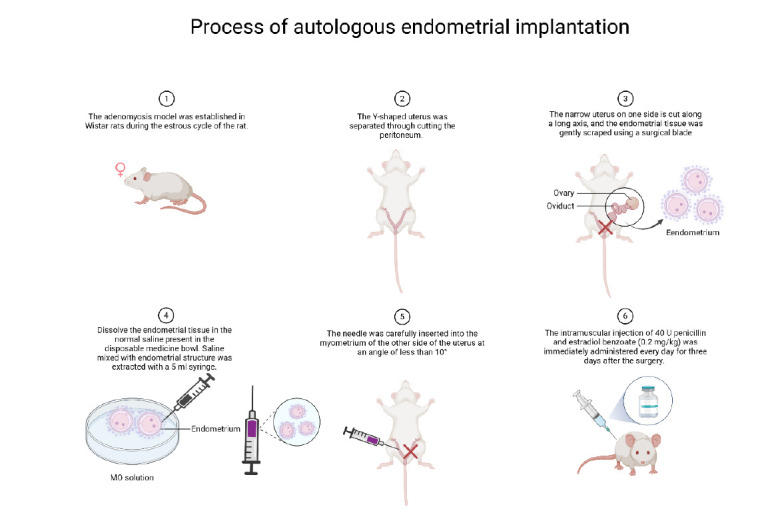 Figure 3
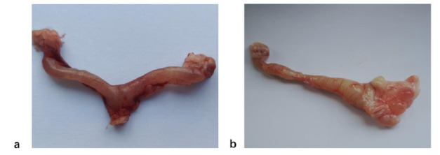 Figure 4
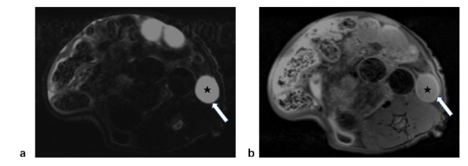 Figure 5
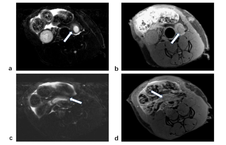 Figure 6
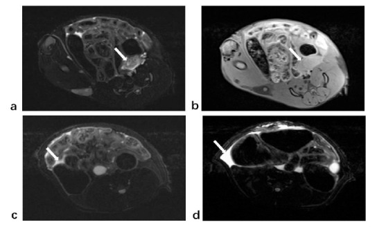 Figure 7
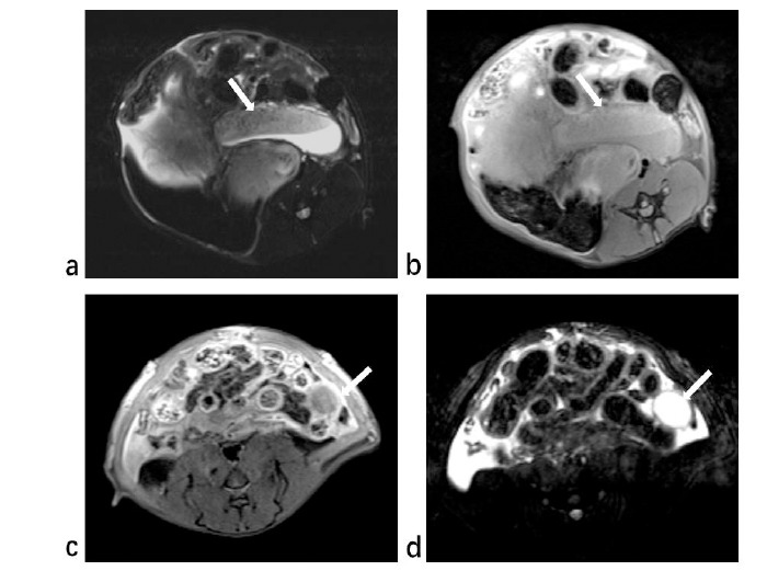 Figure 8
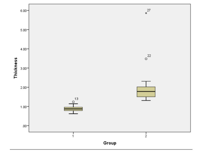 Figure 9
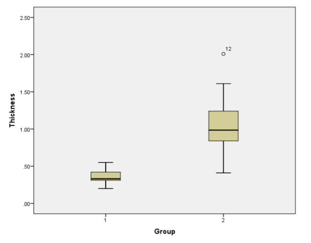 Figure 10
Peer Reviews
No public reviews on file for this paper yet. If you reviewed it on a platform where reviews are public (OpenReview, ICLR, NeurIPS, ICML), you can paste yours below so the community can read it here.
Videos
No videos yet. Explain this paper in a talk, walkthrough, or lecture? Add one.
Taxonomy
TopicsEndometriosis Research and Treatment · Endometrial and Cervical Cancer Treatments · Uterine Myomas and Treatments