Congenital Intramuscular Cavernous Hemangioma: A Rare and Underreported Entity
Hari Vignesh, Pola Govardhan Kumar, Sundeep Selvamuthukumaran, Sreedevi B.V, Mahesh K.G

TL;DR
This paper reports a rare case of a cavernous hemangioma in a five-year-old girl located in an unusual muscle area.
Contribution
The novelty lies in documenting an intramuscular cavernous hemangioma in an uncommon anatomical location.
Findings
The tumor was located in the left infra-axillary region of a five-year-old female.
Biopsy confirmed the diagnosis of intramuscular cavernous hemangioma.
This case emphasizes the rarity of such tumors in deep muscle tissue.
Abstract
Hemangiomas are common pediatric vascular tumors characterized by endothelial cell proliferation. They usually appear shortly after birth and are more frequently observed in females. Deep-seated forms, such as cavernous hemangiomas, are more common, while those arising within muscle tissue are relatively rare. They are most commonly located in the head and neck. We present the case of a five-year-old female with a long-standing swelling in the left infra-axillary region. Biopsy confirmed an intramuscular cavernous hemangioma. This case highlights an uncommon anatomical location for this tumor type.
Genes, proteins, chemicals, diseases, species, mutations and cell lines named across the full text — each resolved to its canonical identifier and authoritative record.
Click any figure to enlarge with its caption.
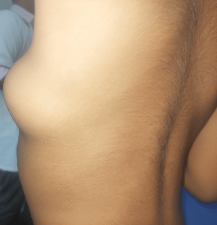 Figure 1
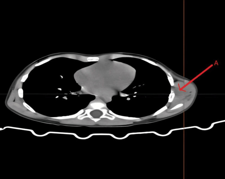 Figure 2
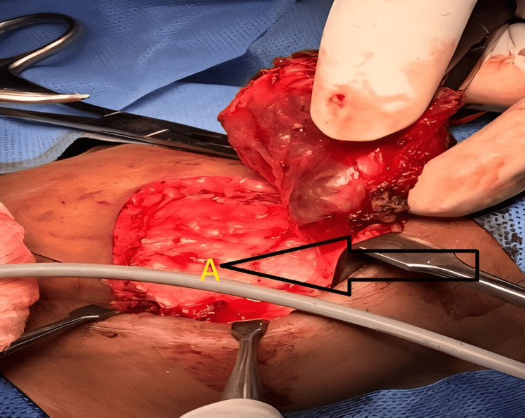 Figure 3
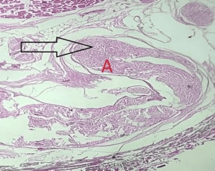 Figure 4
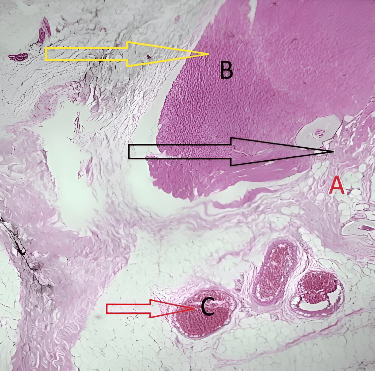 Figure 5
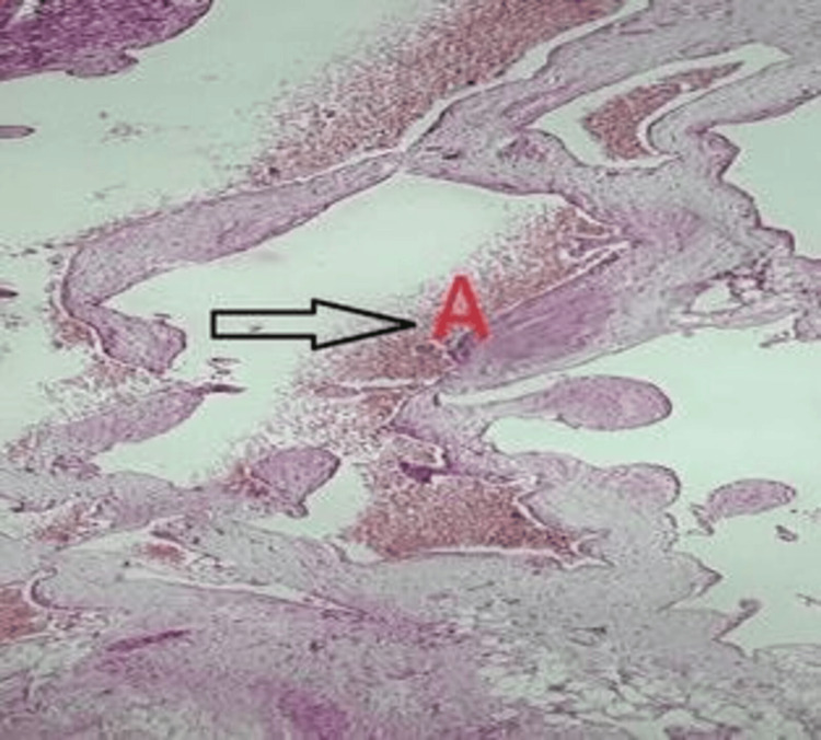 Figure 6
Peer Reviews
No public reviews on file for this paper yet. If you reviewed it on a platform where reviews are public (OpenReview, ICLR, NeurIPS, ICML), you can paste yours below so the community can read it here.
Videos
No videos yet. Explain this paper in a talk, walkthrough, or lecture? Add one.
Taxonomy
TopicsVascular Malformations and Hemangiomas · Vascular Tumors and Angiosarcomas · Central Venous Catheters and Hemodialysis