Pixel-level annotations of context region for trustworthy diagnosis of Breast Lymph Node Metastasis from Histopathological WSI
Richa Malviya Dutta, Arif Ahmed Sekh, Prarthana Raghuram, Krishna Kiran, Shirin Dasgupta, Subhamoy Mandal, Debi Prosad Dogra, Pranab K. Dan

TL;DR
This paper introduces a dataset of breast lymph node metastasis images with precise pixel-level annotations by experts to improve trustworthy automated cancer diagnosis.
Contribution
The paper presents a rare, expert-verified dataset with pixel-level annotations for context regions in breast lymph node metastasis WSI.
Findings
A two-stage annotation protocol was used to create high-quality pixel-level labels for 73 Camelyon16 training images.
Annotations include diagnostically significant regions, background tissue, and artefacts, aiding in reliable patch preparation for metastasis detection.
The dataset supports the development of trustworthy automated diagnosis systems by providing critical context regions for cancer detection.
Abstract
There is a dearth of annotated images in digital pathology, and annotations are pivotal for supervised automated diagnosis. This work aims to create a set of data on breast lymph node metastatic whole slide images (WSI) that is truly valuable as it is annotated by the domain experts in a very precise and intensive manner. The annotations provided here are of rare kind that has pixel-level labels divided into three categories; first is based on the diagnostically significant tissue regions, second is background tissue and the third is artefacts present in breast lymph node metastatic WSI. High level annotations, as is provided in this work, for areas apart from the metastatic region is crucial for diagnosis as this is the context region which is critical for cancer diagnosis. Breast lymph node metastasis is a severe medical condition that requires significant efforts by pathologists to…
Genes, proteins, chemicals, diseases, species, mutations and cell lines named across the full text — each resolved to its canonical identifier and authoritative record.
Click any figure to enlarge with its caption.
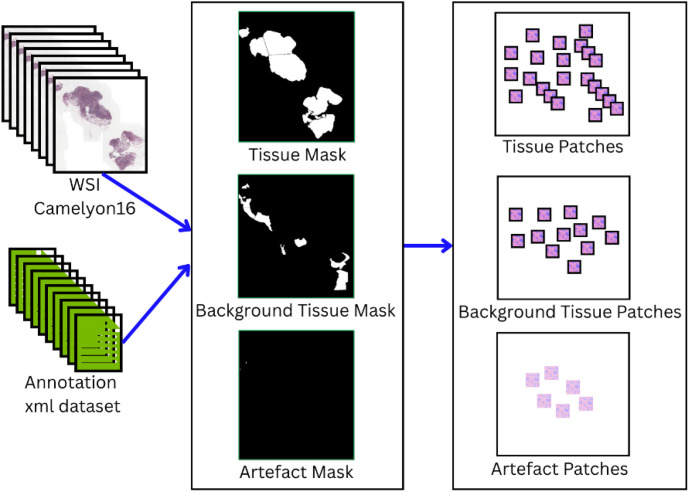 Figure 1
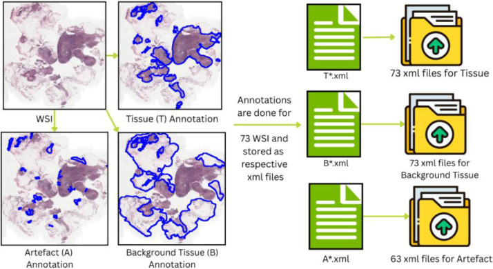 Figure 2
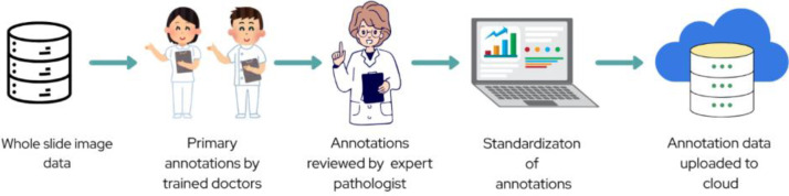 Figure 3
Peer Reviews
No public reviews on file for this paper yet. If you reviewed it on a platform where reviews are public (OpenReview, ICLR, NeurIPS, ICML), you can paste yours below so the community can read it here.
Videos
No videos yet. Explain this paper in a talk, walkthrough, or lecture? Add one.
Taxonomy
TopicsAI in cancer detection · Digital Imaging for Blood Diseases · Breast Lesions and Carcinomas