Vascular graph network for ovarian lesion classification using optical-resolution photoacoustic microscopy
Yixiao Lin, Lukai Wang, Ian S. Hagemann, Lindsay M. Kuroki, Brooke E. Sanders, Andrea R. Hagemann, Cary Siegel, Matthew A. Powell, Quing Zhu

TL;DR
This paper introduces a vascular graph network to classify ovarian lesions using microscopic imaging, improving accuracy in distinguishing cancer from benign conditions.
Contribution
The novel vascular graph network (VGN) leverages vascular structure for lesion classification with high accuracy using minimal tissue sampling.
Findings
VGN achieved 79.5% accuracy in diagnosing ovarian cancer.
The model provided stable predictions from small sampling areas (3 mm× 0.12 mm).
Five-class classification accuracy reached 73.4% using vascular graph data.
Abstract
Diagnosing ovarian lesions is challenging because of their heterogeneous clinical presentations. Some benign ovarian conditions, such as endometriosis, can have features that mimic cancer. We use optical-resolution photoacoustic microscopy (OR-PAM) to study the differences in ovarian vasculature between cancer and various benign conditions. In this study, we converted OR-PAM vascular data into vascular graphs augmented with physical vascular properties. From 94 ovarian specimens, a custom vascular graph network (VGN) was developed to classify each graph as either normal ovary, one of three benign pathologies, or cancer. We demonstrated for the first time that, by leveraging the intrinsic similarity between vascular networks and graph constructs, VGN provides stable predictions from sampling surface areas as small as 3 mm× 0.12 mm. In diagnosing cancer, VGN achieved 79.5 % accuracy and…
Genes, proteins, chemicals, diseases, species, mutations and cell lines named across the full text — each resolved to its canonical identifier and authoritative record.
Click any figure to enlarge with its caption.
 Figure 1
 Figure 2
 Figure 3
 Figure 4
 Figure 5
 Figure 6
 Figure 7
 Figure 8
 Figure 9
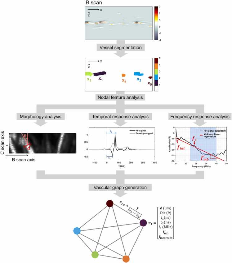 Figure 10
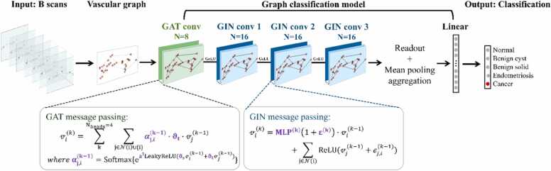 Figure 11
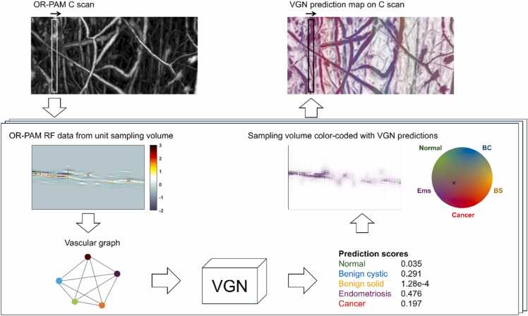 Figure 12
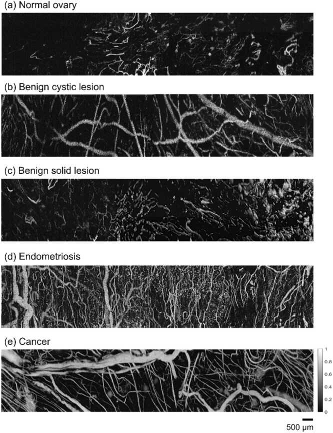 Figure 13
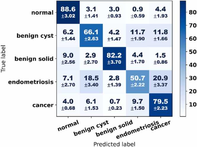 Figure 14
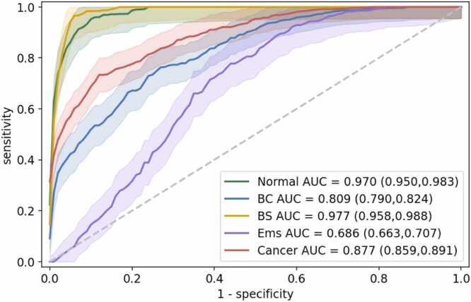 Figure 15
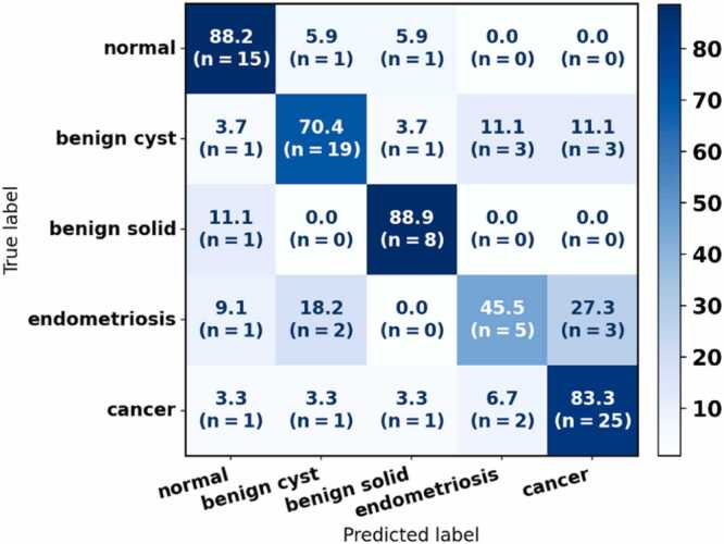 Figure 16
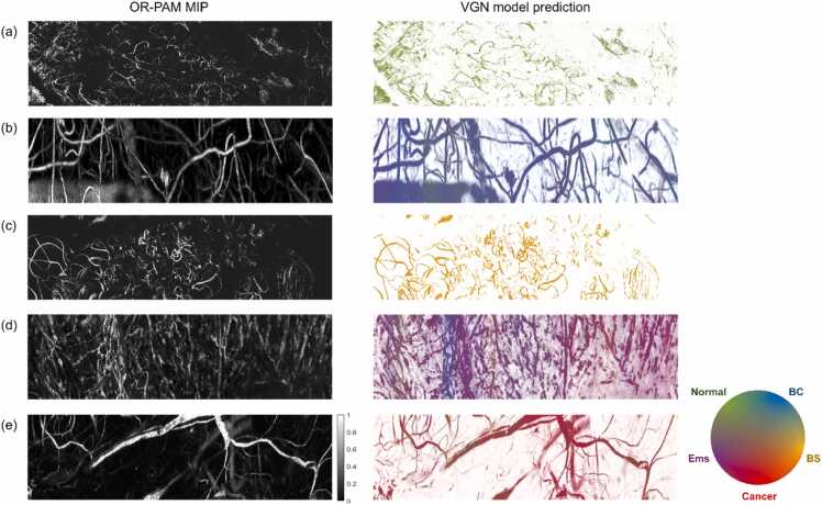 Figure 17
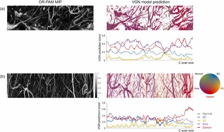 Figure 18
Peer Reviews
No public reviews on file for this paper yet. If you reviewed it on a platform where reviews are public (OpenReview, ICLR, NeurIPS, ICML), you can paste yours below so the community can read it here.
Videos
No videos yet. Explain this paper in a talk, walkthrough, or lecture? Add one.
Taxonomy
TopicsPhotoacoustic and Ultrasonic Imaging · Ultrasound and Hyperthermia Applications · Infrared Thermography in Medicine