Precision, prognosis, and clinical performance of rounded and trabecular segmentation of cine cardiovascular magnetic resonance
George Joy, James C. Moon, Karan Punjabi, Mohammed Alzahir, Jessica Artico, Hunain Shiwani, Iain Pierce, Anish Bhuva, Dhruv Thakur, Hui Xue, Peter Kellman, Erik Schelbert, Thomas A. Treibel, Charlotte Manisty, Rhodri H. Davies

TL;DR
This study compares two automated methods for analyzing heart images and finds both are accurate and useful for clinical decisions.
Contribution
The study introduces an automated method for trabecular segmentation in cardiac MR images with low error rates and high precision.
Findings
Trabecular segmentation showed 4% higher ejection fraction compared to rounded segmentation in healthy subjects.
ML models for both segmentation methods had error rates below 4% and outperformed clinicians in ejection fraction precision.
Both segmentation approaches showed similar prognostication ability for heart failure and mortality outcomes.
Abstract
Measurements of cardiac size and function drive clinical decisions. Left ventricular (LV) metrics can be derived from cardiac MR images by delineating the blood pool and myocardium, by either drawing a rounded contour to approximate the compacted myocardial border, or by delineating the papillary muscles and trabeculae (trabecular segmentation). There is no consensus as to which is best, particularly in the emergent AI era. We developed machine-learning (ML) approaches for both and compared them for clinically important metrics (error rate, precision, and prognosis). Separate ML models were developed for rounded and trabecular segmentation, using U-net models trained on 1923 subjects (mixed pathology, multiple scanners, multiple centers). Blood and myocardial volumes for each segmentation method were compared on 4118 healthy UK biobank subjects. Model segmentation quality was evaluated…
Genes, proteins, chemicals, diseases, species, mutations and cell lines named across the full text — each resolved to its canonical identifier and authoritative record.
Click any figure to enlarge with its caption.
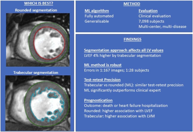 Figure 1
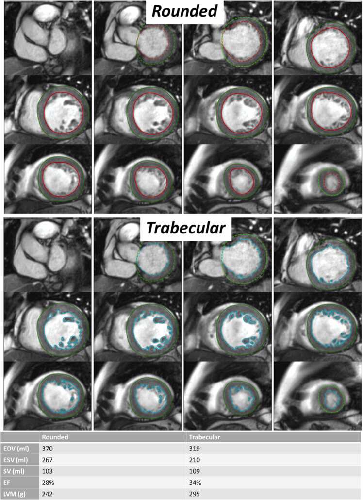 Figure 2
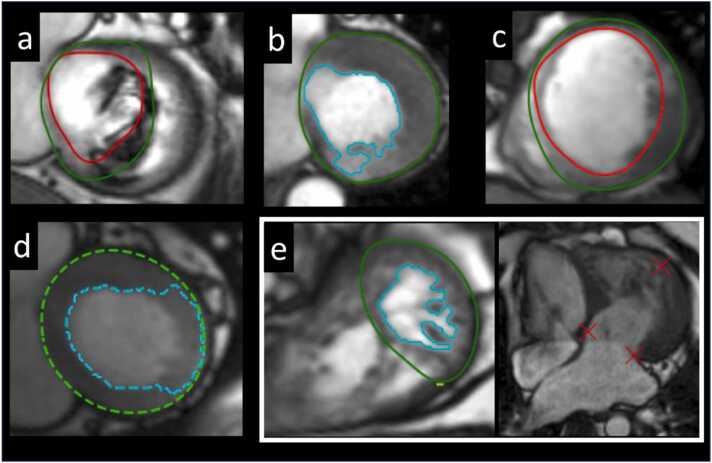 Figure 3
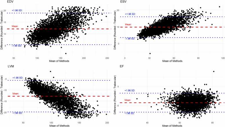 Figure 4
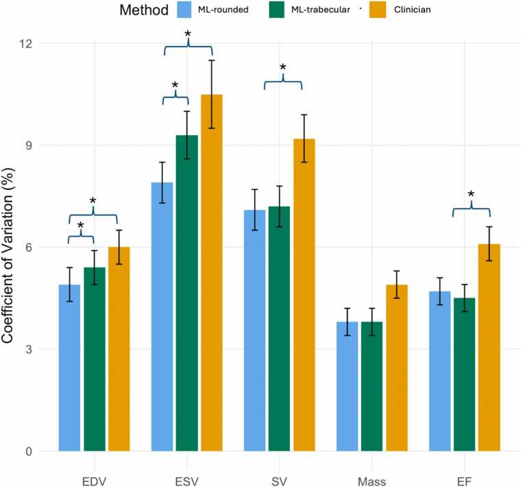 Figure 5
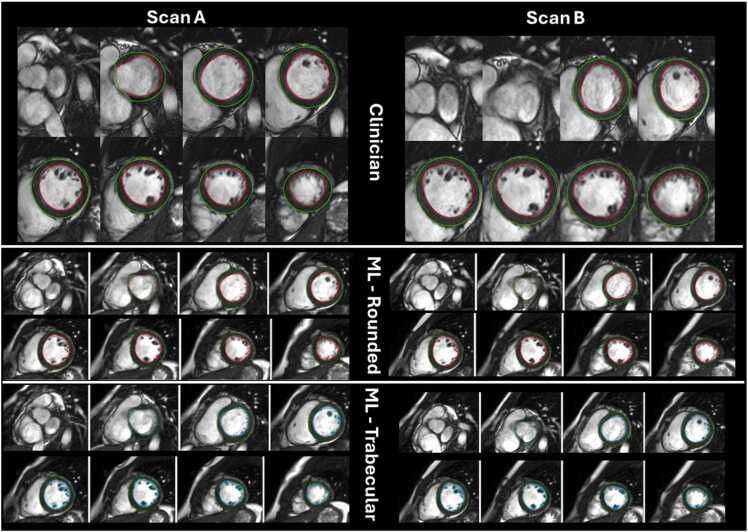 Figure 6
Peer Reviews
No public reviews on file for this paper yet. If you reviewed it on a platform where reviews are public (OpenReview, ICLR, NeurIPS, ICML), you can paste yours below so the community can read it here.
Videos
No videos yet. Explain this paper in a talk, walkthrough, or lecture? Add one.
Taxonomy
TopicsMedical Image Segmentation Techniques · Cardiac Imaging and Diagnostics · Advanced MRI Techniques and Applications