Variability, asymmetry and sexual dimorphism in craniofacial anomalies in Loeys-Dietz syndrome 2: geometric morphometric analysis in mice
Katelin R. Devine, Sarah Lynn, Priyam Jani, Cyrus Keyvanfar, Bikash Lamichhane, Ashleigh S. Hanner, Catharine Dietrich, Rachel S. Chung, Pamela A. Frischmeyer-Guerrerio, Konstantinia Almpani, Olivier Duverger, Janice S. Lee

TL;DR
This study uses 3D analysis to show craniofacial abnormalities in a mouse model of Loeys-Dietz syndrome 2, revealing variability, asymmetry, and sexual dimorphism.
Contribution
The study reveals sexual dimorphism and high variability in craniofacial anomalies in a mouse model of LDS2 using 3D geometric morphometric analysis.
Findings
Craniofacial shape in Tgfbr2G357W/+ mice deviates from wild-type littermates with high variability and asymmetry.
Features like cranial doming and abnormal condylar shape are consistent with LDS patient phenotypes.
Sexual dimorphism is observed, with more severe features in female mice compared to males.
Abstract
Loeys-Dietz syndrome is a rare connective tissue disorder characterized by life-threatening aortic aneurysm and distinctive craniofacial anomalies. It is caused by mutations along the transforming growth factor beta (TGF-β) signaling pathway (LDS1-6). We previously showed that craniofacial anomalies varied among LDS subtypes and that LDS2, caused by mutations in the TGFBR2 gene, exhibited the most severe and variable phenotype. In this study, we performed a thorough qualitative and quantitative analysis of craniofacial anomalies in a mouse model for LDS2, through micro computed tomography and 3D geometric morphometric analysis at multiple postnatal stages. We show that craniofacial shape in Tgfbr2G357W/+ mice strongly deviates from their WT littermates from an early age and exhibit high variability and evidence of left–right asymmetry despite the pure genetic background. Cranial doming,…
Genes, proteins, chemicals, diseases, species, mutations and cell lines named across the full text — each resolved to its canonical identifier and authoritative record.
Click any figure to enlarge with its caption.
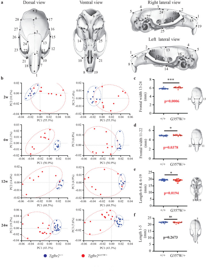 Figure 1
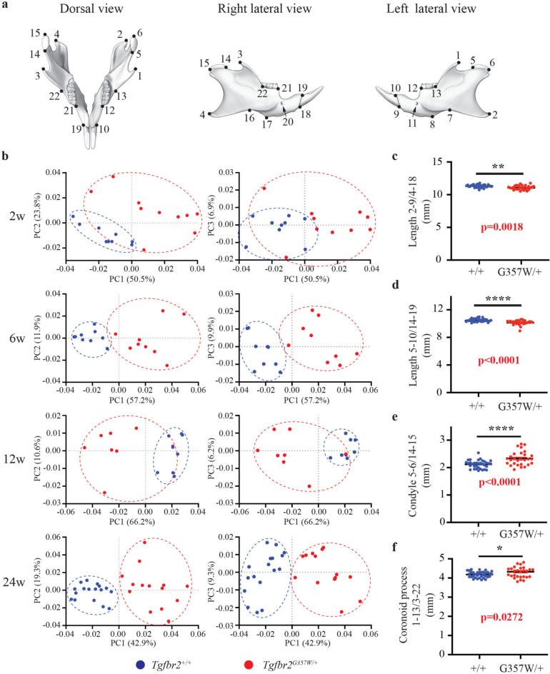 Figure 2
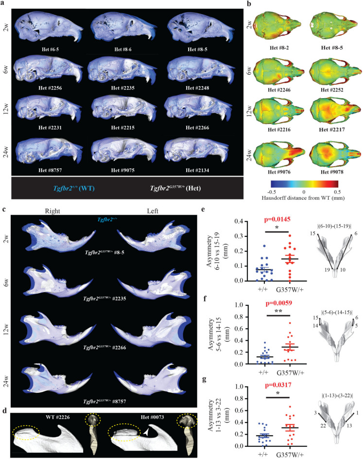 Figure 3
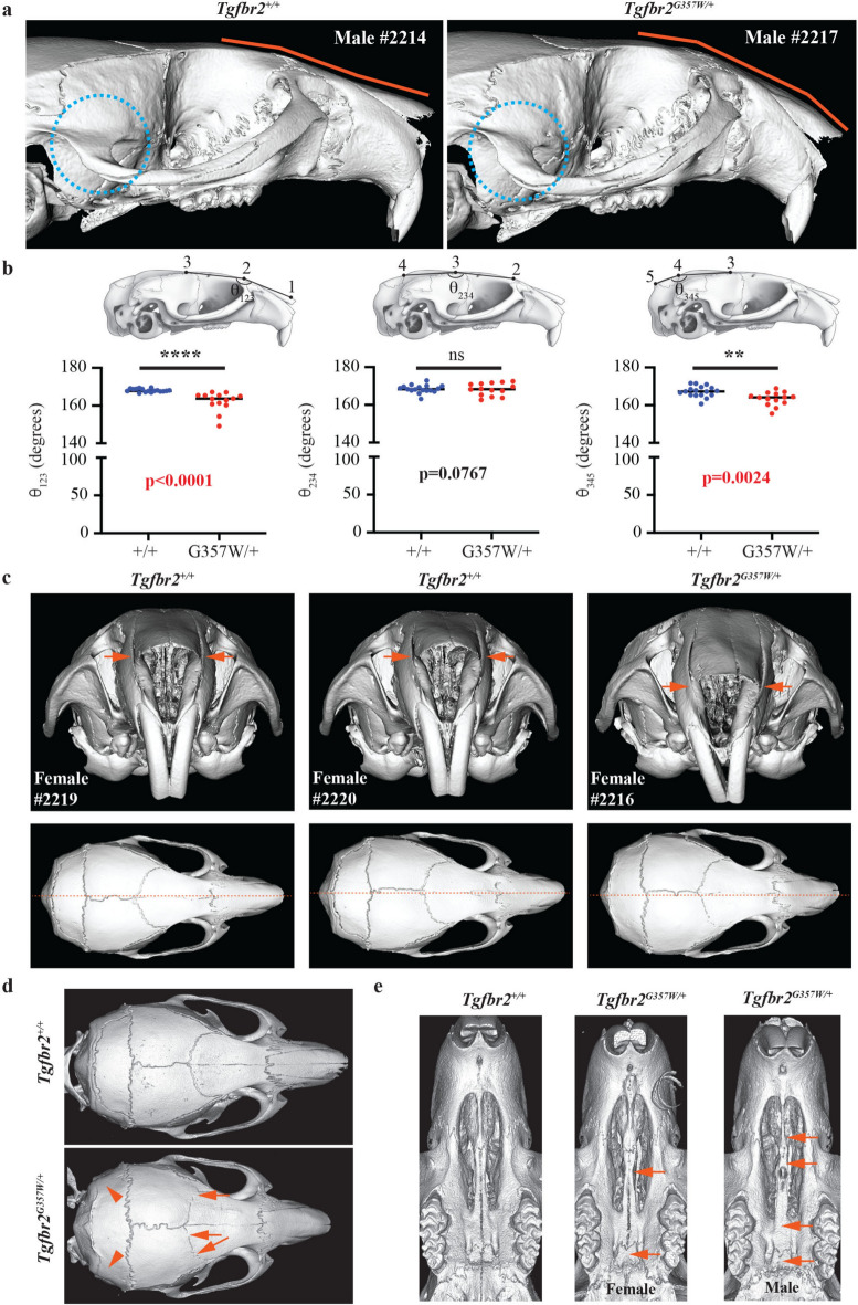 Figure 4
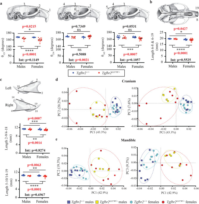 Figure 5
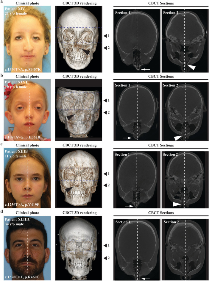 Figure 6
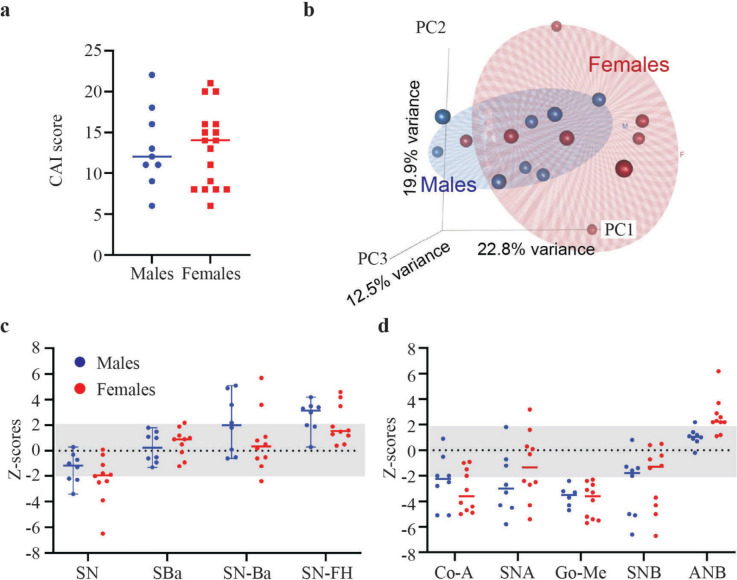 Figure 7
Peer Reviews
No public reviews on file for this paper yet. If you reviewed it on a platform where reviews are public (OpenReview, ICLR, NeurIPS, ICML), you can paste yours below so the community can read it here.
Videos
No videos yet. Explain this paper in a talk, walkthrough, or lecture? Add one.
Taxonomy
TopicsConnective tissue disorders research · Aortic aneurysm repair treatments · Aortic Disease and Treatment Approaches