The Diagnostic Value of Confocal Laser Endomicroscopy in Brain Tumours When Performed by Blinded, Untrained Neuropathologists
Ilona Iff, Marielena Gutt‐Will, Theoni Maragkou, Andrea Mathis, Kathleen Seidel, David Capper, Elisabeth G. Hain, Jenny Meinhardt, Regina Von Manitius, Carsten Dittmayer, Simone Schmid, Ekkehard Hewer, Andreas Raabe, Philippe Schucht

TL;DR
This study evaluates how well untrained neuropathologists can use confocal laser endomicroscopy (CLE) to diagnose brain tumors, finding it less accurate than traditional histology methods.
Contribution
The study provides the first assessment of CLE's baseline diagnostic accuracy by untrained neuropathologists in a blinded setting.
Findings
Untrained neuropathologists correctly identified neoplastic tissue in 70.7% of cases using CLE.
H & E staining showed higher accuracy, correctly identifying neoplastic tissue in 87.6% of cases.
Training in CLE image interpretation is recommended to improve diagnostic accuracy.
Abstract
Achieving maximal and safe tumour resection is a key goal in brain tumour surgery. Confocal laser endomicroscopy (CLE) enables real‐time visualisation of the tissue microstructure at a cellular level, potentially helping neurosurgeons distinguish non‐neoplastic from neoplastic tissue. The core aim of this study was to determine the baseline diagnostic accuracy that can be achieved with CLE alone, when assessed by neuropathologists without prior CLE training and without any additional clinical or contextual information, and to compare these findings to standard haematoxylin and eosin (H & E)‐based histology in a blinded setting. CLE images and corresponding H & E‐stained slides from 100 brain tumour patients treated at the University Hospital Bern over a 22‐month period were analysed. Five blinded neuropathologists with no prior CLE experience independently evaluated the data sets.…
Genes, proteins, chemicals, diseases, species, mutations and cell lines named across the full text — each resolved to its canonical identifier and authoritative record.
Click any figure to enlarge with its caption.
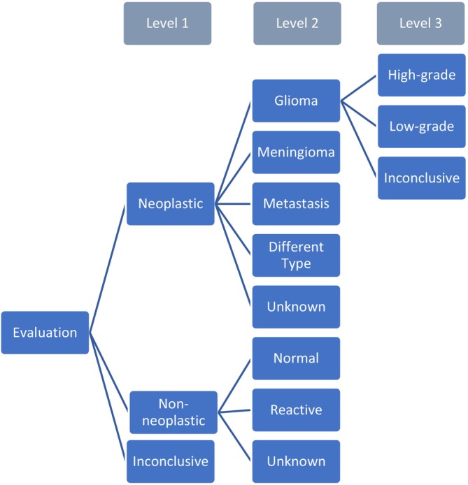 Figure 1
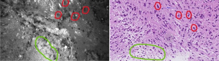 Figure 2
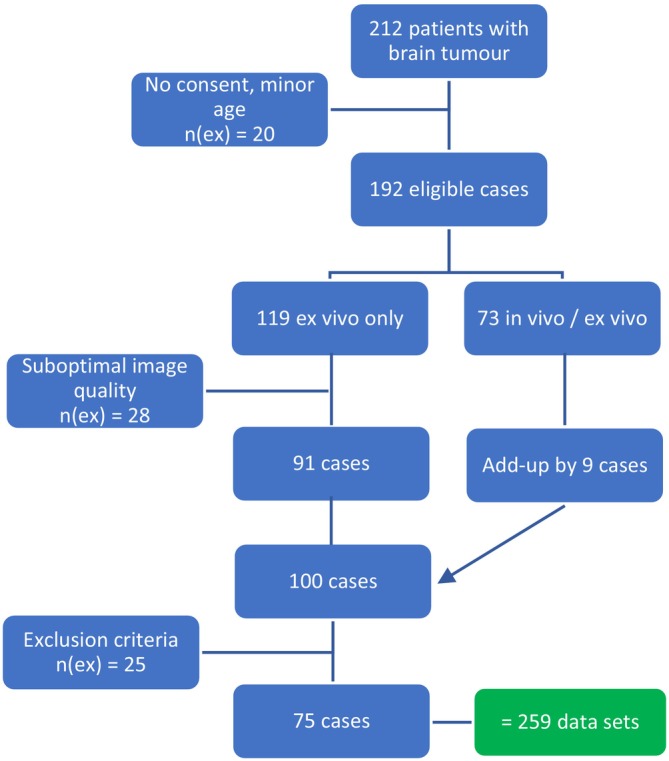 Figure 3
Peer Reviews
No public reviews on file for this paper yet. If you reviewed it on a platform where reviews are public (OpenReview, ICLR, NeurIPS, ICML), you can paste yours below so the community can read it here.
Videos
No videos yet. Explain this paper in a talk, walkthrough, or lecture? Add one.
Taxonomy
TopicsGlioma Diagnosis and Treatment · Esophageal Cancer Research and Treatment · Cerebrospinal fluid and hydrocephalus