Association of oncogene mutations with clinical and histopathological characteristics in patients with metastatic melanoma
Julia Amaral Quintiliano, Adauto Ferreira Nunes, Luiza Pinheiro-Hubinger-Stauffer

Abstract
Genes, proteins, chemicals, diseases, species, mutations and cell lines named across the full text — each resolved to its canonical identifier and authoritative record.
Click any figure to enlarge with its caption.
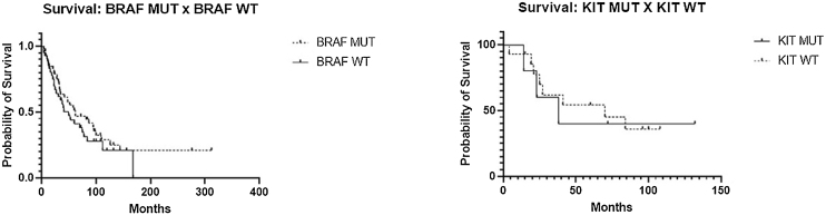 Figure 1
Peer Reviews
No public reviews on file for this paper yet. If you reviewed it on a platform where reviews are public (OpenReview, ICLR, NeurIPS, ICML), you can paste yours below so the community can read it here.
Videos
No videos yet. Explain this paper in a talk, walkthrough, or lecture? Add one.
Taxonomy
TopicsMelanoma and MAPK Pathways · Cutaneous Melanoma Detection and Management · Cancer Genomics and Diagnostics
Dear Editor,
Melanoma is recognized as a highly aggressive form of skin cancer and exhibits the highest mutation rates of all solid tumors. Among the genetic alterations identified, mutations in specific oncogenes, particularly in the BRAF gene, are the most prevalent.1 Characterizing the genetic profile of tumors is essential, as several approved and investigational therapies are effective only in the presence of specific mutagenic alterations.2
The clinical and epidemiological investigation of factors associated with metastatic melanoma can offer valuable insights into disease progression and inform preventive strategies and targeted therapeutic approaches. In this context, we retrospectively evaluated the mutational status of the BRAF, PDGFRA, and C-KIT genes in patients with metastatic melanoma treated at a Brazilian tertiary oncology hospital, correlating these findings with epidemiological and clinical parameters.
Medical records of 94 patients diagnosed with melanoma and treated between 2015 and 2022 at Hospital Amaral Carvalho (HAC), Jaú, São Paulo, Brazil, were reviewed. Clinical and histopathological data were collected. Exon 15 of the BRAF gene (codon 600 and adjacent regions) was sequenced in all cases. In selected samples, additional analyses for C-KIT and PDGFRA mutations were performed based on physician requests.
DNA was extracted using the QIAAMP DNA FFPE Tissue Kit (QIAGEN). PCR amplification employed primers previously described by Qiu et al.3 and Braggio et al.4 DNA sequencing was conducted on an ABI PRISM 3100 Genetic Analyzer-3130xl system.
Descriptive and inferential statistical analyses were performed using Pearson’s chi-square test or Fisher’s exact test, binomial logistic regression (variables included age, gender, patient status, primary tumor site, histological subtype, ulceration, Breslow thickness, and mitotic index), and Kaplan-Meier survival analysis. Cases with missing data were excluded from specific analyses. A significance level of 5% was adopted. This study was approved by the Ethics Committees of the Lauro de Souza Lima Institute and Hospital Amaral Carvalho (Protocol 58842922.0.3001.5434).
Table 1 summarizes the demographic and tumor-specific characteristics of the cohort. The most frequent site of metastasis was the regional lymph nodes (61.7%), followed by the lungs (13.8%), bones (6.4%), liver (4.3%), and other locations (13.8%) (Supplementary Table S1).Table 1. Clinical characteristics of the patients and anatomopathological variables of the primary tumor.Table 1. VariableCategoryn%GenderFemale4547.9Male4952.1Age at diagnosis<40 years old1718.140‒59 years old3739.4>60 years old4042.5Primary tumor siteHead or neck1819.1Upper limb099.6Trunk2627.7Lower limb3335.1Unidentified088.5Histological subtypeSuperficial extensive2930.9Nodular2627.6Acral1111.7Lentigo maligno011.1Others033.2No information2425.5Breslow index<1 mm1313.81.01 – 2 mm066.42.01 – 4 mm2223.4>4 mm3133.0No information2223.4UlcerationYes4446.8No5053.2Mitotic index<1 mitosis/mm^2^1515.9>1 mitosis/mm^2^4547.9No information3436.2LDH at diagnosisLow1313.8Normal4143.6High1010.6No information3032.0LDH at metastasisLow055.3Normal2930.9High1718.1No information4345.7Patient StatusAlive3234.0Death6266.0Overall survival<5-years4572.6>5-years1727.4Observed survival>5-years2475.0<5-years after diagnosis825.0
Among the 94 patients, 41 (43.6%) exhibited wild-type BRAF (BRAF-WT) and 53 (56.4%) harbored BRAF mutations: 48 (51%) had the V600E mutation, 4 (4.3%) had the V600 K mutation, and 1 (1.1%) had the L597Q mutation.
Nineteen patients underwent analysis for C-KIT mutations: 14 (73.7%) were wild-type, and 5 (26.3%) presented mutations (two in exon 17, and one each in exons 13, 11, and 9). Among five acral melanoma biopsies analyzed for C-KIT, 2 (40%) harbored mutations (in exons 9 and 13). The remaining C-KIT–mutated tumors were classified as nodular, polypoid-nodular, or superficial extensive melanomas.
Twelve patients were analyzed for PDGFRA mutations, all of whom were wild-type.
The association between BRAF mutational status and clinical-pathological variables is presented in Table 2. BRAF mutations were significantly associated with younger age at diagnosis (p = 0.0085).Table 2. Relationship between clinical-pathological variables and the presence of BRAF mutation.Table 2. VariableCategoryBRAF-MUTBRAF-WTTotalp-value^a,b^Logistic RegressionOR (95% CI)p-valueGenderFemale261945Male2722490.7938bAge at diagnosis, years<401403170.946 (0.910–0.983)0.00440‒592314370.0085b>60162440Primary tumor siteHead or neck110718Upper limbs040509Trunk1709260.3891bLower limbs151833Histological typeSuperficial extensive1910290.161 (0.0276–0.935)d0.042dOthers1823410.074bBreslow index<1 mm0904132.490 (0.964–6.432)0.060>1 mm3227590.3709aUlcerationYes222244No2615410.2126bMitotic index, mitosis/mm^2^<1080412>12226480.3334aLDH at diagnosis, U/LUp to 480302454Above 480060410>0.9999aLDH at metastasis, U/LUp to 480191524Above 4801106170.5461bcSurvival<5-years242145>5-years2615410.3439baFisher analysis.bChi-Square analysis.cThree patients with BRAF mutation and five BRAF-WT patients who were diagnosed with the disease less than 5-years ago and are still being monitored were excluded.dSignificance was found in univariate analysis for the acral subtype and BRAF-WT.
In univariate logistic regression (n = 56), younger age was associated with BRAF mutation (p = 0.004, OR = 0.946, 95% CI: 0.910–0.983). The acral subtype was associated with wild-type tumors and superficial extensive cases with the BRAF mutation (p = 0.042, OR = 0.161, 95% CI: 0.0276–0.935). Breslow thickness was marginally related to WT tumors (p = 0.060, OR = 2.490, 95% CI: 0.964–6.432). In multivariate analysis, younger age remained the only independent factor associated with BRAF mutation (p = 0.003). No statistically significant associations were observed for C-KIT mutations (Table 3).Table 3. Relationship between clinical-pathological variables and presence of c-KIT mutation.Table 3. VariableCategoryKIT-MUTKIT-WTTotalp-valueGenderFemale040610Male0108090.3034aAge at diagnosis<60-years-old000606>60-years-old0508130.1280aPrimary tumor siteHead/neck/trunk020406Upper and lower limbs031013>0.9999aHistological typeSuperficial extensive010405Others0406100.6004aBreslow index<1 mm010203>1 mm040711>0.9999aUlcerationYes050914No0004040.2778aMitotic index<1 mitosis/mm^2^000202>1 mitosis/mm^2^0508130.5238aLDH at diagnosisUp to 480 U/L040610Above 480 U/L0100010.4545aLDH at metastasisUp to 480 U/L010607Above 480 U/L000202>0.9999aSurvival<5-years030609>5-years020709>0.9999aOne KIT-WT patient who was diagnosed with the disease less than 5-years ago and is still being monitored was excluded.aFisher analysis.
At the time of data collection, the median survival time was 62-months for patients with BRAF-mutated tumors and 50-months for BRAF-WT patients. The Hazard Ratio (HR) for death in the BRAF-WT group was 1.331. For KIT-mutated patients, the median survival was 38-months, compared to 70-months in KIT-WT patients (HR = 1.145) (Fig. 1).Fig. 1. Survival curve BRAF-MUT × BRAF-WT and c-KIT-MUT × c-KIT-WT.Fig. 1
A slight male predominance was observed, and the onset of melanoma was predominantly from the fifth decade of life onwards, consistent with previous reports.5 Although the lower limbs were the most frequently affected primary site in this cohort, prior studies have more commonly reported the trunk and lower limbs.5 This discrepancy may reflect cultural or behavioral differences in sun exposure patterns.
Survival analysis revealed that 72.6% of deceased patients (n = 62) had a survival time of less than 5-years, while 75% of survivors (n = 32) remained alive beyond five years. Prognostic factors influencing melanoma survival include tumor thickness, ulceration, mitotic index, and metastatic burden.6
The most prevalent histological subtype was superficial extensive melanoma (30.9%), followed by the nodular subtype (27.6%). These findings may be attributable to the association between these subtypes and chronic or intermittent sun exposure. Additionally, more than 60% of patients exhibited a high Breslow index, 50% had a mitotic rate >1 mm^2^, and approximately one-third presented with tumors >4 mm in thickness ‒ all features linked to poor prognosis.6
Younger age was significantly correlated with BRAF mutation, in line with previous studies.7 The superficial extensive and nodular subtypes also demonstrated a higher frequency of BRAF mutations. Although not statistically significant, primary tumors located on the trunk and lower limbs tended to exhibit more BRAF mutations than those on the upper limbs or head and neck.
While the majority of patients with lower Breslow thickness and lower mitotic index harbored BRAF mutations, the prognostic relevance of BRAF status remains controversial, as some studies report no consistent correlation between BRAF mutations and histopathological parameters.8
KIT mutations were more frequent in female patients aged over 60-years. All KIT-mutated tumors exhibited ulceration and a mitotic index >1 mm^2^, both unfavorable prognostic markers.6 Notably, KIT mutations were identified in 40% of acral melanoma cases, a subtype typically classified as “non-solar” melanoma, potentially associated with mechanical trauma rather than UV radiation. Acral melanomas generally demonstrate a lower frequency of point mutations but a higher rate of gene copy number variations.9
The median 5-year survival was higher in patients with BRAF mutations and slightly lower in those without mutations. Reports indicate that patients with BRAF mutations treated with selective BRAF inhibitors can achieve a 5-year survival rate of up to 60% in some cases.10
The median 5-year survival was higher in patients without c-KIT mutations. Of the 19 patients tested for KIT mutations in this study, only 5 (26.3%) had mutations in this gene. Previous studies have reported that less than 10% of melanoma cases involve KIT mutations.11 Treatment with KIT inhibitors typically demonstrates lower therapeutic efficacy compared to other selective inhibitors, which may be associated with decreased survival.2
In this study, BRAF mutations were found to be associated with younger age and the superficial spreading subtype. No significant correlation was observed between KIT mutations and the examined variables. Our findings provide important insights into the clinical, pathological, and molecular characteristics of melanoma, offering valuable contributions to the development of future research focused on preventive and therapeutic strategies.
ORCID ID
Quintiliano JA: 0009-0003-9488-1880
Nunes AF: 0000-0003-3473-9252
Pinheiro-Hubinger-Stauffer L: 0000-0001-7377-7652
Financial support
This research did not receive any specific grant from funding agencies in the public, comercial, or not-for-profit sectors.
Authors' contributions
Julia Amaral Quintiliano: Data collection, analysis and interpretation; statistical analysis; article writing.
Adauto Ferreira Nunes: Data interpretation; critical review of important intellectual content.
Luiza Pinheiro-Hubinger-Stauffer: Study conception and design; collection, analysis and interpretation of data; statistical analysis; research guidance; approval of the final version of the manuscript.
Research data availability
The entire dataset supporting the results of this study was published in this article.
Conflicts of interest
None declared.
The reference list from the paper itself. Each links out to its DOI / PubMed record.
- 1Hodis E.Watson I.R.Kryukov G.V.Arold S.T.Imielinski M.Theurillat J.P.A landscape of driver mutations in melanoma Cell.15020122512632281788910.1016/j.cell.2012.06.024PMC 3600117 · doi ↗ · pubmed ↗
- 2Melis C.Rogiers A.Bechter O.van den Oord J.J.Molecular genetic and immunotherapeutic targets in metastatic melanoma Virchows Arch.47120172812932835748910.1007/s 00428-017-2113-3 · doi ↗ · pubmed ↗
- 3Lv J.Dai B.Kong Y.Shen X.Kong J.Acral melanoma in chinese: a clinicopathological and prognostic study of 142 cases Sci Rep.620163143210.1038/srep 31432 PMC 499286027545198 · doi ↗ · pubmed ↗
- 4Qiu T.Lu H.Guo L.Huang W.Ling Y.Shan L.Detection of BRAF mutation in Chinese tumor patients using a highly sensitive antibody immunohistochemistry assay Sci Rep.52015921110.1038/srep 09211 PMC 436382825784606 · doi ↗ · pubmed ↗
- 5Fernandes N.C.Calmon R.Maceira J.P.Cuzzi T.Silva C.S.C.Melanoma cutâneo: estudo prospectivo de 65 casos An Bras Dermatol.8020052534
- 6Balch C.M.Gershenwald J.E.Soong S.J.Thompson J.F.Atkins M.B.Byrd D.R.Final version of 2009 AJCC melanoma staging and classification J Clin Oncol.272009619962061991783510.1200/JCO.2009.23.4799 PMC 2793035 · doi ↗ · pubmed ↗
- 7Saint-Jean M.Quereux G.Nguyen J.M.Peuvrel L.Brocard A.Vallee A.Younger age at the time of first metastasis in BRAF-mutated compared to BRAF wild-type melanoma patients Oncol Rep 3220148088142492683610.3892/or.2014.3265 · doi ↗ · pubmed ↗
- 8Jung J.E.Falk T.M.Bresch M.Matias J.E.F.Böer A.BRAF mutations in cutaneous melanoma: no correlation with histological prognostic factors or overall survival J Bras Patol Med Lab.462010487493