Loop-mediated isothermal amplification combined with a Pyrococcus furiosus argonaute system for the rapid detection of goose astrovirus
Fosheng Yang, Zhong Liu, Jianqiang Ye, Quan Xie, Chang Wu, Chengjun Jiang, Huangsheng Wu, Qianlang Gu, Deping Song, Fanfan Zhang

TL;DR
This paper introduces a new method for quickly and accurately detecting goose astrovirus using a combination of RT-LAMP and a Pyrococcus furiosus Argonaute system.
Contribution
The novel contribution is combining RT-LAMP with PfAgo for specific and sensitive detection of goose astrovirus.
Findings
The assay detected as few as 10 copies of GAstV per microliter.
The method showed no cross-reactivity with other viruses and bacteria.
Clinical sample results matched those of qPCR, confirming its accuracy.
Abstract
Goose astrovirus (GAstV) is a newly emerged viral pathogen in goose, characterized by a high incidence and significant mortality rates. This etiology has repeatedly occurred in coastal areas of China, rapidly spreading to inland provinces in recent decade, and thus imposed huge economic losses on China’s goose industry. Therefore, it is essential to establish a rapid, accurate and sensitive method for GAstV diagnosis. In this study, we employed reverse transcription loop-mediated isothermal amplification (RT-LAMP) assay combined with a Pyrococcus furiosus Argonaute (PfAgo) system, providing a simple and precise approach for GAstV. Specific primers and guide DNA (gDNA) were designed to target conserved regions of the GAstV genome. The assay achieved a detection limit of 10 copies/μL in preliminary validation assays when targeting conserved regions of the viral genome with optimized…
Genes, proteins, chemicals, diseases, species, mutations and cell lines named across the full text — each resolved to its canonical identifier and authoritative record.
Click any figure to enlarge with its caption.
 Figure 1
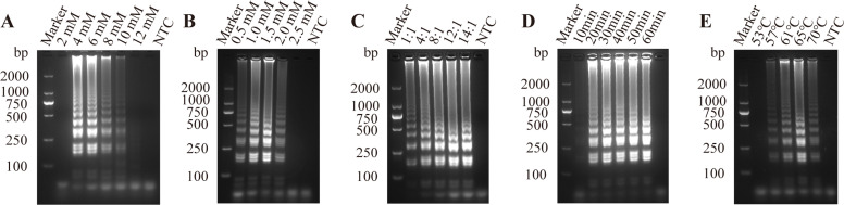 Figure 2
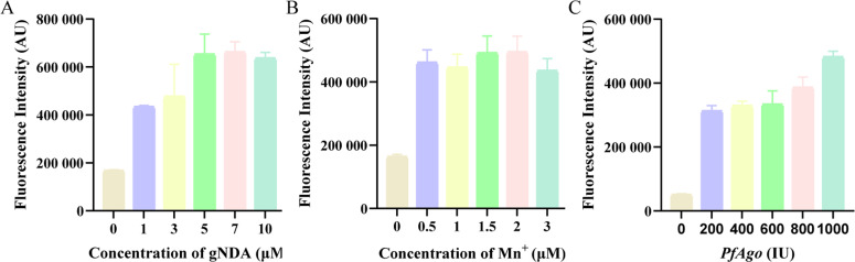 Figure 3
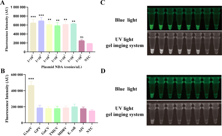 Figure 4
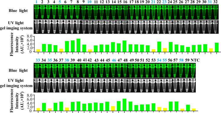 Figure 5
Peer Reviews
No public reviews on file for this paper yet. If you reviewed it on a platform where reviews are public (OpenReview, ICLR, NeurIPS, ICML), you can paste yours below so the community can read it here.
Videos
No videos yet. Explain this paper in a talk, walkthrough, or lecture? Add one.
Taxonomy
TopicsViral gastroenteritis research and epidemiology · Respiratory viral infections research · Virus-based gene therapy research
Introduction
GAstV is a newly emerged viral pathogen that causes novel gosling gout disease with two established genotypes (GAstV-I and GAstV-II) (Xu et al., 2023; Zhang et al., 2022). GAstV is the pathogen responsible for novel gosling gout disease, characterized by urate deposition in internal organs and joints, presenting typical gout symptoms (Wang et al., 2022). Current epidemiological data confirm GAstV-II as the predominant etiological agent responsible for gout outbreaks in geese (Zhu and Sun, 2022). GAstVs are non-enveloped viruses with a positive-sense, single-stranded RNA genome of 7.1 to 7.3 Kb (Yuan et al., 2019), which includes a 5′-untranslated region (UTR), three open reading frames (ORFs; ORF1a, ORF1b, and ORF2), a 3′UTR, and a poly(A) tail (Zhang et al., 2018a). ORF1a and ORF1b are highly conserved sequences encoding important non-structural proteins (NSPs) involved in viral replication and transcription (Cortez et al., 2017; Fu et al., 2022).
Since 2017, outbreaks of gout associated with goose astrovirus infection have been reported across China. This virus primarily affects goslings within 20 days of age, leading to growth retardation, younger goslings exhibiting higher mortality (An et al., 2020). The disease has spread rapidly nationwide, causing substantial economic losses (Wang et al., 2021). To date, studies on GAstV remain preliminary, and there is no effective treatment or vaccine. The primary focus of disease management is on outbreak surveillance and containment. Therefore, rapid diagnosis is critical for early surveillance and control of GAstV outbreak risks.
Diagnostic approaches for GAstV include virus isolation, enzyme-linked immunosorbent assay (ELISA), reverse transcription-polymerase chain reaction (RT-PCR), reverse transcription-quantitative PCR (RT-qPCR), RT-LAMP, and combined reverse transcription-enzymatic recombinase amplification (RT-ERA) with the Cas12a. Zhang et al. first discovered that GAstV could be isolated and propagated in LMH cell, a chicken liver cell line (Zhang et al., 2018b). However, isolating GAstV is difficult and time-consuming. Serological methods are fast and easy to operate but cannot timely and accurately reflect GAstV infection (He et al., 2023; Zhang et al., 2023). RT-PCR and RT-qPCR have been established for the detection of GAstV, usually with good accuracy and specificity, but they require cumbersome and precise equipment (Liu et al., 2025a; Yi et al., 2022; Yin et al., 2020). Furthermore, in recent years, with rapid advances in isothermal amplification technologies, such as LAMP, recombinase polymerase amplification (RPA), and recombinase-aided amplification (RAA) have been employed for the detection of various pathogens (Fan et al., 2020; Mao et al., 2022; Yu et al., 2019). However, the result is limited by non-specific amplification and false-positive. Currently, CRISPR-based rapid detection methods have been developed to mitigate false-positive results (Li et al., 2018; Liu et al., 2023; Yang et al., 2022). However, their application remains constrained by the requirement for protospacer adjacent motif (PAM) sequences adjacent to the target sites.
Argonaute (Ago)-based detection methods are increasingly recognized as a promising platform (Kropocheva et al., 2022). These methods activate nucleases through nucleic acid targeting, generating detection signals with high specificity without being limited by specific target sequences (Zhao et al., 2024). PfAgo is a prokaryotic argonaute (pAgo) from Pyrococcus furiosus, is commonly employed for nucleic acid detection (Wang et al., 2023; Ye et al., 2022). In this study, a rapid detection method for GAstV was established by integrating LAMP and PfAgo protein technologies. This method can effectively reduce the false positives by the specifically recognition and cleavage of LAMP products via PfAgo, and improve the detection limit of PfAgo through isothermal amplification (Xun et al., 2021; Yu et al., 2025). In the LAMP-PfAgo analytical method established in this research, PfAgo is a prokaryotic Argonaute (pAgo) from Pyrococcus furiosus (Swarts et al., 2015). As a nucleic-acid-guided endonuclease, it specifically cleaves the target between the 10th and 11th nucleotide bases from the 5′ to 3′ ends of aligned gDNA under the guidance of three short 5′-phosphorylated single-strand DNA, and producing a 16 nucleotide 5′-phosphorylated single-stranded DNA (ssDNA) fragment. The cleavage generates a ssDNA fragment that functions as a secondary gDNA to guide PfAgo for secondary cleavage, leading to the split of the quenchers from the fluorophore (He et al., 2019). Finally, the fluorescence signal can be detected with a real-time fluorescent qPCR detector or fluorescence spectrometer. Probe and gDNA sequences were designed between the FIP and BIP of the LAMP primer. This design ensures that the sequences recognized by PfAgo are derived from the amplification of taret sequences rather than non-specific amplification in the LAMP reaction. To prevent aerosol contamination during the reaction, the LAMP amplification system was added to the bottom of the reaction tube, and the PfAgo cleavage system was added to the lid of the tube. After the completion of the LAMP amplification reaction, a simple centrifugation step was performed to mix the LAMP and PfAgo detection systems, and the results can be visualized under UV light. Therefore, the single-tube LAMP-PfAgo detection platform minimizes aerosol contamination and enables rapid and sensitive detection of GAstV. In this study, we developed a single-tube assay for detecting GAstV that integrates LAMP with the PfAgo system. This method is completed within 60 minutes with high sensitivity and specificity, providing a rapid, reliable and convenient method for GAstV detection.
Materials and methods
Ethics statement
All samples were collected on commercial goose farms by veterinarians during routine diagnostic sampling after permission from the farm owner. No specific permits from an animal ethics committee were required.
viruses and clinical samples
The GAstV strain JXGZ/2021 (GenBank accession number: OL762473) was provided by the Institute of Animal Husbandry and Veterinary Medicine, Jiangxi Academy of Agricultural Sciences. Viruses isolated in this study were archived at the Institute of Animal Husbandry and Veterinary Medicine, Jiangxi Academy of Agricultural Sciences, including: GPV, GoCV, TMUV, and E. coli. Viral genomic RNA was extracted using RNAiso Plus (Takara, Beijing, China) according to the manufacturer's instructions, and cDNA synthesis was performed with PrimeScript RT Master Mix (Takara, Beijing, China). The resulting cDNA/DNA templates were stored at −80°C until further use.
Construction of recombinant plasmid
The target fragments were amplified by PCR, purified using a Gel Extraction Kit (Omega Bio-tek, Guangzhou, China), and cloned into the pMD-19T vector (Takara, Beijing, China). The recombinant plasmids were transformed into Top10 competent cells (Tolo Bio, Hong Kong, China). DNA sequencing was performed by Sangon Biotech (Shanghai, China). Plasmid extraction used with a TIAN prep Mini Plasmid Kit (TIANGEN, Beijing, China). The concentration was measured on a NanoDrop 2000 spectrophotometer (Thermo Fisher Scientific, Waltham, MA, USA). The plasmid copy number was calculated using the following formula: copies/μL = (6.02 × 10^23^) × (ng/μL × 10^-9^) / (length of DNA × 660). Serial 10-fold dilutions were performed to obtain plasmid concentrations ranging from 1 × 10^6^ to 1 × 100 copies/μL.
Primer design
The conserved ORF1b gene region of GAstV was targeted to design LAMP primers using the online primer software Primer Explorer V5 (https://primerexplorer.eiken.co.jp/e/), including an external primer pair (F3 and B3), an internal primer pair (FIP and BIP), and two loop primers (LB and LF). Additionally, three gDNA sequence and corresponding probes were designed based on PfAgo cleavage specificity, the probes were modified with FAM, and the 5′ ends of the gDNA were phosphorylated. The primers qPCR-F and qPCR-R were utilized for qPCR targeting GAstV (Xu, 2019). The designed primers, gDNAs, and probes were synthesized by Sangon Biotech (shanghai China). The sequence information for the primers and gDNAs is listed in Table 1.Table 1. Sequences of primers, gDNAs and probe used in this study.Table 1. LabelSequence (5′−3′)F3GCAGGACCAGAATGAGAAB3TCTTATGATGGTTGGACAGGAAFIPTCCACCAAAAAAGGGTGTCCATGAAGCAACAGACAGAACGBIPACAGGTTTTTTGTAGAGACGGATTCGCATCTGTCGTATYCGCLFCAACTTGTGCAGCCCGCLBGGACGCGTTATGATGGTACGgDNA1GAATACATCAGCGAGTgDNA2AGATACTCGCTGATGTgDNA3CACCACCAATGAGCCTProbeFAM-GAGCCTAGATACTCGCT-BHQ1qPCR-FTGAAGCAACAGACAGAACGGqPCR-RGGACAGGAAAAAGTAACGCA
Optimization of LAMP reaction conditions
LAMP reactions were performed following the manufacturer's protocol for Bst 2.0 WarmStart DNA Polymerase (New England Biolabs, Ipswich, MA, USA). Each 25 μL reaction mixture contained 2.5 μL of 10 × Isothermal Amplification Buffer, 6 mM MgSO₄, 1.4 mM dNTP mix (Takara, Beijing, China), 1.6 μM internal primer (FIP/BIP), 0.2 μM external primers (F3/B3), 0.4 μM loop primers (LF/LB), 8 U Bst 2.0 WarmStart DNA Polymerase, and 1 μL template DNA, with nuclease-free water added to achieve a final volume of 25 μL. Reactions were incubated at 65°C for 30 min in a thermal cycler or isothermal heating device. Amplification products were electrophoresed on 1.5% agarose gels in 1 × TAE buffer or directly visualized by adding 1 × SYBR Green I in the reaction tube for diction by a color change. To achieve the best reaction conditions, a systematic optimization process was carried out by adjusting the concentration of MgSO₄(2, 4, 6, 8, 10, 12 mM), the concentration of dNTPs (0.2, 0.4, 0.6, 0.8, 1.0, 1.2, 1.4 mM), internal and external primer concentration ratio (1:1, 4:1, 8:1, 12:1, 14:1), reaction temperature (53, 57, 61, 65, 69°C), and reaction time (10, 20, 30, 40, 50, 60 min).
Optimization of the LAMP-PfAgo reaction system
First, LAMP reaction mixture was dispensed into the tube bottoms. Then PfAgo system was added, which included 2 μL of 10 × reaction buffer, 1.0 mM Mn²⁺, 2.0 μM of each gDNA, and 3 μL PfAgo (200 U/μL), and tube caps were affixed to filter paper discs preloaded with 0.25 μM probes. To immerse discs, tubes were centrifuged at 5,000 × g for 1 min after incubation for 30 minutes at 65°C. Tubes were mixed and incubated for 30 min at 95°C. The reaction results were observed in a fluorescence detector or under UV light. To achieve the best reaction conditions, a systematic optimization process was carried out by adjusting the concentration of gDNA (1, 3, 5, 7, 10 μM), MnCl (0.5, 1.0, 1.5, 2.0, 3.0 μM), and PfAgo enzyme (200, 400, 600, 800, 1000 U).
LAMP-PfAgo sensitivity and specificity tests
To evaluate the sensitivity, serially diluted plasmids were used as templates to determine detection limit of the LAMP-PfAgo assay in vitro. For specificity assay, six common pathogens were analyzed. The pathogens included GPV, GoCV, TMUV, MDRV, E. coli. Results were visualized under UV light or blue light conditions after 30 min LAMP-PfAgo reaction at 95°C.
Clinical evaluation of LAMP-PfAgo method
To validate the LAMP-PfAgo assay for GAstV detection, 59 clinical intestinal samples from gout-afflicted geese were collected. All samples underwent concurrent analysis using both LAMP-PfAgo and qPCR for comparative evaluation.
Statistical analysis
Schematic diagrams were created using Adobe Illustrator 2020 (Adobe Inc., San Jose, CA). Data were analyzed by one-way ANOVA using GraphPad Prism 5.0 (GraphPad Software, San Diego, CA). Values are presented as mean ± SEM. Experiments included three technical replicates. Statistical significance is indicated as follows: *P < 0.05, **P < 0.01, ***P < 0.001, and ****P < 0.0001.
Results
Mechanism of the LAMP-PfAgo detection method
Fig. 1 illustrates the workflow for detecting GAstV genomic RNA using the LAMP-PfAgo assay. Briefly, the LAMP amplicons are specifically identified by PfAgo protein under the guidance of 5′-phosphorylated guide DNA (gDNA). Following base pairing between the gDNA and one strand of the LAMP amplicon, PfAgo cleaves the phosphodiester bond between the 10th and 11th nucleotides of the target DNA, thereby generating a newly formed 5′-phosphorylated ssDNA. This ssDNA can subsequently serve as a gDNA to guide PfAgo to cleave the secondary cleavage molecular beacon probe, releasing the fluorescent groups. The fluorescent signal can be detected with a fluorescence detector or visualized under UV or blue light.Fig. 1. Workflow of the RT-LAMP-PfAgo assay for GAstV detection.Fig 1
Optimization of LAMP reaction
To optimize the LAMP reaction system, the concentrations of key components were adjusted, including the concentration of MgSO4, the concentration of dNTPs, internal and external primer concentration ratio (Fig. 2A–C). To further enhance amplification efficiency, reaction time and temperature were optimized (Fig. 2D and E). Taken together, the optimal conditions were determined as follows: 6 mM MgSO4, 1.4 mM dNTP, an internal to external primer concentration ratio of 12:1, a reaction temperature of 65°C, and a reaction time of 30 min.Fig. 2. Optimization of LAMP reaction conditions. Screening for optimal concentrations of (A) MgSO_4_, (B) dNTPs, (C) internal and external primers for LAMP assay; (D) Screening for optimal reaction times and (E) temperature for LAMP assay.Fig 2
Optimization of LAMP-PfAgo reaction
To enhance the LAMP-PfAgo reaction efficiency, the concentrations of key components were optimized, including MnCl₂, gDNA, and PfAgo (Fig. 3A and C). The optimal reaction conditions were determined as follows: 7 μM gDNA, 1.5 mM MnCl2, and 1000 U of PfAgo.Fig. 3. Optimization of components for the LAMP-PfAgo assay. Fluorescence intensity of LAMP-PfAgo reaction at different concentrations of (A) Mn^2+^, (B) PfAgo, and (C) gDNA.Fig 3
Sensitivity and specificity of the LAMP-PfAgo
To evaluate the sensitivity of the LAMP-PfAgo assay, the recombinant plasmid templates serially diluted from 1 × 10⁶ to 1 × 100 copies/μL were tested to determine the detection limit. Results demonstrated a limit of detection (LOD) of 10 copies/μL (Fig. 4A), with visual detection under UV and blue light yielding consistent results (Fig. 4B). Specificity testing of the LAMP-PfAgo assay was performed using common clinical pathogens including GPV, GoCV, and TMUV. The assay produced fluorescence signals only when GAstV was detected, with no cross-reactivity detected with other pathogens (Fig. 4C), with visual detection under UV and blue light yielding consistent results (Fig. 4D).Fig. 4. Evaluation of the sensitivity and specificity of LAMP-PfAgo assay. Endpoint fluorescence values indicating LAMP-PfAgo (A) sensitivity and (B) specificity; Images capture indicating (C) sensitivity and (D) specificity under UV light and blue light. Each experiment was performed in triplicate and statistical analysis was conducted using student’s t-test in GraphPad Prism 8. NTC indicates negative control; ** indicates p < 0.01; *** indicates p < 0.001; **** indicates p < 0.0001; ns indicates no significance.Fig 4
Clinical sample evaluation
To evaluate the clinical applicability of the LAMP-PfAgo assay, the clinical samples suspected of GAstV infection were tested using both RT-LAMP-PfAgo and qPCR assays. Both assays detected 40 positive samples and 30 negative samples. The images captured under UV or blue light were consistent with those of fluorescence detection (Fig. 5). These findings demonstrate that the LAMP-PfAgo method is suitable for the detection of GAstV in clinical diagnostic specimens.Fig. 5. Reliability of the LAMP-PfAgo assay for detecting clinical samples. Samples in colored in blue indicate a negative result. NTC indicates negative control.Fig 5
Discussion
Since 2016, a disease characterized primarily by urate deposition in visceral organs and joints has been observed in several regions of China, including Guangdong, Hunan, Shandong, Jiangsu, Fujian, and Henan (Wan et al., 2018). The pathogen predominantly affects goslings within 20 days of age, with mortality rates reaching up to 50%, causing significant economic losses to the poultry industry in China. Currently, there are no effective detection methods or preventive measures available. Moreover, as most farms are located in remote areas, the risk of disease transmission is indirectly increased. To date, several diagnostic methods have been developed for GAstV, including ELISA, RT-PCR, RT-qPCR, RT-LAMP, and RT-ERA-CRISPR/Cas12a.Although RT-PCR and RT-qPCR are widely used for viral detection, they rely on specialized equipment and involve complex procedures, limiting their applicability for clinical diagnosis. Compared with other rapid detection systems, such as RT-LAMP and RT-ERA-CRISPR/Cas12, standalone RT-LAMP can rapidly amplify nucleic acids, but the results are prone to false positive results due to non-specific amplification. Although RT-ERA-CRISPR/Cas12a system has high sensitivity, it si limited by the high synthetic cost of gDNA and the dependence on the PAM. Therefore, the development of a rapid, accurate, sensitive, portable, and cost-effective diagnostic technology is urgently needed to enable efficient on-site screening.
PfAgo can bind to short gDNA molecules for specific target recognition and exhibits the ability of precise guided cleavage (Hong et al., 2025; Swarts et al., 2015). This capability ensures the targeted elimination of non-specific amplification products, significantly reducing the possibility of false-positive results, providing a new platform for nucleic acid molecular diagnosis, and demonstrating broad application potential (Wu et al., 2023). In recent years, the combined application of PfAgo protein and isothermal amplification technology has pioneered a detection method with high sensitivity, high specificity, and rapidity in the field of nucleic acid detection, demonstrating tremendous advantages in on-site rapid diagnosis (Liu et al., 2024, 2025b; Yu et al., 2024).We combine PfAgo with LAMP to increase the convenience and specificity of detection based on the high sensitivity and specificity of PfAgo cleavage activity, and it can effectively prevent false positive results caused by the amplification process, thereby ensuring the accuracy of the diagnostic results because of its stepwise cleavage specificity. This method was further validated with 30 clinical samples. Our findings demonstrated that the RT-LAMP-pfAgo based assay yielded identical results to those obtained from RT-qPCR tests, indicating that its reliability was comparable to that of the current gold standard. Additionally, the RT-LAMP-pfAgo assay is faster and involves a simpler workflow compared to qPCR.
In summary, the RT-LAMP-pfAgo assay developed in the study can detect GAstV, and offers advantages including operational simplicity, rapidity, and high specificity. The assay is applicable for GAstV detection in epidemiological surveys.
Conclusions
In summary, we have successfully developed a rapid and highly sensitive detection method for detecting GAstV by integrating LAMP with PfAgo. The sensitivity of this LAMP-PfAgo assay is 10 copies/µL, and it does not cross-react with other pathogens. Furthermore, the procedure is straightforward does not require precision instruments, making it suitable for the clinical detection of GAstV.
Disclosures
Author Baobao Xie is employed by Dabeinong Technology Co.,Ltd. of Jiangxi. Author Yangyang Luo is employed by Wen's Foodstuff Group Co., Ltd., Wen's Group Research Institute. The remaining authors declare that the research was conducted in the absence of any commercial or financial relationships that could be construed as a potential conflict of interest.
Data availability statement
The data that support the findings of this study are available from the corresponding authors upon reasonable request.
CRediT authorship contribution statement
Fosheng Yang: Writing – original draft, Software, Methodology, Data curation. Zhong Liu: Validation, Supervision, Methodology, Formal analysis. Jianqiang Ye: Writing – review & editing, Writing – original draft, Funding acquisition. Quan Xie: Resources, Project administration, Methodology, Investigation. Chang Wu: Methodology, Investigation, Formal analysis. Chengjun Jiang: Validation, Data curation, Conceptualization. Huangsheng Wu: Resources, Project administration. Qianlang Gu: Validation, Supervision, Methodology, Conceptualization. Deping Song: Writing – review & editing, Writing – original draft. Fanfan Zhang: Writing – review & editing, Resources, Methodology, Funding acquisition.
The reference list from the paper itself. Each links out to its DOI / PubMed record.
- 1An D.Zhang J.Yang J.Tang Y.Diao Y.Novel goose-origin astrovirus infection in geese: the effect of age at infection Poult. Sci.99920204323433310.1016/j.psj.2020.05.04132867976 PMC 7598121 · doi ↗ · pubmed ↗
- 2Cortez V.Meliopoulos V.A.Karlsson E.A.Hargest V.Johnson C.Schultz-Cherry S.Astrovirus biology and pathogenesis Annu. Rev. Virol 41201732734810.1146/annurev-virology-101416-04174228715976 · doi ↗ · pubmed ↗
- 3Fan X.Li L.Zhao Y.Liu Y.Liu C.Wang Q.Dong Y.Wang S.Chi T.Song F.Sun C.Wang Y.Ha D.Zhao Y.Bao J.Wu X.Wang Z.Clinical validation of two recombinase-based isothermal amplification assays (RPA/RAA) for the rapid detection of African Swine Fever virus Front. Microbiol.112020169610.3389/fmicb.2020.0169632793160 PMC 7385304 · doi ↗ · pubmed ↗
- 4Fu X.Hou Z.Liu W.Cao N.Liang Y.Li B.Jiang D.Li W.Xu D.Tian Y.Huang Y.Insight into the epidemiology and evolutionary history of novel goose astrovirus-associated gout in Goslings in southern China Viruses 1462022130610.3390/v 1406130635746777 PMC 9230684 · doi ↗ · pubmed ↗
- 5He D.Sun M.Jiang X.Zhang S.Wei F.Wu B.Diao Y.Tang Y.Development of an indirect competitive ELISA method based on ORF 2 detecting the antibodies of novel goose astrovirus J. Virol. Methods 311202311464310.1016/j.jviromet.2022.11464336332715 · doi ↗ · pubmed ↗
- 6He R.Wang L.Wang F.Li W.Liu Y.Li A.Wang Y.Mao W.Zhai C.Ma L.Pyrococcus furiosus Argonaute-mediated nucleic acid detection Chem. Commun.55882019132191322210.1039/c 9cc 07339 f 31589231 · doi ↗ · pubmed ↗
- 7Hong M.Wu G.Ren Y.Wu S.Zhu H.Chen Z.Advancements in pathogen detection: argonaute-based nucleic acid detection technology Pathogens 146202555410.3390/pathogens 1406055440559562 PMC 12196298 · doi ↗ · pubmed ↗
- 8Kropocheva E.V.Lisitskaya L.A.Agapov A.A.Musabirov A.A.Kulbachinskiy A.V.Esyunina D.M.Prokaryotic argonaute proteins as a tool for biotechnology Mol. Biol.566202285487310.1134/s 002689332206010336060308 PMC 9427165 · doi ↗ · pubmed ↗