Apical Sparing of Longitudinal Strain and Myocardial Fibrosis in Hypertensive Patients and Spontaneously Hypertensive Rats: Based on Speckle Tracking and Histological Analysis
Chunyan Huang, Yongxin Wu, Meiyan Lin, Yupeng Chen, Shengnan Lin, Liyun Fu, Huimei Huang

TL;DR
This study finds that in hypertension, heart muscle damage and fibrosis mainly occur in the base of the heart, while the tip remains relatively unaffected, linking this pattern to worsening heart function.
Contribution
The study identifies a novel 'basal-damage, apical-sparing' pattern in myocardial strain and fibrosis in hypertensive patients and rats, linking it to diastolic dysfunction and hypertrophy progression.
Findings
Myocardial dysfunction and fibrosis show regional heterogeneity with more damage at the base and sparing at the apex in hypertensive cardiac hypertrophy.
Apical sparing pattern correlates with diastolic dysfunction and hypertrophic progression in hypertension.
Spontaneously hypertensive rats show increased fibrosis in basal segments but not in apical segments compared to controls.
Abstract
This study aimed to investigate regional myocardial strain and fibrosis distribution to analyze the apical sparing pattern and the relation with hypertrophy in hypertension. This study included clinical and experimental animal investigations. Seventy‐three hypertensive patients were divided into two groups: hypertension without left ventricular hypertrophy (HT‐NLVH) and hypertension with LVH (HT‐LVH). Six 16‐week‐old male spontaneously hypertensive rats (SHR) and six age‐matched male Wistar‐Kyoto (WKY) rats were included in this experiment. Echocardiographic measurements were obtained. Myocardial strain indexes, including global longitudinal strain (GLS), the basal, middle, and apical segmental LS (LS‐bas, LS‐mid, LS‐ap), and the proportion of LS‐ap/(LS‐bas + LS‐mid + LS‐ap) (P‐ap) were measured. The histological collagen volume fraction (CVF) and perivascular collagen area (PVCA) of…
Genes, proteins, chemicals, diseases, species, mutations and cell lines named across the full text — each resolved to its canonical identifier and authoritative record.
Click any figure to enlarge with its caption.
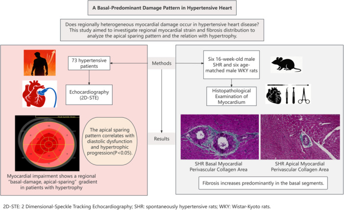 Figure 1
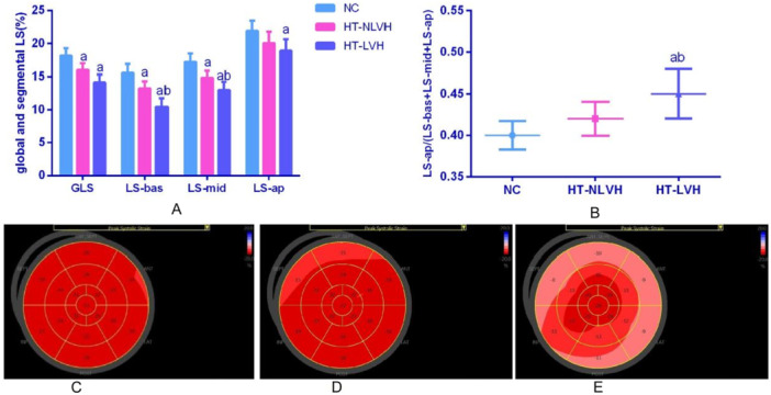 Figure 2
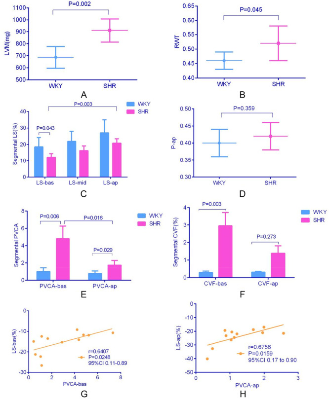 Figure 3
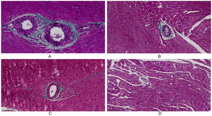 Figure 4
Peer Reviews
No public reviews on file for this paper yet. If you reviewed it on a platform where reviews are public (OpenReview, ICLR, NeurIPS, ICML), you can paste yours below so the community can read it here.
Videos
No videos yet. Explain this paper in a talk, walkthrough, or lecture? Add one.
Taxonomy
TopicsCardiovascular Function and Risk Factors · Cardiovascular Health and Disease Prevention · Cardiac Fibrosis and Remodeling