Combining 3D printing and elastographic characterization: A novel sphenoid wing meningioma simulation model for neurosurgical training
Sifian Al-Hamid, Fränze Grellmann, Julius Reiser, Firat Taskaya, Vanessa M. Swiatek, Klaus-Peter Stein, Amir Amini, Karl Hartmann, Yuzhe Fan, Ali Rashidi, Claus-Dieter Ohl, I. Erol Sandalcioglu, Belal Neyazi

TL;DR
This paper introduces a realistic and cost-effective 3D-printed simulator for training neurosurgeons to remove sphenoid wing meningiomas, validated using advanced imaging techniques.
Contribution
The novel integration of 3D printing and shear wave elastography for creating a biomechanically validated meningioma simulator.
Findings
A 10% cake glaze formulation best mimicked real tumor tissue in elasticity and surgical realism.
Novice participants showed significant improvement in surgical performance across simulation rounds.
The simulator was rated highly for realism, educational value, and motivation by users.
Abstract
High-fidelity simulation models are crucial for advancing neurosurgical training, particularly for complex skull base pathologies such as sphenoid wing meningiomas. This study introduces and validates a novel, cost-effective 3D-printed simulator specifically designed for sphenoid wing meningioma resection. A key innovation of this model is the integration of shear wave elastography (SWE) to enable objective biomechanical validation of tumor-mimicking materials. A patient-specific skull model was created using fused deposition modeling (FDM) 3D printing and paired with custom-molded tumor replicas. In a material validation substudy, 14 tumors made from seven candidate materials were assessed through SWE-based elasticity measurements and blinded evaluations by experienced neurosurgeons, focusing on tactile feedback, anatomical resemblance, and microsurgical handling. The most suitable…
Genes, proteins, chemicals, diseases, species, mutations and cell lines named across the full text — each resolved to its canonical identifier and authoritative record.
Click any figure to enlarge with its caption.
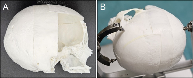 Figure 1
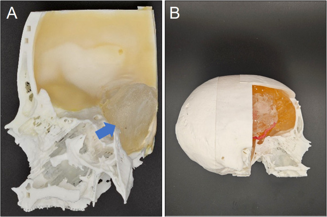 Figure 2
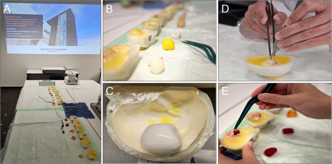 Figure 3
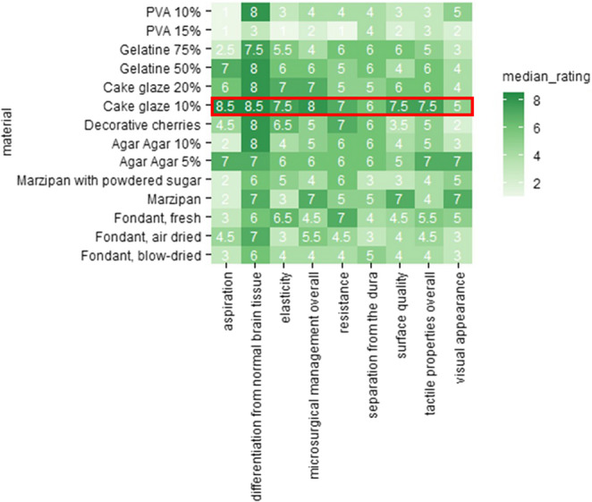 Figure 4
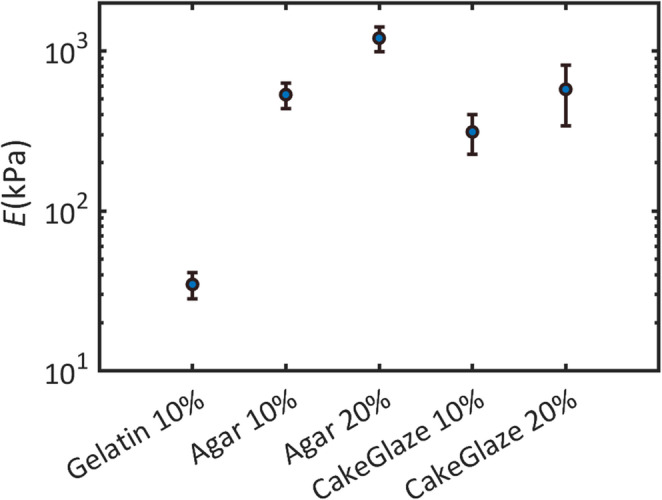 Figure 5
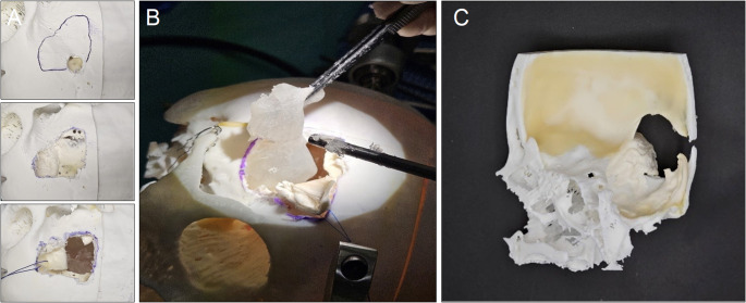 Figure 6
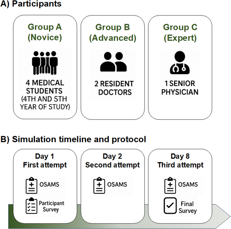 Figure 7
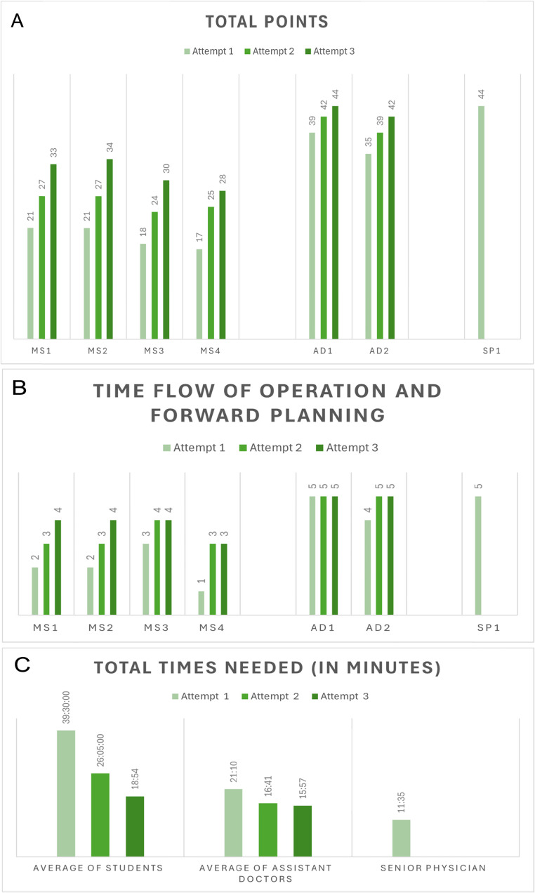 Figure 8
 Figure 9
Peer Reviews
No public reviews on file for this paper yet. If you reviewed it on a platform where reviews are public (OpenReview, ICLR, NeurIPS, ICML), you can paste yours below so the community can read it here.
Videos
No videos yet. Explain this paper in a talk, walkthrough, or lecture? Add one.
Taxonomy
TopicsSurgical Simulation and Training · Cervical and Thoracic Myelopathy · Ultrasound Imaging and Elastography