Combining Correlative Cryogenic Fluorescence and Electron Microscopy and Correlative Cryogenic Super‐Resolution Fluorescence and X‐Ray Tomography—Novel Complementary 3D Cryo‐Microscopy Across Scales to Reveal Nanoparticle Internalization Into Cancer Cells
Pavitra Sokke Rudraiah, Louisa Herbsleb, Michaela Salakova, Henriette Gröger, Anna Maria Steyer, Frauke Alves, Claus Feldmann, Andreas Walter

TL;DR
This paper introduces a new cryogenic microscopy method to study how nanoparticles enter and accumulate in cancer cells, offering insights for drug delivery.
Contribution
A novel 3D cryogenic microscopy workflow combining multiple imaging techniques to study nanoparticle internalization in near-native cells.
Findings
Nanoparticles are internalized within 2 hours and accumulate in endolysosomes over time.
Quantitative analysis shows increased endolysosomal accumulation from 2 to 24 hours.
The workflow provides sufficient contrast and resolution across modalities for detailed nanoparticle tracking.
Abstract
Understanding the intracellular fate of nanoparticles (NPs) is essential for advancing nanomedicine, particularly in targeted drug delivery for cancer therapy. Here, we present a complementary cryogenic microscopy workflow across scales to investigate the uptake and subcellular localization of zirconyl‐containing inorganic–organic hybrid nanoparticles (IOH‐NPs) in murine breast cancer cells. Our approach integrates cryogenic fluorescence microscopy (cryo‐FM), cryo‐focused ion beam scanning electron microscopy (cryo‐FIBSEM), and cryo‐soft X‐ray tomography (cryo‐SXT), enabling molecular specificity, high‐resolution imaging, and volumetric ultrastructural analysis in near‐native cellular states. We demonstrate that the cryogenic workflow provides enough contrast and resolution across all modalities for quantifying the IOH‐NP uptake: NPs are internalized within 2 h of incubation and…
Genes, proteins, chemicals, diseases, species, mutations and cell lines named across the full text — each resolved to its canonical identifier and authoritative record.
Click any figure to enlarge with its caption.
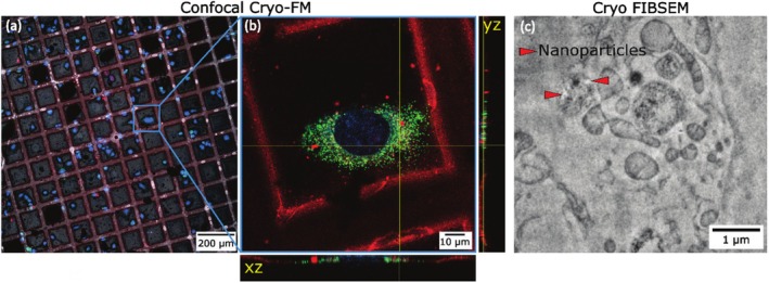 Figure 1
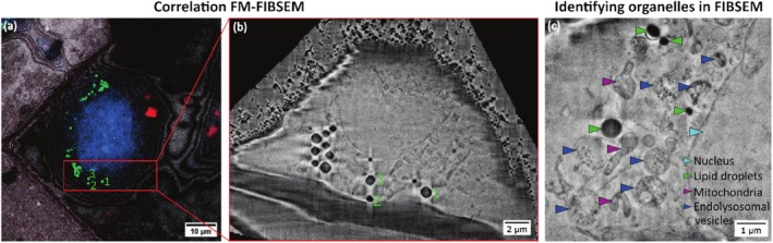 Figure 2
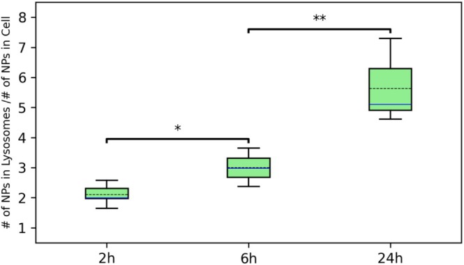 Figure 3
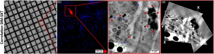 Figure 4
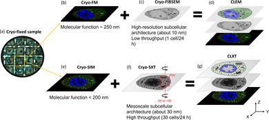 Figure 5
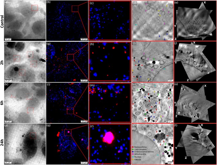 Figure 6
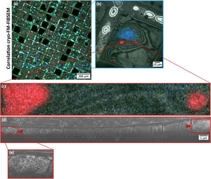 Figure 7
 Figure 8
Peer Reviews
No public reviews on file for this paper yet. If you reviewed it on a platform where reviews are public (OpenReview, ICLR, NeurIPS, ICML), you can paste yours below so the community can read it here.
Videos
No videos yet. Explain this paper in a talk, walkthrough, or lecture? Add one.
Taxonomy
TopicsAdvanced Electron Microscopy Techniques and Applications · Nanoparticle-Based Drug Delivery · Electron and X-Ray Spectroscopy Techniques