Wound Healing Activity of Uncaria callophylla Stem Methanol Extract in Diabetic Rats and Its Phytochemical Profile
Astri Rozanah Siregar

TL;DR
This study shows that a methanol extract from Uncaria callophylla stems helps heal wounds in diabetic rats more effectively than some conventional treatments.
Contribution
The study demonstrates the wound healing efficacy and phytochemical profile of Uncaria callophylla extract in diabetic wound models.
Findings
UCSME ointment significantly accelerated wound closure and improved epithelialization in diabetic rats.
The extract enhanced angiogenesis, collagen synthesis, and inflammatory cell activity in wound healing.
Phytochemical analysis revealed high levels of phenolics and flavonoids with strong antioxidant activity.
Abstract
Impaired wound healing is a major complication in diabetic patients. Currently available wound therapies may cause adverse effects, highlighting the need for safer, natural alternatives. This study evaluated the wound healing potential of Uncaria callophylla Blume ex Korth. stem methanol extract (UCSME) in diabetic rats. Diabetic excision wound models were established and animals were divided into six groups: non-diabetic untreated wounds, diabetic untreated wounds, diabetic wounds treated with UCSME ointment at concentrations of 5%, 10%, and 20%, and diabetic wounds treated with betadine ointment. Wound healing was assessed through wound area reduction, epithelialization time, inflammatory and proliferative cell counts, angiogenesis, collagen deposition, and hydroxyproline content. Phytochemical composition and antioxidant activity were also analyzed. UCSME ointment significantly…
Genes, proteins, chemicals, diseases, species, mutations and cell lines named across the full text — each resolved to its canonical identifier and authoritative record.
Click any figure to enlarge with its caption.
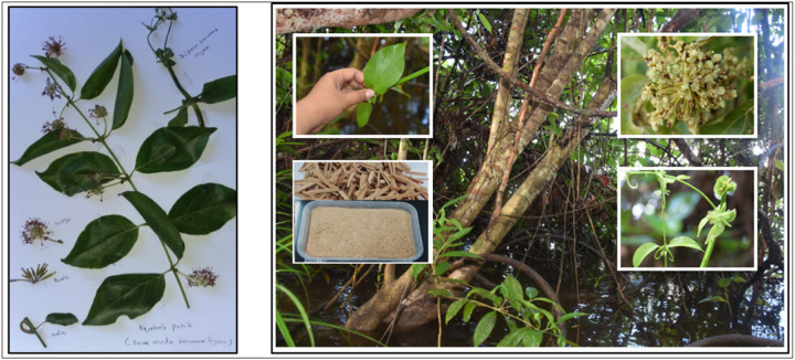 Figure 1
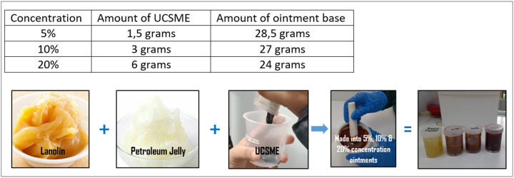 Figure 2
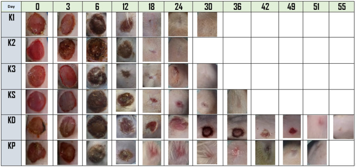 Figure 3
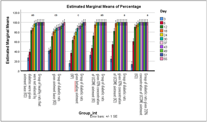 Figure 4
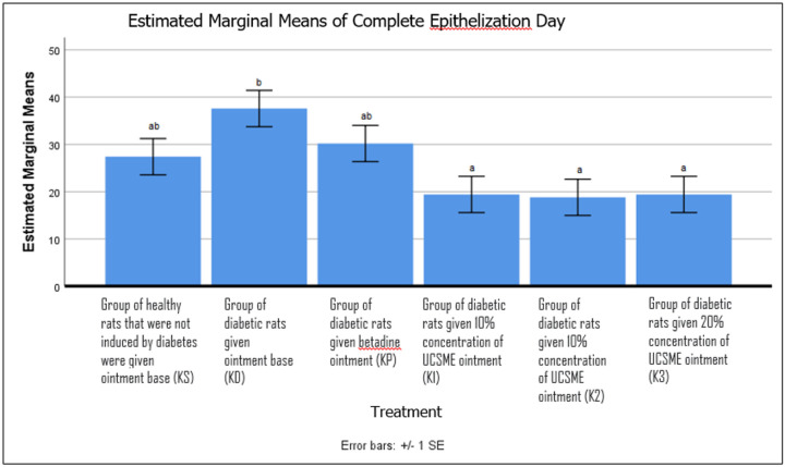 Figure 5
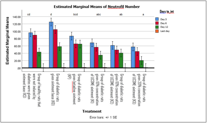 Figure 6
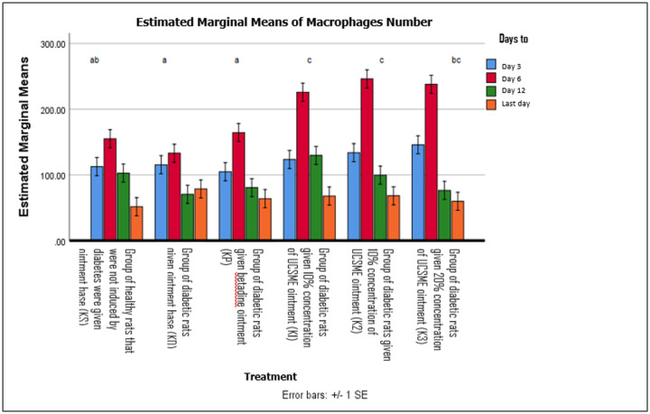 Figure 7
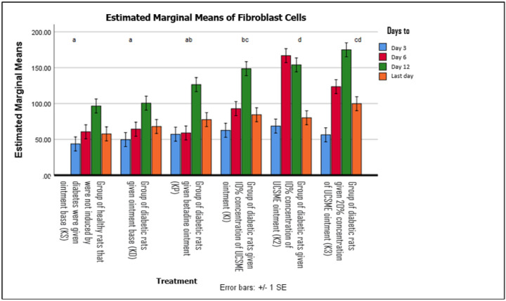 Figure 8
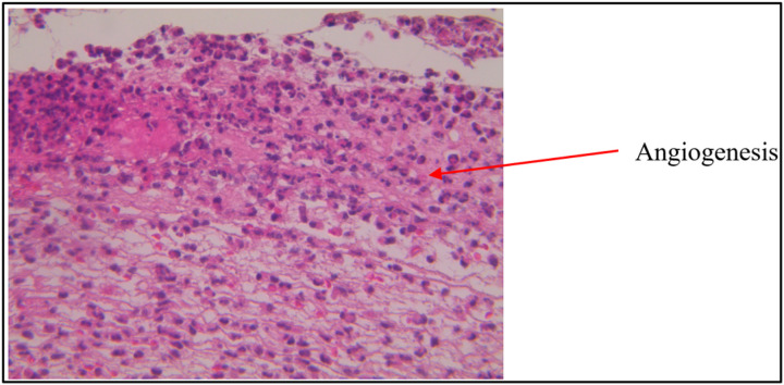 Figure 9
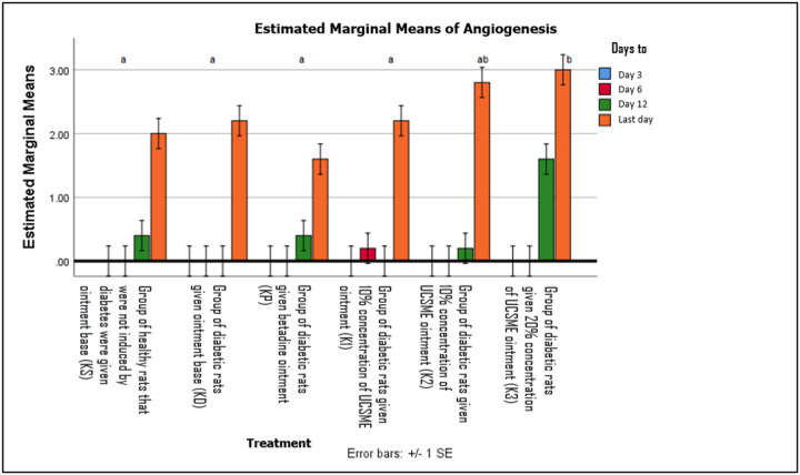 Figure 10
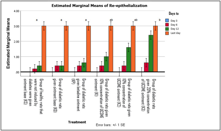 Figure 11
 Figure 12
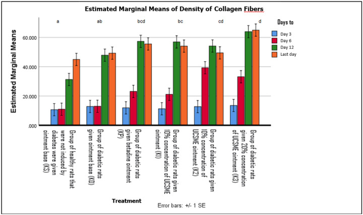 Figure 13
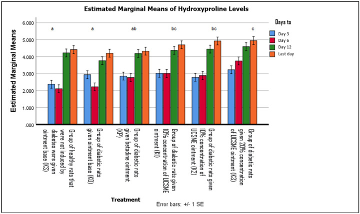 Figure 14
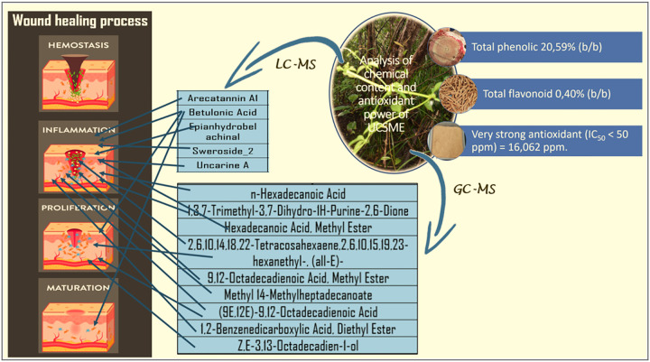 Figure 15
Peer Reviews
No public reviews on file for this paper yet. If you reviewed it on a platform where reviews are public (OpenReview, ICLR, NeurIPS, ICML), you can paste yours below so the community can read it here.
Videos
No videos yet. Explain this paper in a talk, walkthrough, or lecture? Add one.
Taxonomy
TopicsWound Healing and Treatments · Berberine and alkaloids research · Alkaloids: synthesis and pharmacology
INTRODUCTION
Wound healing is a complex biological process involving four overlapping phases: hemostasis, inflammation, proliferation, and maturation. After injury, platelet aggregation prevents bleeding, followed by neutrophil infiltration to remove debris and prevent infection. Neutrophils differentiate into macrophages, which regulate inflammation and tissue repair. During the proliferative phase, keratinocyte migration, angiogenesis, and fibroblast activation lead to granulation tissue formation, while remodeling restores extracellular matrix integrity and closes the wound (Nasiry et al. 2022).
Diabetes mellitus is a rapidly growing global health problem and is frequently associated with impaired wound healing, chronic ulceration, and limb amputation (Okonkwo dan Dipietro 2017). The World Health Organization predicts that the global prevalence of diabetes will reach 693 million by 2045, making diabetic wounds a major clinical and socioeconomic burden. Approximately 25% of diabetic patients experience delayed wound healing, often resulting in infection, amputation, and reduced quality of life (Burgess et al. 2021).
Delayed healing in diabetic wounds is driven by chronic hyperglycemia, oxidative stress, impaired angiogenesis, delayed collagen synthesis, and fibroblast dysfunction. Hyperglycemia causes endothelial dysfunction, reduced capillary density, and impaired nutrient delivery to wound tissue. In addition, oxidative stress and biofilm formation disrupt inflammatory and proliferative responses, leading to chronic non-healing wounds (Tan et al. 2019; Zandifar et al. 2012; Sinaga et al., 2019).
Given the limitations and side effects of current therapies, plant-based treatments with antioxidant and regenerative properties are increasingly explored. Uncaria callophylla Blume ex Korth., called bajakah by local Dayak community in Central Kalimantan, traditionally used in wound treatment, contains bioactive compounds with potential antioxidant and wound-healing activities. Therefore, this study aimed to evaluate the effectiveness of topical Uncaria callophylla stem methanol extract (UCSME) ointment on diabetic wound healing in rats by assessing macroscopic healing, histopathological changes, hydroxyproline content, antioxidant activity, and phytochemical composition.
MATERIAL AND METHODS
Plant Material and Identification
Uncaria callophylla stem samples were collected from tropical peat swamp forests in Central Kalimantan, Indonesia. Botanical identification was conducted at the Bogoriense Herbarium, Research Center for Biology, BRIN (Cibinong, Indonesia), to confirm species authenticity.
Preparation of Uncaria callophylla Stem Methanol Extract (UCSME)
Stems were air-dried at 50 °C, pulverized, and 500 g of powder was macerated in methanol (1:8 w/v) for 72 h with orbital shaking. The filtrate was concentrated using a rotary evaporator to obtain a viscous extract, which was stored at 5 °C until use.
Experimental Animals and Ethical Approval
Male Sprague Dawley rats (180–200 g) were obtained from Iratco Laboratory (Bogor, Indonesia). All experimental procedures were approved by the Iratco Laboratory Ethics Committee (No. 4.2.004–1/KEHI/I/2023 and 4.2.005–1/KEHI/I/2023).
Induction of Diabetes
After a 14-day acclimatization period, diabetes was induced by a single intraperitoneal injection of streptozotocin (65 mg/kg BW) as done by other researchers (Furman 2021 and Ozay et al. 2013). Rat with fasting blood glucose levels >200 mg/dL after 10 days were considered diabetic and included in the study (Furman, 2021).
Excision Wound Model
Rats were anesthetized with ketamine-xylazine (1:1; 5 mg/kg BW). A full-thickness excision wound (2 cm diameter) was created on the dorsal surface using a sterile biopsy punch as done by other researchers with slight modifications (Sahoo et al. 2015; Nagar et al. 2016; Tuhin et al. 2017; Nasiry et al. 2022).
Ointment Formulation and Treatment
UCSME ointments (5%, 10%, and 20% w/w) were prepared using a petroleum jelly-lanolin base (1:1) as used by other researchers (Ozay et al. 2013 and Yassine et al. 2020). Treatments were applied topically three times daily until complete wound closure. Betadine ointment served as the reference treatment.
Wound Healing Assessment
Wound area reduction was measured every three days using transparent tracing and millimeter paper, and expressed as percentage reduction. Complete epithelialization time was recorded as the number of days required for total wound closure without scab (Nagar et al. 2016).
Histopathological and Biochemical Analyses
Wound tissues were excised on days 3, 6, and 12, fixed in 10% neutral buffered formalin, and stained with hematoxylin-eosin or Masson’s trichrome as done by other researchers (Zandifar et al. 2012; Gautam and Goel 2013; Nasiry et al. 2022). Neutrophils, macrophages, fibroblasts, and capillary density were quantified microscopically as done by other researchers (Chen et al. 2017; Nasiry et al. 2022; Haestidyatami et al. 2019; Stryker et al. 2019). Re-epithelialization was scored using a standardized scale. Collagen density was analyzed using ImageJ software. Hydroxyproline content was determined by HPLC following acid hydrolysis as done by other researchers (Karimi et al. 2013 and Roodbari et al. 2004).
Phytochemical and Antioxidant Analyses
Total phenolic content was measured using the Folin–Ciocalteu method, while total flavonoid content was determined by the aluminum chloride colorimetric method. Antioxidant activity was assessed using the DPPH assay, and IC_50_ values were calculated.
LC-MS/MS and GC-MS Analyses
Chemical profiling of UCSME was performed using UPLC-QToF-MS (ESI positive and negative modes) and GC-MS. Compounds were identified by comparison with spectral libraries and expressed as relative abundances.
Statistical Analysis
Data were analyzed using one-way ANOVA followed by Tukey HSD post-hoc test (p < 0.05). Data homogeneity was confirmed using Levene’s test.
Results
Plant Identification
Botanical identification conducted at the Bogoriense Herbarium (BRIN, Cibinong, Indonesia) confirmed that the plant material used in this study was Uncaria callophylla Blume ex Korth. (family Rubiaceae).
Characteristics and Yield of UCSME
The Uncaria callophylla stem methanol extract (UCSME) appeared as a thick, sticky, blackish-green gel. Maceration of 100 g dried stem powder yielded 14.61 g of extract, corresponding to a yield of 14.61%. This value exceeds the minimum standard for herbal extracts (>12%) set by the Indonesian Herbal Pharmacopoeia, indicating efficient extraction of bioactive compounds.
Macroscopic Evaluation of Wound Healing
Macroscopic observation of wound healing progression is shown in Figure 3. Diabetic rats treated with UCSME ointment at concentrations of 5% (K1), 10% (K2), and 20% (K3) exhibited faster wound healing compared with untreated diabetic rats (KD) and diabetic rats treated with betadine ointment (KP). Notably, wound contraction and drying were evident as early as day 6 in all UCSME-treated groups.
By day 12, wounds in the 20% UCSME group (K3) were markedly smaller than those in untreated diabetic rats, betadine-treated diabetic rats, and even healthy control rats (KS). In contrast, wounds in the betadine-treated group remained relatively larger throughout the observation period. Overall, topical application of UCSME ointment at all tested concentrations accelerated macroscopic wound closure in diabetic rats more effectively than betadine and ointment base controls.
Wound Area Reduction and Complete Epithelialization Time
Two-way ANOVA analysis showed that treatment groups and observation time significantly affected wound area reduction (p < 0.05), with homogeneous data distribution (Levene’s test, p > 0.05). From day 3 to day 24, topical application of UCSME ointment at concentrations of 5%, 10%, and 20% significantly accelerated wound contraction compared to untreated diabetic rats (KD). However, no significant differences were observed among the three UCSME concentrations, indicating comparable effectiveness in reducing wound area (Figure 4)
Similarly, UCSME ointment at all tested concentrations significantly shortened the time required for complete epithelialization compared to KD (p < 0.05). No significant differences were observed between UCSME-treated groups and the betadine-treated group (KP), demonstrating comparable efficacy in accelerating epithelial closure (Figure 5).
Inflammatory Cell Response
Topical application of UCSME ointment increased neutrophil counts during the inflammatory phase. Concentrations of 10% and 20% significantly increased neutrophil numbers compared to KD and showed no significant difference from healthy controls (KS) and KP. The 5% UCSME ointment also significantly increased neutrophil counts compared to KD (Figure 6).
Macrophage numbers were significantly higher in all UCSME-treated groups compared to KD, KP, and KS (p < 0.05). Macrophage counts peaked on day 6 in all groups and declined thereafter, corresponding to wound progression. The highest macrophage numbers were observed in the 10% and 20% UCSME groups during the proliferative phase (Figure 7)
Fibroblast Proliferation and Angiogenesis
UCSME ointment at all concentrations significantly increased fibroblast numbers compared to KD and KS. Fibroblast counts peaked on day 12, with the highest values observed in the 20% UCSME group, followed by the 10% and 5% groups (Figure 8).
Angiogenesis was first observed after day 6. On day 12, angiogenesis was present in all groups except KD, with the highest angiogenesis scores observed in diabetic rats treated with 20% UCSME (Figure 9 and 10).
Re-epithelialization, Collagen Deposition, and Hydroxyproline Content
Re-epithelialization began on day 6 in all groups. UCSME ointment at 20% significantly enhanced re- epithelialization compared to KD, KP, and KS, while 5% and 10% concentrations showed similar effects (Figure 11).
Collagen fiber density was significantly increased by UCSME ointment at 10% and 20% compared to KD and KS, with the 20% concentration showing the strongest effect. Collagen density peaked on day 3 and day 12 in the 20% group and on day 6 in the 10% group (Figure 12 dan 13).
Hydroxyproline levels were significantly higher in the 20% UCSME group compared to KP, KD, and KS. However, no significant differences were observed among UCSME concentrations, indicating similar effectiveness in enhancing collagen synthesis (Figure 14).
Total Phenolic, Flavonoid Content, and Antioxidant Activity of UCSME
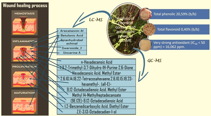
Spectrophotometric analysis using the Folin-Ciocalteu method showed that UCSME contained a high level of total phenolic compounds, amounting to 20.59% (w/w) gallic acid equivalents (GAE). Quantification was based on a gallic acid calibration curve with a strong linear relationship (R^2^ = 0.9986), indicating good analytical accuracy.
Total flavonoid content of UCSME, determined using the aluminum chloride colorimetric method with quercetin as the standard, was 0.40% (w/w). The quercetin calibration curve exhibited good linearity, confirming reliable flavonoid quantification.
Antioxidant activity of UCSME, assessed by the DPPH free radical scavenging assay, demonstrated very strong antioxidant capacity, with an IC50 value of 16.06 ppm, which falls within the category of highly potent antioxidants (IC_50_ < 50 ppm). Ascorbic acid, used as a reference antioxidant, showed an IC_50_ of 4.38 ppm.
These findings indicate that UCSME is rich in phenolic and flavonoid compounds and possesses strong antioxidant activity, which may contribute to its observed wound-healing effects through modulation of oxidative stress.
LC-MS/MS AND GC-MS Analysis of UCSME
LC-MS/MS analysis identified five major secondary metabolites in UCSME, namely Arecatannin A1, Betulonic acid, Epianhydrobelachinal, Sweroside-2, and Uncarine A. These compounds are known to possess biological activities related to anti-inflammatory, antioxidant, vasodilatory, cytoprotective, and tissue-regenerative functions, supporting their potential role in diabetic wound healing.
GC-MS analysis revealed the presence of 24 phytochemical constituents, of which nine compounds showed high similarity indices (≥90%). The dominant compounds included n-hexadecanoic acid(16.63%), 1,3,7-trimethyl-3,7-dihydro-1H-purine-2,6-dione (13.07%), hexadecanoic acid methyl ester, and several unsaturated fatty acid derivatives. These compounds are associated with anti-inflammatory, enzyme inhibitory, antioxidant, and tissue-protective activities.
Discussion
Diabetic wounds represent one of the most severe complications of diabetes mellitus, characterized by delayed healing due to hyperglycemia-induced vascular dysfunction, immune dysregulation, oxidative stress, and impaired tissue regeneration (Burgess et al. 2021 and Spampinato et al. 2020). These multifactorial disturbances limit the effectiveness of conventional wound therapies and justify the exploration of plant-based alternatives rooted in ethnomedicine. Uncaria callophylla Blume ex Korth., traditionally used by the Dayak Ngaju community of Central Kalimantan for chronic wound treatment, represents such a promising candidate.
In the present study, topical application of Uncaria callophylla stem methanol extract (UCSME) ointment at concentrations of 5%, 10%, and 20% significantly improved wound healing in diabetic rats compared with untreated and betadine-treated controls. Even the lowest concentration (5%) effectively reduced wound area and shortened epithelialization time, indicating that UCSME exhibits strong biological activity at relatively low doses. Histological analyses further confirmed enhanced fibroblast proliferation, collagen deposition, and epithelial regeneration in UCSME-treated wounds.
The acceleration of wound contraction observed from day 6 onward corresponds to the proliferative phase of healing, during which fibroblast migration, extracellular matrix synthesis, and angiogenesis dominate (Velnar et al. 2009 and Yates, 2017). UCSME treatment promoted fibroblast activity and collagen synthesis, with peak fibroblast numbers and collagen density occurring between days 6 and 12, reflecting efficient progression from inflammation to proliferation.
UCSME also modulated the inflammatory response by increasing neutrophil and macrophage infiltration during early healing stages. Neutrophils facilitate debris clearance, while macrophages coordinate tissue repair through cytokine and growth factor release, thereby stimulating angiogenesis and fibroblast function (Yang et al. 2025). Angiogenesis was most pronounced in the 20% UCSME group, although all concentrations enhanced vascularization compared to controls, ensuring improved oxygen and nutrient delivery to wound tissue.
Re-epithelialization was significantly accelerated in UCSME-treated groups, with the strongest effect observed at 20%, while 5% and 10% concentrations showed comparable efficacy. This suggests a largely dose-independent effect, potentially enhanced by the moist wound environment created by the ointment base, which is known to support epithelial regeneration (Karimi et al. 2013).
Phytochemical profiling revealed that UCSME contains diverse bioactive compounds, including betulonic acid, uncarine A, sweroside-2, arecatannin A1, and several fatty acid derivatives. These compounds possess antioxidant, anti-inflammatory, antimicrobial, and antidiabetic properties, which collectively contribute to wound repair. High phenolic (20.59% GAE) and flavonoid (0.40% QE) contents, along with strong antioxidant activity (IC_50_ = 16.06 ppm), support the role of UCSME in mitigating oxidative stress, a key factor impairing diabetic wound healing.
Enhanced collagen fiber density and hydroxyproline levels in UCSME-treated wounds further indicate improved collagen maturation and tensile strength, essential for stable wound closure. Among the tested formulations, the 10% UCSME ointment provided the most balanced therapeutic effect, achieving robust healing responses without the potential risks associated with higher concentrations. In contrast, betadine ointment showed inferior outcomes, likely due to its cytotoxic effects on fibroblasts and collagen synthesis.
Overall, UCSME promotes diabetic wound healing by modulating inflammation, enhancing angiogenesis, stimulating fibroblast activity, accelerating epithelialization, and improving collagen deposition. These findings validate the traditional use of Uncaria callophylla and highlight its potential as a natural, multi-target therapeutic agent for diabetic wound management.
Topical application of Uncaria callophylla stem methanol extract (UCSME) ointment effectively accelerates wound healing in diabetic rats. UCSME treatment significantly enhances wound contraction, shortens complete epithelialization time, promotes angiogenesis, accelerates re-epithelialization, stimulates collagen synthesis, increases neutrophil infiltration during the inflammatory phase, and elevates macrophage and fibroblast numbers during the proliferative phase. These findings demonstrate that UCSME positively modulates multiple stages of the wound healing process.
Phytochemical analysis revealed that UCSME contains high levels of bioactive compounds, including total phenolics (20.99% w/w) and flavonoids (0.40% w/w), which are closely associated with its strong antioxidant activity (IC_50_ = 16.06 ppm). LC-MS/MS analysis identified five major secondary metabolites—betulonic acid, epianhydrobelachinal, uncarine A, sweroside-2, and arecatannin A1, while GC-MS analysis revealed nine dominant compounds with high spectral quality, including fatty acids, alkaloids, and phenolic derivatives known to possess antioxidant, anti-inflammatory, antimicrobial, and tissue-regenerative properties.
Overall, the wound healing efficacy of UCSME is attributed to the synergistic action of its diverse secondary metabolites, particularly phenolic and flavonoid compounds with strong antioxidant capacity. These results scientifically validate the traditional use of Uncaria callophylla in wound management and highlight UCSME as a promising natural therapeutic candidate for the treatment of diabetic wounds.
Supplementary Material
Supplementary Files
This is a list of supplementary files associated with this preprint. Click to download.
The reference list from the paper itself. Each links out to its DOI / PubMed record.
- 1Annals of Pharmacology and Pharmaceutics Evaluation of the Wound Healing Properties of Jasminum Mesnyi H in Diabetic Rats. 2017. 2(9):1–3.
- 2Burgess JL, Wyant WA, Abujamra BA, Kirsner RS, Jozic I. 2021. Diabetic wound-healing science. Med. 57(10).doi:10.3390/medicina 57101072. · doi ↗
- 3Chen Y, Yu Q, Xu C-B. 2017. Original Article A convenient method for quantifying collagen fibers in atherosclerotic lesions by Image J software. Volume ke-10.
- 4Farmasetika M. 2019. Efektivitas Suplementasi Ekstrak Daging Ikan Bujuk (Channa Lucius) dalam Mempercepat Penyembuhan Luka Diabetik. 4(Suppl 1):195–200.
- 5Furman BL. 2021. Streptozotocin-Induced Diabetic Models in Mice and Rats. Curr. Protoc. 1(4).doi:10.1002/cpz 1.78. · doi ↗
- 6Gautam MK, Goel RK. 2013. Wound healing effect of Ocimum sanctum leaves extract in diabetic rats. Int. J. Medical, Heal. Biomed. Bioeng. Pharm. Eng. 7(9):224–227.
- 7Haestidyatami VL, Sugiritama IW, Linawati NM. 2019. Pengaruh ekstrak krim Morinda citrifolia terhadap jumlah fibroblas pada penyembuhan luka tikus Wistar. Intisari Sains Medis. 10(3).doi:10.15562/ism.v 10i 3.487. · doi ↗
- 8Karimi M, Parsaei P, Asadi SY, Ezzati S, Boroujeni RK, Zamiri A, Rafieian-Kopaei M. 2013. Effectsof Camellia sinensis ethanolic extract on histometric and histopathological healing process of burn wound in rat. Middle East J. Sci. Res. 13(1):14–19.doi:10.5829/idosi.mejsr.2013.13.1.66118. · doi ↗