Safeguarding genome integrity: Polo-like kinase Cdc5 and phosphatase Cdc14 orchestrate Topoisomerase II-mediated catenane resolution in mitosis
Lucia F Massari, Alice Finardi, Clara Visintin, Erika Calabrese, Ambra Dondi, Rosella Visintin

TL;DR
This study reveals how two enzymes, Cdc5 and Cdc14, work together to untangle DNA during cell division, ensuring accurate chromosome separation and genome stability.
Contribution
The study identifies a novel coordination mechanism between Cdc5 and Cdc14 in regulating Topoisomerase II to resolve DNA intertwines during mitosis.
Findings
Cdc14 and Cdc5 regulate Topoisomerase II through SUMOylation and phosphorylation to resolve DNA catenanes.
The collaboration between Cdc5 and Cdc14 ensures timely removal of sister chromatid linkages during mitosis.
This mechanism preserves chromosome segregation and genome integrity during cell division.
Abstract
Resolution of sister chromatid intertwines in mitosis is vital for safeguarding genome integrity. While cohesin, condensin, the polo-like kinase Cdc5, and the phosphatase Cdc14 have all been implicated in this process, the underlying molecular mechanisms remain elusive. Our study unveils a coordination between spindle elongation and the timely resolution of DNA linkages, orchestrated by Cdc14 and Cdc5. We show that Cdc14 and Cdc5 collaborate to facilitate the resolution of DNA catenanes, the predominant species of DNA intertwines in mitosis, by regulating Topoisomerase II (Top2) function, both through SUMOylation and phosphorylation. Our findings contribute to unraveling a mechanism wherein Cdc5 and Cdc14 work together to ensure the timely removal of all sources of linkages between sister chromatids, thereby preserving the integrity of chromosome segregation and genome stability.…
Genes, proteins, chemicals, diseases, species, mutations and cell lines named across the full text — each resolved to its canonical identifier and authoritative record.
Click any figure to enlarge with its caption.
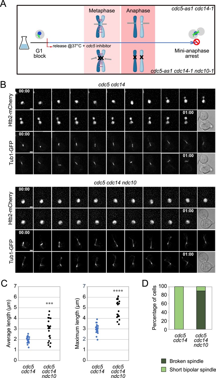 Figure 1
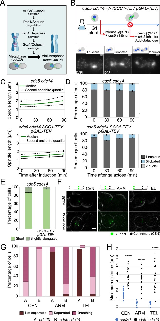 Figure 2
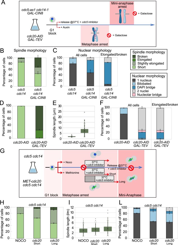 Figure 3
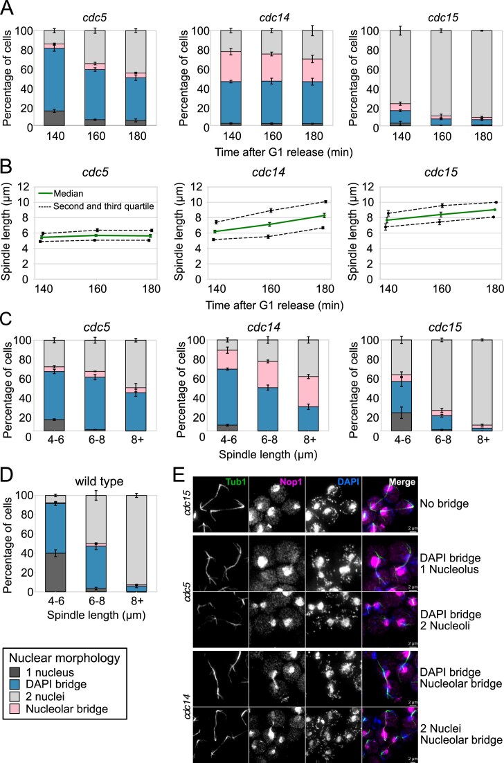 Figure 4
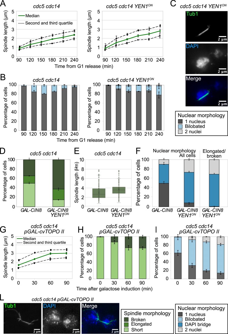 Figure 5
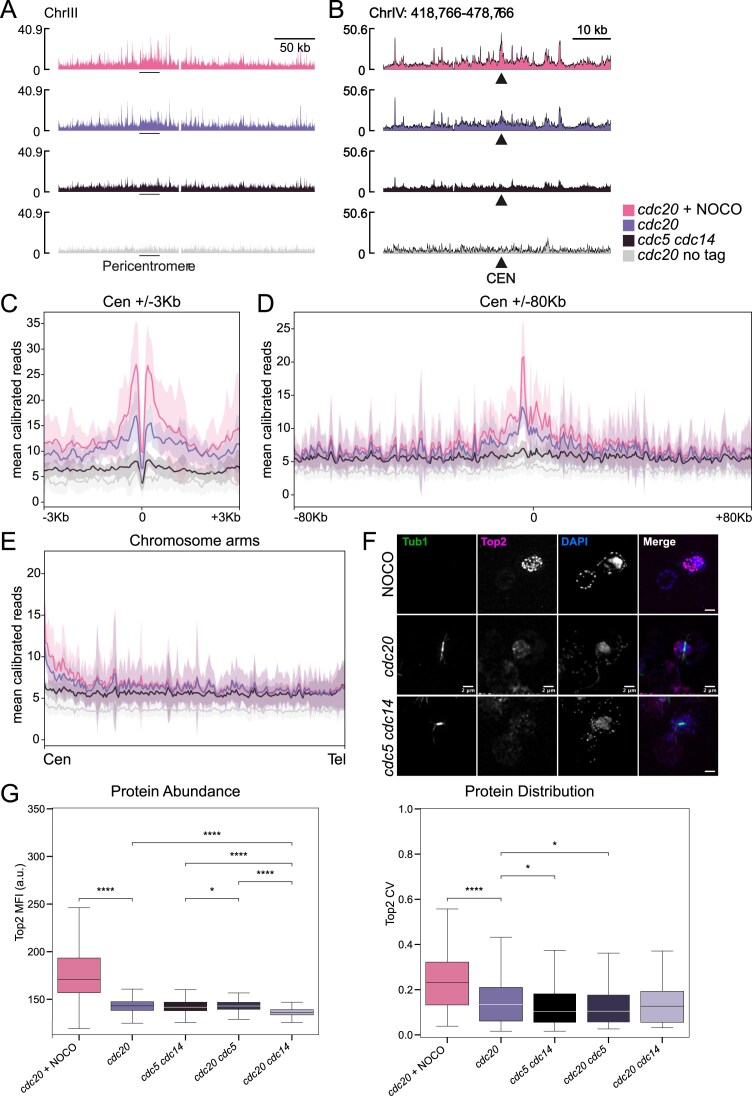 Figure 6
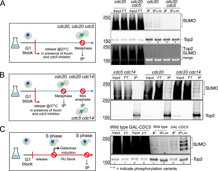 Figure 7
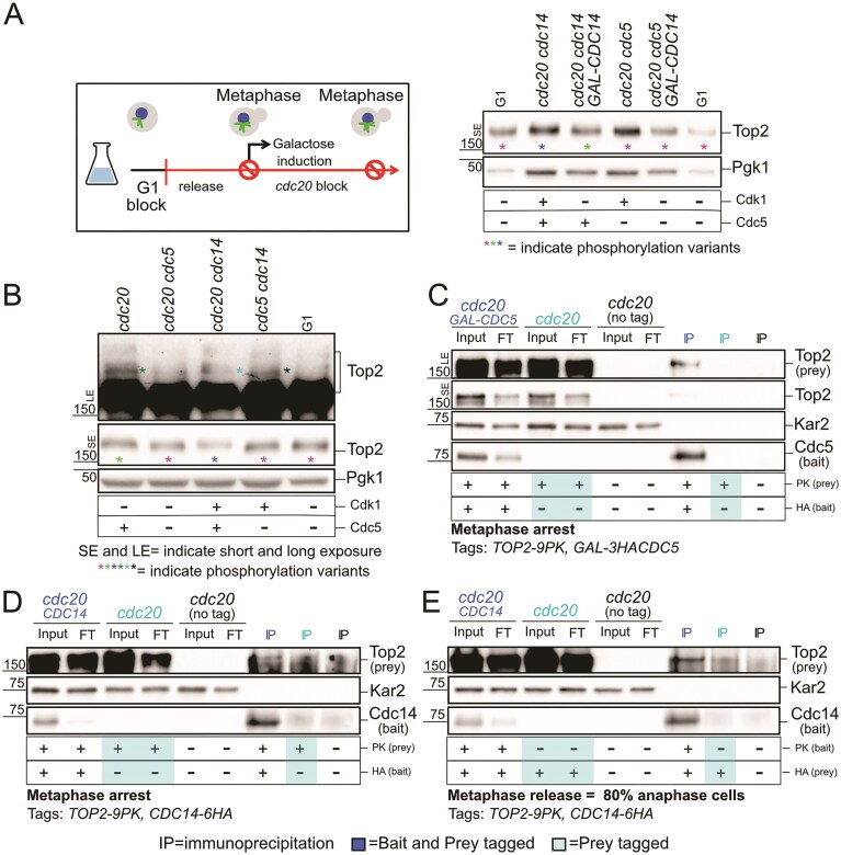 Figure 8
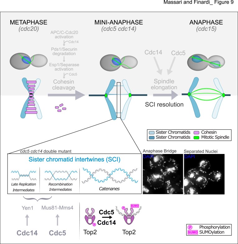 Figure 9
- —Italian Association for Cancer Research10.13039/501100005010
- —Italian Ministry of Health10.13039/501100003196
- —Howard Hughes Medical Institute10.13039/100000011
- —FIRC-AIRC
- —European School of Molecular Medicine
- —Italian Ministry of Health10.13039/501100003196
Peer Reviews
No public reviews on file for this paper yet. If you reviewed it on a platform where reviews are public (OpenReview, ICLR, NeurIPS, ICML), you can paste yours below so the community can read it here.
Videos
No videos yet. Explain this paper in a talk, walkthrough, or lecture? Add one.
Taxonomy
TopicsMicrotubule and mitosis dynamics · DNA Repair Mechanisms · Genomics and Chromatin Dynamics
Introduction
During mitosis, the accurate distribution of the newly replicated genetic material relies on a tightly-regulated process called chromosome segregation. To ensure proper partitioning, the duplicated chromosomes, referred to as sister chromatids, must be paired and recognized as sisters. Cohesin, a ring-shaped protein complex belonging to the structural maintenance of chromosomes (SMC) family, plays a pivotal role in holding sister chromatids together from their synthesis until their separation at anaphase (reviewed in [1]), with cohesin cleavage marking anaphase entry [2, 3].
Besides cohesin, sister chromatids are also connected by DNA linkages called sister chromatid intertwines (SCIs), which arise as byproducts of DNA replication and can be classified as nonreplicated DNA segments, recombination intermediates, or double-stranded catenanes (reviewed in [4]). To ensure accurate chromosome segregation, all sources of cohesion between sister chromatids must be removed. While most SCIs are resolved in S phase, some persist in mitosis and, in anaphase, appear as threads of DNA spanning between the two segregating DNA masses. Such threads are referred to as anaphase bridges [4]. Failure to adequately address these residual linkages can lead to double-stranded DNA breaks during cytokinesis. Even a single unresolved anaphase bridge can cause gross chromosomal rearrangements and genome instability, a hallmark of cancer development [5].
The complete resolution of SCIs is linked to cell cycle events and requires the organization of chromatin into chromosomes, the bipolar attachment of sister chromatids onto the mitotic spindle, and the activity of specialized enzymes. Chromosome formation consists in the non-overlapping compaction of each DNA molecule, which results in the individualization of sister chromatids. Individualization supports the resolution of SCIs by confining their presence to a small region at the interface between the chromatids, thus enhancing the accessibility of the enzymes deputed to their resolution. This process is achieved through the organization of chromatin into loops that are formed by the extruding activity of SMC complexes. In vertebrates the SMC complex carrying out chromosome formation is condensin [6–8], while cohesin, by holding sister chromatids in close proximity, counteracts catenane resolution [9]. Indeed, in vertebrates catenanes persist up to metaphase only at the centromere, the only region that still retains cohesin in this stage [10]. In the budding yeast Saccharomyces cerevisiae, instead, chromosomes are folded mainly by cohesin [11], that has the ability to introduce links both between sister DNA molecules, to achieve cohesion, and within the same molecule, to form loops. Nevertheless, also in budding yeast cohesin counteracts SCI resolution. It has been shown that SCIs can be fully resolved only after cohesin has been cleaved at anaphase onset and sister chromatids are allowed to separate [12, 13].
SCI resolution is facilitated also by the bipolar attachment of sister chromatids onto the spindle, the microtubule structure responsible for pulling them apart. Biorientation and the tension that results from the pulling force exerted by the spindle on centromeres result in the delocalization of condesin from the centromere to chromosome arms, where it can promote SCI resolution [14, 15]. It is not known whether anaphase spindle elongation contributes to SCI resolution as well.
Finally, a set of enzymes is dedicated to SCI resolution. Non-replicated DNA patches and recombination intermediates can be cleaved by the mitotic nucleases Yen1 and Mus81–Mms4 [16, 17]. Double-stranded catenanes disentanglement relies instead on the action of Type II Topoisomerases (Top2 in budding yeast). Top2 activity is essential for maintaining genome integrity and indeed top2 mutants are not viable [18–20]. Top2 facilitates replication fork progression at termination, facilitates transcription, allows chromatid individualization and compaction and is necessary for catenane resolution [21, 22].
From coordinating the spatial and temporal aspects of key mitotic events to regulating individual enzymatic activities, post-translational modifications (PTMs) exert critical roles [23]. These include phosphorylation, ubiquitination, and SUMOylation (conjugation with small ubiquitin-like modifier). In budding yeast, two major regulators of mitotic events are the Polo-like kinase Cdc5 and the Cdk-counteracting phosphatase Cdc14. Both proteins are integral components of two interconnected signaling networks governing late mitotic events, the FEAR (Cdc14 Early Anaphase Release) and the MEN (Mitotic Exit Network) pathways (reviewed in [24, 25]). The FEAR network facilitates the timely release of Cdc14 from its nucleolar inhibitor Cfi1/Net1 at anaphase onset [26–28]. It orchestrates events including sister chromatid separation, nuclear movement, nucleolar segregation [24], and anaphase spindle elongation [29]. The MEN pathway instead activates Cdc14 during late anaphase and is crucial for Cdk inactivation, leading to mitotic exit and entry into a new G1 phase [25].
Cdc5 and Cdc14 have also been implicated in SCI resolution. Cdc5 phosphorylates and activates the endonuclease Mus81–Mms4 in G2/M; while in anaphase, Cdc14 activates the endonuclease Yen1 through dephosphorylation (reviewed in [30]). Cdc5 indirectly influences the SUMOylation of Top2 in metaphase by inactivating the SUMO protease Ulp2 [31]. It has been suggested that SUMOylation of Top2 directs its recruitment to chromatin [32, 33]. Moreover, Cdc5 also promotes condensin activity, the establishment of a bipolar spindle and timely cohesin cleavage.
Although several actors implicated in SCI resolution are known, the intricate molecular mechanisms that govern the disentanglement of DNA during mitosis and their interaction with other mitotic processes remain largely uncharacterized. In this study, we investigate the roles of Cdc5 and Cdc14 in this process. We provide evidence that Cdc5 and Cdc14 orchestrate the timely resolution of the three classes of DNA linkages. We show that during mitosis, Cdc5 and Cdc14 facilitate the resolution of DNA catenanes by influencing Top2 localization, likely by impacting its SUMOylation and phosphorylation status. This function of Cdc5 and Cdc14 operates in parallel with their role in modulating spindle elongation. By coupling SCI resolution with spindle elongation, these two proteins ensure that sister chromatid segregation occurs together with the removal of all sources of linkages, thereby preserving genomic integrity.
Materials and methods
Yeast strains and growth conditions
All yeast strains were W303 (K699) derivatives (Supplementary Table S1). Cell cycle arrest and synchronization followed standard protocols [34]. Unless specified, cells were grown at 23°C in Yeast Extract Peptone (YEP) medium with 2% glucose, raffinose, or galactose. G1, S-phase, and metaphase arrests were induced with 5 µg/ml α-factor (Genscript), 10 mg/ml hydroxyurea (Sigma), and 15 µg/ml nocodazole (Sigma, dissolved in DMSO), respectively. To release from the arrests, cells were washed with 10 volumes of fresh medium lacking the pheromone/drug and released into appropriate conditions of medium and temperature (see figure legends for details relative to specific experiments). When pertinent, drug(s) were added and restrictive conditions applied. Temperature-sensitive alleles were inactivated at 37°C. Analog-sensitive cdc5-as1 and cdc15-as1 strains were inhibited with 5 µM CMK (custom-made, Accendatech; [35]) or 1NM-PP1 analog 9 (A603003; Toronto Research Chemicals). The CDC20-AID allele was inactivated with 500 µM auxin (I5148;Sigma), and MET-CDC20 was repressed with 8 mM methionine.
Indirect in situ immunofluorescence
Immunofluorescence (IF) was performed as described in [36]. Briefly, 1 ml of culture at OD_600_ = 0.4–0.6 was fixed overnight in 3.7% formaldehyde/0.1 M potassium phosphate buffer (pH 6.4) at 4°C. Cells were washed three times in 0.1 M potassium phosphate buffer and once in sorbitol buffer (1.2 M sorbitol, 0.1M K_2_HPO_4,_ 0.33 M citric acid, pH 5.9), then digested with 0.1 mg/ml Zymolyase 100T for ∼30 min at 30°C. Spheroplasts were washed, adhered to poly-L-lysine–coated slides, and sequentially treated with cold methanol (−20°C, 3 min) and acetone (−20°C, 10 s). Speroplasts were incubated 90 min with primary antibodies diluted in PBS/BSA (1% BSA, 0.04 M K₂HPO_4_, 0.01 M KH₂PO_4_, 0.15 M NaCl, and 0.1% NaN₃); washed; incubated 90 min with fluorophore-conjugated secondary antibodies; washed and covered with 4′,6-diamidino-2-phenylindole (DAPI) mounting solution (90% glycerol, 10% KPBS, 100 mg p-phenylenediamine, 5 μg DAPI).
Primary antibodies were rat anti–α-tubulin YOL34 (1:100, AbD Serotec), mouse anti-Nop1 (1:2000, EnCore), and mouse anti-PK SV5-Pk1 (1:500, AbD Serotec). Secondary antibodies were donkey anti-rat (FITC 1:100, Jackson; Cy3 1:100, Jackson) and donkey anti-mouse (FITC 1:100, Jackson; Cy3 1:500, Jackson). Slides were sealed with nail polish and analyzed by fluorescence microscopy.
Microscopy and image analysis
Cell cycle progression was monitored by categorizing ≥100 cells per sample as interphase, metaphase, or anaphase based on spindle morphology. For spindle length and nuclear morphology, images were acquired on an upright Olympus AX70 Provis microscope with a ×100/1.40 oil UPlanSApo∞/0.17/FN 26.5 objective and a Photometrics CoolSnap 12-bit B&W camera using MetaMorph 7.5.6.0 software (MDS Analytical Technologies), and analyzed with Fiji. For anaphase bridge and spindle measurements, images were collected on a DeltaVision Elite deconvolution system (Applied Precision) equipped with an Olympus IX71 inverted microscope, UPlanSApo ×100 oil objective (NA 1.4), and a CoolSnap HQ2 CCD camera. Z-stacks (12 × 0.5 µm) were acquired with FITC, TRITC, and DAPI filters, deconvoluted with SoftWoRx, and analyzed with Fiji to assess nuclear and nucleolar morphology and measure spindle length. Anaphase cells were scored for DNA and/or nucleolar bridges using 4′,6-diamidino-2-phenylindole (DAPI) and Nop1 staining. Intensity thresholds were defined in wild-type controls lacking bridges (4000 a.u. for DAPI and 5200 a.u. for Nop1). Cells exceeding the DAPI threshold were classified as DAPI-bridge positive, those exceeding the Nop1 threshold as nucleolar-bridge positive, and cells above both thresholds as double-bridge positive. Cells below both thresholds were scored negative.
For Top2 localization, cells stained for tubulin, Top2-PK, and DNA (DAPI) were imaged on a Nikon Eclipse Ti2 microscope with a spinning-disk X-Light V3 module (CrestOptics), solid-state lasers (Lumencor Celesta), a Kinetix sCMOS camera, and a ×100/1.49 NA oil objective. Z-stacks (25 planes, 0.2 µm spacing) were acquired and deconvoluted using the Blind algorithm in NIS. Briefly, images were resampled to obtain isotropic voxels, ensuring geometric accuracy for 3-D morphometry. The DAPI channel was then denoised (Gaussian blur with σ = 2) and segmented by four-level Multi-Otsu thresholding. The resulting segmented nuclei were filtered to based on their volume to remove debris and aggregates. The nuclear masks were then expanded to enclose the Top2 signal (5 px corresponding to 325 nm). From each expanded mask, the mean and standard deviation of the Top2 signal intensity were calculated. The coefficient of variation for each mask was calculated by dividing the standard deviation by the mean. At least 250 cells per strain were analyzed. Statistical significance was assessed using the Kruskal–Wallis test followed by Mann–Whitney U test for post-hoc pairwise comparisons (Supplementary Tables S3 and S4).
Live-cell imaging
Cells were grown in SCD medium supplemented with 2% adenine and released from G1 arrest into Y04C microfluidic plates (CellASIC). Imaging was performed on a DeltaVision microscope as previously described. To avoid UV-induced DNA damage checkpoint activation, live imaging began once cells reached metaphase, identified by spindle morphology and nuclear positioning. For spindle elongation (Fig. 1, and Supplementary Figs S2 and S3) and nuclear/nucleolar segregation (Fig. 1, and Supplementary Figs S1 and S2), 8 z-stacks (0.6 µm step) were acquired every 3 min using FITC and Cherry filters with a ×100 oil objective. For GFP-dot separation (Fig. 2, and Supplementary Figs S4 and S5), 15 z-stacks (0.4 µm step) were acquired every 3 min under the same conditions. Reference images were collected in DIC. Stacks were deconvoluted with SoftWoRx software, and spindle length and chromosome locus distances were measured in x,y,z using Fiji with a custom plug-in (Spindle Z manual tracker). Representative images are shown as maximum-intensity projections.
*cdc5 cdc14 cells are defective in sister chromatid separation. (A–D) cdc5-as1 cdc14-1 (Ry6591) and cdc5-as1 cdc14-1 ndc10-1 (Ry6589) cells carrying a HTB2-Cherry and a GFP-TUB1 fusion were synchronously released from a G1 block in conditions restrictive for cdc5, cdc14, and ndc10. A schematic of the rationale and experimental setup is shown (A). Cells were imaged starting from metaphase every 3 min for 3 h; n = 20 cells for each strain. Representative images of the first hour of imaging are shown (B); scale bars = 2 μm. Average and maximum spindle length for each cell analyzed are shown (C). NOTE: each dot represents one cell. The average spindle length in cdc5 cdc14 ndc10 cells (mean = 3.0 μm, S.D. = 1) was higher than in cdc5 cdc14 cells (mean = 2.0 μm, S.D. = 0.3); ***P < 0.001. The maximum spindle length in cdc5 cdc14 ndc10 (mean = 4.8 μm, S.D. = 0.8) was higher than in cdc5 cdc14 cells (mean = 3.0 μm, S.D. = 0.5); ***P < 0.0001. Percentage of cells that either maintained a short bipolar spindle or broke the spindle during the time-lapse (D).
*Cohesin does not contribute to the sister chromatid separation defect of cdc5 cdc14 mutants. (A) Schematic representation of the signaling cascade leading to cohesin cleavage, the critical step for anaphase entry. (B–E) cdc5-as1 cdc14-1 (Ry1602) and cdc5-as1 cdc14-1 SCC1-TEV GAL-TEV (Ry2795) cells were synchronously released from a G1 block in YPR in conditions restrictive for cdc5 and cdc14. At the cdc5 cdc14 arrest (3h30 after release), galactose was added to induce TEV protease expression. Samples were collected at the indicated time points after induction and analyzed through indirect IF (anti-Tub1, DAPI) to monitor spindle length and nuclear morphology. The schematic diagram of the experimental setup and images representative of nuclear morphology categories are shown; scale bars = 2 μm (B). Spindle length: Median (solid green line), second and third quartile (dotted lines), and s.e.m. (error bars) are shown (C). Nuclear and spindle morphology: Mean and s.e.m. (error bars) are shown (D and E). Three independent experiments are shown, n = 100 cells counted for each time point in each experiment. (F–H) CDC20-AID and cdc5-as1 cdc14-1 cells carrying a CENXV-GFP (Ry7519 and Ry5815), a HIS3-GFP (Ry7411 and Ry5824) or a TELXV-GFP (Ry7481 and Ry7098) fusion were synchronously released from a G1 block into conditions restrictive for cdc20, cdc5, and cdc14. Cells were imaged starting from metaphase every 3 min for 3 h; n = 20 cells were analyzed in each condition. Representative images are shown; scale bars = 2 μm (F). Distance between GFP dots. According to the distance between GFP dots throughout the time-lapse, cells were assigned to the following categories: Cells in which the distance between dots consistently remained below 2 μm = Not separated; Cells in which the distance between dots exceeded 2 μm, but subsequently returned to zero = Breathing; Cells in which the distance between dots exceeded 2 μm and never went back to zero = Separated (G). Maximum distance between GFP dots for each cell analyzed. The distance was higher in cdc5 cdc14 cells compared to cdc20 cells at every locus analyzed (H); ***P < 0.0001.
Immunoblot analysis
Cells were treated with cold 5% TCA for 10 min, pelleted, and washed with acetone. Pellets were resuspended in 50 mM Tris–HCl (pH 7.5), 1 mM EDTA, 1 mM p-nitrophenyl phosphate, 50 mM DTT, 1 mM PMSF, and 2 μg/ml pepstatin, lysed with glass beads, and boiled in 1× SDS sample buffer.
Proteins were detected with the following primary antibodies: mouse anti-PK (SV5-Pk1, AbD Serotec; 1:5000) for Top2-9PK, rabbit anti-Smt3 (gift from Dr. Branzei; 1:2000) for SUMOylated species, and mouse anti-Pgk1 (A-6457, Molecular Probes; 1:5000) as a loading control. Secondary antibodies were HRP-conjugated goat anti-rabbit IgG (Bio-Rad, 170-6515; 1:5000) and HRP-conjugated goat anti-mouse IgG (Bio-Rad, 170-6516; 1:10 000). Signals were visualized by chemiluminescence (ECL; GE Healthcare).
Phos-tag gels were prepared with 40 μM Phos-tag AAL-107 (Fujifilm Wako) and 10 mM MnCl₂ in 6% acrylamide gels.
Immunoprecipitation of Top2
Cells were lysed as in [31]. After resuspending the pellet in the SDS-containing solution, the lysate was boiled and clarified by centrifugation. The supernatant was diluted 1:10 with NP40 buffer and incubated with SV5-Pk1 monoclonal mouse anti-PK antibody, used at 1:200 dilution, and Pierce protein-G agarose beads (Thermo Scientific). The beads were washed with NP40. The beads were resuspended in 3× SDS sample buffer to elute the proteins.
Co-immunoprecipitation
A 50 ml culture was grown to OD_600_ ≈ 0.5, harvested at 3000 rpm for 5 min at 4°C, and resuspended in 150 µl of NP-40 buffer with protease and phosphatase inhibitors. Cells were lysed using FastPrep (4 × 45 s, maximum speed, 4°C), and lysates clarified at 13 000 rpm for 10 min. Protein concentration was determined by Bradford, and 500 µg extract (50 µl) was used per IP, adjusted to equal volumes with NP-40 buffer. For immunoprecipitation, 10 µl of anti-V5 magnetic beads (Top2-PK, SAE0203, Sigma) or 15 µl of Pierce^™^ anti-HA magnetic beads (Cdc14-HA or Cdc5-HA, 88837, ThermoFisher) were added and rotated 2 h at 4°C. Beads were washed five times with NP-40 buffer and eluted in 30 µl of 3× Laemmli buffer, boiled 5 min, centrifuged, and supernatants analyzed by SDS–PAGE.
Calibrated Chromatin ImmunoPrecipitation and sequencing (ChIP-Seq)
Chromatin ImmunoPrecipitation and sequencing (ChIP-Seq) was performed as described in [37] with modifications from [38, 39]. For each condition, 200 ml of S. cerevisiae cultures (TOP2-PK9) arrested in metaphase were fixed in 20 ml of fixing solution (6 ml 37% formaldehyde, 143 mM NaCl, 1.43 mM EDTA, and 71.4 mM HEPES-KOH pH 7.5) for 80 min at room temperature, quenched with 0.125 M glycine for 5 min, pelleted, and washed twice in 10 ml of ice-cold TBS (20 mM Tris-HCl pH 7.5, 150 mM NaCl) and once in 10 ml of ice-cold 1× FA lysis buffer (50 mM HEPES-KOH pH 7.5, 140 mM NaCl, 1 mM EDTA, 1% Triton X-100, and 0.1% Na deoxycholate) added with 0.1% SDS. Pellets were snap-frozen in liquid nitrogen and stored at –80°C.
For calibration, S. cerevisiae pellets were mixed 1:1 with S. pombe (rad21-V5:KANMX6) pellets, prepared as follow. S. pombe cells were grown in YES media (0.5% yeast extract, 3% glucose, 2% agar 225 mg/l each of adenine, histidine, leucin, uracil, and lysine hydrochloride) at 30°C until reaching OD_600_ = 0.4–0.6. The culture was fixed, quenched, harvested, and washed like the S. cerevisiae one. S. cerevisiae and S. pombe cell pellets were thawed on ice, and each S. cerevisiae pellet was combined with one S. pombe pellet in 400 μl of 1× FA* lysis buffer (FA buffer added with 1× Roche EDTA-free protease inhibitors and 1 mM PMSF) with 0.5% SDS. Cells were lysed with silica beads (FastPrep) and chromatin was sheared by sonication (Bioruptor Plus, 2 × 20 cycles, 30 s ON/OFF, high power). Soluble, sheared, chromatin was collected after centrifugation, pooled, and split into input and IP samples. Inputs were diluted 1:50 in TE and stored overnight at 4°C. For IPs, 1 ml of extract was incubated either with 15 µl of protein A Dynabeads (Invitrogen) or 10 µl of V5 antibody (Bio-Rad) overnight at 4°C. Beads were washed as follow: twice in cold 1 ml of 1× FA lysis buffer/0.1%SDS; twice in cold 1 ml of 1× FA lysis buffer/0.1%SDS + 360 mM NaCl (total: 500 mM NaCl); twice in 1 ml of cold 10 mM Tris–HCl, pH 8.0, 250 mM LiCl, 0.5% NP-40, 0.5% Na-deoxycholate, and 1 mM EDTA; once in 1 ml of cold TE pH 8. Inputs and IPs were decrosslinked overnight with proteinase K at 65°C, purified with the Wizard kit (Promega), and stored in 35 µl of water at –20°C.
DNA libraries were prepared from 2 ng DNA as in [38]. DNA ends were blunted and phosphorylated (NEB Quick Blunting kit), <100 bp fragments removed (AMPure XP beads), and dA tails added (NEB Klenow exo-). Each sample was then ligated with a different NEXTflex-6 DNA Barcode (PerkinElmer) using the Quick Ligation kit (NEB), unligated adapters removed and libraries PCR-amplified (Phusion polymerase, NEB) using primers specific for NextFlex adapters (5′-AATGATACGGCGACCACCGAGATCTACAC; 5'-CAAGCAGAAGACGGCATACGAGAT). Fragments (150–300 bp) were size-selected with AMPure XP, quantified by Qubit, and quality-checked by Bioanalyzer (Agilent) using the 2100 Bioanalyzer High Sensitivity DNA kit (Agilent, Santa Clara, CA).
INPUT and IP libraries for each sample were pooled at a 15:85 ratio and sequenced on an Illumina MiniSeq with a 150-cycle high-output kit.
Data analysis was performed as in [37]. Reads were mapped to both the S. pombe calibration genome and the S. cerevisiae sacCer3 genome. Occupancy ratios [OR = (INPUT_cal_ × IP_exp_) / (INPUT_exp_ × IP_cal_)] were calculated and used to normalize read counts.
Region-specific plots were generated with the Spark package (https://github.com/harbourlab/SparK). Mean calibrated ChIP-seq plots (all chromosome pileups) were generated and visualised with computeMatrix and plotProfile in deepTools (ref: https://academic.oup.com/nar/article/44/W1/W160/2499308). In plots showing 3Kb flanking the centromere, reads were binned at 50 bp, in plots showing 80 kb flanking the centromere, at 500 bp. In plots showing the chromosome arm from cen to tel, each arm was averaged to 10 kb and binned to 50 bp. Centromere coordinates used are listed in Supplementary Table S2.
Statistical analysis
Depending on the experiment and as indicated in the corresponding figure legends, P-values were determined by unpaired or paired Student’s t-test after arcsine transformation as appropriate for the data obtained from a count and expressed as percentages. A P-value of <0.05 was considered statistically significant (^∗^P < 0.05; ^∗∗^P < 0.01, ^∗∗∗^P < 0.001, and ^****^P < 0.0001). Averages ± s.e.m. (standard error of the mean) of three independent experiments are reported.
Repeatability of the experiments
Each experiment in the manuscript has been repeated at least three independent times. The results were highly reproducible.
Results
Mini-anaphase arrested cdc5 cdc14 cells are defective in sister chromatid separation
To investigate how DNA linkages are removed during mitosis, we analyzed budding yeast cells lacking both the Polo-like kinase Cdc5 (cdc5-as1, henceforth cdc5) [35] and the Cdk-counteracting phosphatase Cdc14 (cdc14-1, henceforth cdc14) [40]. The cdc5-as1 cdc14-1 double mutant strain (henceforth cdc5 cdc14) arrests in “mini-anaphase,” a state characterized by a short, stable bipolar mitotic spindle and unseparated nuclei despite cohesin cleavage, due to defective spindle elongation [29], and, potentially, to linkages between sister chromatids. To further characterize the mini-anaphase arrest of cdc5 cdc14 cells, we performed live-cell imaging with Htb2-mCherry and GFP-Tub1 to capture chromosome segregation dynamics under restrictive conditions (Fig. 1A). In stark contrast to the results obtained through fixed-sample indirect immunofluorescence (IF), which showed a single undivided nuclear mass [29], live imaging revealed highly dynamic nuclei that often appeared bilobated (Fig. 1B). Such transient morphology, overlooked in population-based assays, likely reflects nuclei initiating separation and reuniting due to spindle defects, or being compressed while passing through the bud neck. The bilobated phenotype can also originate from the nucleus and nucleolus residing in two different cellular compartments, as already documented for mutants associated with the G2/M DNA damage checkpoint and with the FEAR network [41, 42]. Indeed, the nucleolus, which contains ribosomal DNA (rDNA) repeats, is inadequately stained by DAPI, but it can be effectively labeled with Htb2-Cherry. The latter enabling clear visualization of both the nucleus and the nucleolus. To discriminate among these possibilities, we examined cdc5 cdc14 cells carrying Htb2-Cherry and Cfi1-GFP fusions, the latter marking the nucleolus. In all cells examined, the nucleus migrated into the daughter cell, while the nucleolus remained in the mother (Supplementary Fig. S1A and B). During its passage through the bud-neck, the nucleus appeared bilobated due to compression (Supplementary Fig. S1A). Therefore, such morphology reflects both nuclear compression at the bud neck and the spatial separation of nucleus and nucleolus, consistent with known roles of Cdc14 and Cdc5 in nucleolar segregation [43–45]. Importantly, this phenotype does not represent chromosome segregation, confirming that cdc5 cdc14 cells are defective in this process. Previous data suggested that the chromosome segregation defect of cdc5 cdc14 cells is independent of spindle elongation [29]. Disrupting chromosome attachment to the spindle by inactivating the kinetochore protein Ndc10, ndc10-1 (Fig. 1A) [46], altered spindle morphology in double mutant cells. Unlike cdc5 cdc14 cells, which formed short and stable spindles, ndc10 cdc5 cdc14 mutants developed fragile spindles that often broke during attempted elongation [29]. To further characterize this phenotype, we analyzed nuclear morphology and spindle dynamics in ndc10, cdc5 cdc14, and cdc5 cdc14 ndc10 cells. In line with the absence of microtubule attachment to chromosomes, both ndc10 and cdc5 cdc14 ndc10 cells displayed a single nucleus that remained in the mother cell throughout the experiment (Supplementary Fig. S2A and Fig. 1B). Spindle measurements supported the persistence of residual cohesion in cdc5 cdc14 cells. In this strain, the spindles remained stable and short, with an average length ranging from 1.2 to 2.8 μm (mean of 2 μm) and a maximum length ranging from 2 to 4 μm (mean of 3.0 μm) (Fig. 1B and C, and Supplementary Fig. S3A). Instead, in cdc5 cdc14 ndc10 cells the spindles were longer, reaching a maximum length of 6 μm (mean of 4.8 μm) and an average length ranging from 1.2 to 4.8 μm (mean of 3 μm) (Fig. 1B and C, and Supplementary Fig. S3B). Furthermore, spindles in this mutant displayed increased fragility, collapsing in 90% of the analyzed cells during the experiment (Fig. 1D and Supplementary Fig. S3B). Consistent with the spindle elongation defect observed in cdc5 cdc14 mutants, cdc5 cdc14 ndc10 cells failed to achieve full spindle elongation, unlike ndc10 cells, which successfully elongated their spindles up to 14 μm (mean of 9 μm), and completed the cell cycle (Supplementary Fig. S2B). Together, these results indicate that unresolved cohesion between sister chromatids persists in cdc5 cdc14 cells. This residual linkage counteracts the pulling forces of a compromised spindle, preventing full chromatid separation.
Linkages other than cohesin contribute to the sister chromatid separation defect of cdc5 cdc14 cells
The residual cohesion observed in cdc5 cdc14 cells could arise either from uncleaved cohesin or from SCIs. Although separase Esp1 is active and cleaves bulk cohesin in these cells (Fig. 2A) [29], it remained possible that a fraction of cohesive cohesin escaped cleavage. To address this, we applied two complementary strategies: ectopic cohesin cleavage and direct analysis of cohesion at specific chromosomal loci.
For ectopic cleavage, we overexpressed the Tobacco Etch Virus (TEV) protease [47] under the galactose-inducible GAL1-10 promoter (GAL-TEV) in strains carrying a TEV-cleavable allele of the SCC1 subunit of cohesin (scc1Δscc1-TEV268, henceforth SCC1-TEV) [3]. If residual cohesin were responsible for cohesion, TEV induction in cdc5 cdc14 cells should mimic the phenotype of cdc5 cdc14 ndc10 cells, where chromatids no longer restrain spindle elongation (Fig. 1). After synchronization and TEV induction at the cdc5 cdc14 block (Fig. 2B and Supplementary Fig. S4A), we measured spindle length and categorized cells into three groups based on nuclear morphology—with a single nucleus, a separating nuclear mass (bilobated), or with two divided nuclei (Fig. 2B)—in cdc5 cdc14 and cdc5 cdc14 SCC1-TEV GAL-TEV cells. Consistent with our previous report [29], cdc5 cdc14 cells maintained short and stable spindles (1–2.5 μm) (Fig. 2C) and largely undivided nuclei (Fig. 2D). The shorter spindle length in this experiment, as compared to the one in Fig. 1, is attributed to the use of a less rich media, containing raffinose instead of glucose. Remarkably, ectopic cohesin cleavage (Supplementary Fig. S4A) did not elicit spindle elongation nor induce nuclear division in cdc5 cdc14 SCC1-TEV GAL-TEV cells (Fig. 2C and D) with spindles remaining short and stable (Fig. 2E). Thus, anaphase cohesion in cdc5 cdc14 cells does not originate from residual cohesin.
We next monitored cohesion at individual chromosomal loci using the TetR-GFP/tetO system [48]. Distinct chromosomal regions exhibit different behaviors depending if they are bound by cohesin or not. When bound by cohesin (e.g. in metaphase), sister telomeres and chromosome arms remain tightly associated, and, when labelled, appear as one fluorescent dot. Conversely, at the centromeres the tension exerted by the mitotic spindle counteracts the cohesive function of cohesin, leading to a phenomenon known as “centromere breathing”, where sister centromeres continuously separate (two dots) and rejoin (one dot) over distances shorter than 2 μm [49–51]. We examined a centromere (CENXV), a chromosome arm (HIS3), and a telomere (TELXVr), all on the long arm of chromosome XV ([52] and Fig. 2F), comparing cdc20 metaphase-arrested cells (with intact cohesin) to cdc5 cdc14 cells (Fig. 2A). Chromosome XV was chosen due to its lack of specialized sequences and its length (it is one of the longest yeast chromosomes), which could exacerbate a sister chromatid separation defect. The cdc20 metaphase arrest was obtained by depleting Cdc20, the activating subunit of the anaphase-promoting complex/cyclosome (APC/C) (Fig. 2A) with the CDC20-AID allele, that allows conditional degradation of Cdc20 upon auxin treatment [53]. In the absence of Cdc20, the APC/C cannot be activated and cells remain arrested in metaphase with uncleaved cohesin and high Cdk activity [54]. In designing the experiment, we planned to visualize spindle pole bodies (SPBs) to monitor mitosis and reference GFP dot positioning. However, integrating a fluorescently tagged SPB component into the cdc5 cdc14 background either led to lethality or caused synthetic interactions that disrupted normal cell cycle progression and SPB dynamics, preventing our analysis. To distinguish coordinated breathing (elastic motion) from the random movement of two dots, for a preliminary analysis, we used the SPC42-mScarlet allele—this allele significantly slows down cell cycle progression, but preserves spindle pole body dynamics (Supplementary Fig. S4B). After confirming that we were not observing random movements, we investigated chromosome behavior in the absence of a SPB reference. As expected [49, 50], cdc20 cell showed centromeric breathing (two dots oscillating within ∼2 µm) (Fig. 2F and G, and Supplementary Fig. S5A), while arms and telomeres were largely undivided (Fig. 2F and G, and Supplementary Fig. S5B and C). In contrast, cdc5 cdc14 cells displayed extensive separation: centromeres split up to 5–6 µm without rejoining (Fig. 2F and G, and Supplementary Fig. S5A), and arms and telomeres displayed significant separation (Fig. 2F and G, and Supplementary Fig. S5B and C), reaching distances up to 4–5 μm and 5–6 μm, respectively (Fig. 2H). These patterns are incompatible with stable cohesin-mediated cohesion. Notably, some cdc5 cdc14 cells showed a breathing-like rejoining of arms and telomeres even after separation >2 µm (Supplementary Fig. S5B and C), suggesting either stochastic movement, chromatin reorganization, or persistence of non-cohesin linkages. If the latter is true, then these linkages may be more prominent on chromosome arms than on telomeres.
Together these results indicate that in cdc5 cdc14 cells: (i) centromeres and pericentromeric regions lack cohesin or other linkages; (ii) most cohesin is cleaved and removed genome-wide; and (iii) chromosome arms and telomeres likely retain SCI-derived linkages.
Cell lacking Cdc5 and Cdc14 activities accumulate mitotic SCIs
To determine whether cohesin-independent cohesion in cdc5 cdc14 cells derives from SCIs, we examined the presence of DNA anaphase bridges [4]. To visualize anaphase bridges in cdc5 cdc14 cells we forced spindle elongation, hence chromosome segregation, by overexpressing the Kinesin 5 motor protein Cin8 [55]. The phospho-null constitutively active cin8-3A allele [56, 57] was previously shown to rescue spindle elongation in these cells [29]. Here, we expressed CIN8 under the galactose-inducible GAL1-10 promoter, allowing induction after cells had reached the cdc5 cdc14 arrest (Fig. 3A). As readouts, we analysed spindle length, nuclear morphology, and anaphase bridges by indirect immunofluorescence (IF). Since Cdc5 and Cdc14 are required for the segregation of the nucleolus [43–45], to exclude the possibility that bridges reflected only rDNA segregation defects, we stained the nucleolar marker Nop1 [58]. Threads decorated only by Nop1 were classified as nucleolar bridges. Instead, DAPI-stained threads were scored as anaphase/DAPI bridges, regardless of the presence of Nop1 (Supplementary Fig. S6A).
Cdc5 and Cdc14 promote the mitotic resolution of SCIs. (A–C) Forcing spindle elongation in cdc5 cdc14 cells originates anaphase bridges. cdc5-as1 cdc14-1 (Ry1602) and cdc5-as1 cdc14-1 GAL-CIN8 (Ry5956) cells were synchronously released from a G1 block in YPR in conditions restrictive for cdc5 and cdc14. At the cdc5 cdc14 arrest (3h30 after release), galactose was added to induce Cin8 overexpression. A schematic of the rationale and experimental setup is shown (A). Samples were collected 120 min after induction and analyzed through indirect IF (anti-Tub1, DAPI, and anti-Nop1) to monitor spindle (B) and nuclear and nucleolar morphologies (C), respectively. (D–F) Ectopic cohesin cleavage in metaphase-arrested cells leads to spindle elongation without forming anaphase bridges. CDC20-AID (Ry4852) and CDC20-AID SCC1-TEV GAL-TEV (Ry4931) cells were synchronously released from a G1 block in YPR in conditions restrictive for cdc20. At the metaphase arrest (3 h after release), galactose was added to induce TEV protease expression. Samples were collected 120 min after induction and analyzed through indirect IF (anti-Tub1, DAPI) to monitor spindle morphology (D), spindle lenght (E), and nuclear morphology (F). (G–L) Cdc14 and/or Cdc5 activity requires tension establishment. cdc5-as1 cdc14-1 (Ry1602) and cdc5-as1 cdc14-1 MET-CDC20 (Ry3203) cells were synchronously released from a G1 block into a metaphase arrest by adding the depolymerizing drug, nocodazole (NOCO), and methionine (cdc20), respectively. When the arrest was complete Cdc5 and Cdc14 were inhibited for 45 min before release maintaining restrictive conditions (NOCO and cdc20 short). Half culture of cells carrying the MET-CDC20 allele were kept arrested for an additional 30 min (cdc20 long) before inactivating Cdc5 and Cdc14 and next treated as above. A schematic of the rationale and experimental setup is shown (G). Samples were collected at the indicated time after release and analyzed through indirect IF (anti-Tub1 and DAPI) to monitor spindle morphology (H), spindle length (I), and nuclear morphology (L). Mean and s.e.m. (error bars) from three independent experiments are shown. n = 200 cells were analyzed for each condition in each experiment.
Following galactose induction, spindles in cdc5 cdc14 cells remained short and stable, whereas in cdc5 cdc14 GAL-CIN8 cells they elongated and broke in ~60% of cases (Fig. 3B and Supplementary Fig. S6B). Nuclear morphology was unchanged in cdc5 cdc14 cells but separation occurred concurrently with spindle elongation in cdc5 cdc14 GAL-CIN8 cells (Fig. 3C). Notably, 73% of cells with elongated or broken spindles displayed DAPI bridges, confirming the persistence of DNA linkages in cdc5 cdc14 cells at the terminal arrest (Fig. 3C and Supplementary Fig. S6A). Nucleoli remained undivided in both strains and did not contribute to bridging.
Cdc5 and/or Cdc14 are required for the mitotic resolution of SCIs
The persistence of DNA linkages in cdc5 cdc14 cells raised the question of whether these are a normal feature of mini-anaphase or a direct consequence of lacking Cdc5 and/or Cdc14 activity. Since mini-anaphase cannot be assayed independently of the cdc5 cdc14 mutant, at least in the W303 background, we examined wild-type cells arrested in metaphase and forced into segregation by ectopic cohesin cleavage [3]. If spindle elongation alone allows segregation without anaphase bridges, SCI resolution must require Cdc5 and/or Cdc14. To test this, we arrested cdc20 and cdc20 SCC1-TEV GAL-TEV cells in metaphase, induced TEV protease, and analyzed samples 120 min later (Fig. 3A). Because Cdc14 is activated sequentially by FEAR and then MEN, and MEN activation requires spindle pole body entry into the daughter cell [25], we considered the possibility that MEN might still contribute in cdc20-arrested cells, in which the nucleus migrates into the daughter compartment. Although MEN has been reported to remain inactive in this mutant [59], we included cdc20 cdc15 double mutants to ensure that any potential MEN contribution was excluded (reviewed in [60]). Following cohesin cleavage (Supplementary Fig. S7A) and consistent with prior studies [3], spindle elongated and collapsed in the majority (∼99%) of cdc20 GAL-TEV and cdc20 cdc15 GAL-TEV cells (Fig. 3D and Supplementary Fig. S7B), with spindles reaching an average length of 6–7 μm (Fig. 3E and Supplementary Fig. S7C). Remarkably, only 27% of the cells with elongated and broken spindles displayed DAPI-positive bridges, compared with 73% in cdc5 cdc14 GAL-CIN8 cells (Fig. 3F and Supplementary Fig. S7D versus Fig. 3C). Thus, the metaphase biochemical environment can resolve DNA intertwines once cohesin is cleaved and spindles elongate, whereas mini-anaphase without Cdc5 and Cdc14 cannot. To exclude incomplete cohesin cleavage as the cause of the segregation defect in cdc5 cdc14 cells, we compared Scc1 processing in cdc20 SCC1TEV GAL-TEV and cdc5 cdc14 SCC1TEV GAL-TEV strains, analyzing samples side by side. In both cases, full-length Scc1 was nearly undetectable 120 minutes after galactose induction (Supplementary Fig. S7E). Yet, only cdc20 cells completed chromatid separation and bridge resolution, while cdc5 cdc14 cells remained unsegregated despite similar residual Scc1 levels (Fig. 3D–F versus Fig. 2C and D). These results indicate that the segregation failure in cdc5 cdc14 cells does not stem from incomplete cohesin cleavage but rather from persistent DNA intertwines. Together, these findings demonstrate that full SCI resolution requires Cdc5 and/or early Cdc14 activity, independently of MEN function.
Mitotic disentanglement of DNA linkages requires Cdc5, Cdc14, and an active bipolar spindle during both metaphase and anaphase
In previous experiments, Cdc5 and Cdc14 were inactivated from G1 onward. To pinpoint when these proteins promote SCI resolution, we tested their inactivation specifically in metaphase and simultaneously asked whether their role is mediated through the control of spindle dynamics [29, 61]. Cells were arrested in metaphase using an allele of CDC20 under the control of the methionine-repressible promoter (MET-CDC20), the sole mutant allele of CDC20 viable in the cdc5 cdc14 double mutant background [29], either with or without a bipolar spindle (the latter by nocodazole treatment—NOCO). Once arrests were established, Cdc5 and Cdc14 were inactivated for 45 min before release into anaphase (Fig. 3G) in restrictive conditions. Spindle and nuclear morphologies were evaluated 120 min post metaphase release (Fig. 3H–L).
Both conditions produced a mini-anaphase–like phenotype. In nocodazole-treated cells, ∼80% showed a single nucleus and a short/stable spindle, whereas cells released from a cdc20 arrest (referred to as cdc20 short arrest) displayed a phenotype resembling cdc5 cdc14 ndc10 mutants: ∼40% formed slightly elongated spindles (mean 3.8 µm) and ∼50% segregated their DNA (Fig. 3H–L). These different outcomes are most likely explained by differences in the tension exerted on chromatin, rather than by spindle checkpoint activation in nocodazole-treated cells [62], since Securin degradation and Scc1 cleavage confirmed SAC inactivation after nocodazole release (Supplementary Fig. S7F). To assess whether metaphase activity alone is sufficient, we extended the cdc20 arrest for 30 min before inactivating Cdc5 and Cdc14 (Fig. 3G). This “long arrest” improved outcomes—60% of cells elongated spindles to ∼4 µm and segregated DNA (Fig. 3H–L; Supplementary Fig. S7G)—but still failed to mimic cdc5 cdc14 ndc10 mutants (∼80% of cells with broken spindles), suggesting that SCI resolution requires Cdc5 and Cdc14 activity also from anaphase onward.
We next compared anaphase phenotypes of cdc5, cdc14, and cdc15 single mutants. Inactivation of either Cdc5 or Cdc14 increased anaphase bridges relative to cdc15 cells, but with distinct profiles: cdc14 mutants exhibited a lower fraction of cells with anaphase bridges but retained nucleolar bridges, while cdc5 mutants accumulated chromosomal bridges, particularly at early anaphase when spindles were short (Fig. 4A, B, and E). Quantification of bridges relative to spindle length across mutants and synchronized wild-type cells revealed a correlation between bridge resolution and spindle length (Fig. 4C and D). However, both cdc5 and cdc14 mutants retained bridges even when spindles exceeded 8 µm, whereas cdc15 and wild-type cells did not (Fig. 4C and E, and Supplementary Fig. S8A). Thus, spindle elongation facilitates SCI removal but is insufficient without Cdc5 or Cdc14. This aligns with the findings that Cin8 overexpression in cdc5 cdc14 cells elongates spindles but fails to resolve bridges.
Anaphase bridges resolution requires spindle elongation and Cdc5 and Cdc14 activities. (A–E) Wild-type (Ry1), cdc5-as1 (Ry2446), cdc14-1 (Ry1573), and cdc15-as1 (Ry1112) cells were synchronously released from a G1 block in conditions restrictive for cdc5, cdc14, and cdc15. Samples were collected at the indicated times after release and analyzed through indirect IF (DAPI, anti-Nop1, and anti-tubulin) to monitor nuclear and nucleolar morphologies (A) and spindle length (B). n = 100 cells counted for each time point in each experiment. Spindles were measured and cells were assigned to the indicated categories according to nuclear and nucleolar morphology. n = 100 anaphase cells (spindle > 4 μm) were analyzed at each time point. For wild-type cells we chose samples from 80 to 120 min after release (D), while for cdc5, cdc14, and cdc15 cells we analyzed samples 140, 160, and 180 min after release (C). Representative images for each strain are shown; scale bars = 2 μm (E). Mean and s.e.m. (error bars) from three independent experiments are shown.
Together, these results demonstrate that SCI resolution requires Cdc5 and Cdc14 both in metaphase and anaphase. In anaphase, they act in parallel with regulating spindle elongation, with Cdc5 primarily promoting chromosomal bridge resolution and Cdc14 nucleolar segregation.
DNA catenanes hinder sister chromatid separation in cdc5 cdc14 mutants
To understand how Cdc5 and Cdc14 promote the resolution of DNA linkages, we sought to identify which SCI species persist in their absence. Sister chromatid can be linked by non-replicated DNA, recombination intermediates, or double-stranded catenanes [4]. Because replication of difficult regions can extend into anaphase [63], and Cdc5 and Cdc14 regulate nucleases acting on replication and recombination intermediates, we first tested whether these structures underlie the segregation defect. Both replication and recombination intermediates can be resolved by Yen1. We therefore introduced a constitutively active allele, YEN1^ON^ [64], into cdc5 cdc14 cells. YEN1^ON^ modestly improved spindle elongation and nuclear division (Fig. 5A–C), similar to the effect of overexpressing wild-type Yen1 (Supplementary Fig. S8B and C). In cdc5 cdc14 GAL-CIN8 cells, YEN1^ON^ allowed further spindle elongation and nuclear separation, but reduced anaphase bridges only slightly (Fig. 5D–F). As ∼65% of cells still retained bridges, recombination and replication intermediates likely contribute to—but do not primarily account for—the persistence of DNA linkages in this mutant.
Cdc5 and Cdc14 promote catenane resolution in mitosis. (A–C) cdc5-as1 cdc14-1 (Ry1602) cells and cdc5-as1 cdc14-1 YEN1ON-MYC (Ry5272) cells were synchronously released from a G1 block in conditions restrictive for cdc5 and cdc14. Samples were collected at the indicated time points after release and analyzed through indirect IF (anti-Tub1 and DAPI) to monitor spindle length (A) and nuclear morphology (B). Representative images for the cdc5-as1 cdc14-1 YEN1ON-MYC are shown; scale bars: 2 μm (C). (D–F) cdc5-as1 cdc14-1 GAL-CIN8 (Ry5956) and cdc5-as1 cdc14-1 GAL-CIN8 YEN1ON-MYC (Ry7220) cells were synchronously released from a G1 block in YPR in conditions restrictive for cdc5 and cdc14. At the cdc5 cdc14 arrest (3h30 after release), galactose was added to induce Cin8 overexpression (schematic diagram in Fig. 3A) Samples were collected at the indicated time to monitor spindle morphology (D), spindle lenght (E), and nuclear morphology (F). (G–L) cdc5-as1 cdc14-1 (Ry1602) and cdc5-as1 cdc14-1 GAL-cvTOPOII (Ry5156) cells were synchronously released from a G1 block in YPR in conditions restrictive for cdc5 and cdc14. At the terminal arrest (3h30 after release), galactose was added to induce cv-TopoII overexpression. At the indicated time points spindle length (G) and morphology (H) and nuclear morphology (I) were assessed. Representative images for the cdc5-as1 cdc14-1 GAL-cvTOPOII are shown; scale bars: 2 μm (L). Three independent experiments are shown. n = 100 cells counted at each time point in each experiment. Median (solid line), second and third quartile (dotted lines) and s.e.m. (error bars) are shown (A and G). Mean and s.e.m. (error bars) are shown for the remaining graphs.
We next examined the contribution of DNA catenanes, whose resolution requires type II topoisomerases (Top2 in S. cerevisiae). Overexpression of yeast Top2 only marginally improved spindle and nuclear phenotypes of cdc5 cdc14 cells (Supplementary Fig. S9A–C).
By contrast, overexpression of the viral type II topoisomerase from Paramecium bursaria Chlorella virus (cv-Topo II), which lacks the C-terminal regulatory domain [65] nearly fully rescued nuclear division and produced elongated but fragile spindles (Fig. 5G–L), similar to those in the cdc5 cdc14 ndc10 mutant. Notably, cv-Topo II had no effect on cdc20 metaphase-arrested cells, where cohesin remains uncleaved (Supplementary Fig. S9D and E), indicating that its rescue activity depends on the absence of cohesin.
These findings strongly suggest that unresolved DNA catenanes are the major form of SCI in the cdc5 cdc14 mutant and that they are sufficient to prevent chromatid separation and spindle elongation in these cells.
Cdc5 and Cdc14 regulate Top2 recruitment to chromatin and nuclear localization
The complete resolution of DNA catenanes during mitosis requires the coordinated action of a bipolar spindle, condensin, and Topoisomerase II. Given that cdc5 cdc14 mutants are defective in both spindle elongation [29], and topoisomerase function, we asked whether impaired condensin activity also contributes to their terminal phenotype [15, 43, 44]. Because condensin cannot easily be reconstituted in vivo and overexpressed, we tested its contribution indirectly. If condensin were inactive in cdc5 cdc14 cells, the presence of the smc2-8 loss-of-function allele of condensin should not alter the ability of cv-Topo II to rescue the segregation defect in these cells. Instead, cv-Topo II failed to rescue the SCI resolution defect of cdc5 cdc14 smc2 cells (Supplementary Fig. S9F and G), indicating that condensin retains sufficient function in the double mutant.
We therefore next investigated how Cdc5 and Cdc14 influence Top2 function. Because Top2 activity relies on its localization within the nucleus and recruitment to chromatin [15, 32, 66], we tested whether these processes were disrupted in cdc5 cdc14 cells by analyzing Top2 distribution through calibrated ChIP-seq and fluorescence imaging.
We examined Top2 localization with ChIP-seq in cdc5 cdc14 cells at their terminal arrest. As controls we used a condition in which Top2 localization is known (wild-type cells arrested in metaphase without a bipolar spindle—NOCO) and an intermediate condition in which the other player in catenane resolution, condensin, already displays significant changes (wild-type cells with a bipolar spindle*—cdc20* arrest). Previous studies showed that the tension exerted by the bipolar attachment of sister centromeres onto the metaphase spindle is sufficient to delocalize condensin from the centromere to chromosome arms and that Top2 levels increase on chromosome arms upon release from metaphase [15, 67]. As before, we arrested cells in metaphase by inactivation of Cdc20 (CDC20-AID) and disrupted the spindle by treatment with nocodazole. Our ChIP-seq results indicate that Top2 is enriched at the centromere in the absence of a bipolar spindle, as shown both on an individual chromosome (full chromosome III in Fig. 6A, zoom-in on the pericentromere in Fig. 6B) and in a pile-up plot averaging all 16 yeast chromosomes, centered on the centromere (Fig. 6C). This enrichment diminishes with the establishment of tension (Fig. 6A–C), similarly to what happens to condensin [15]. However, we did not see a corresponding increase of Top2 levels along chromosome arms, neither looking at individual chromosomes (Fig. 6A), nor by averaging all chromosomes in the 160 kb flanking the centromere (Fig. 6D), nor by averaging all chromosome arms from centromere to telomere (Fig. 6E). Strikingly, cdc5 cdc14 cells displayed markedly reduced Top2 occupancy at both centromeres and chromosome arms (Fig. 6), suggesting a defect in Top2 recruitment to DNA.
*Cdc5 and Cdc14 recruit Top2 to chromatin. (A–D) Calibrated Top2 ChIP-seq in CDC20-AID osTIR (Ry4852) and CDC20-AID osTIR TOP2-PK9 (Ry8315) cells arrested in metaphase with and without a spindle and in arrested cdc5-as1 cdc14-1 TOP2-PK9 cells (Ry7998). Cultures were arrested in G1 and released in presence of auxin (cdc20, cdc20 NOCO, and cdc20 no tag), nocodazole (cdc20 NOCO) or DMSO (cdc20, cdc20 no tag, and cdc5 cdc14) or in restrictive conditions for cdc5-as1 (cdc5 cdc14) or for cdc14 (37°, all cultures). Cultures were harvested when they reached the metaphase (2.30 h after G1 release, cdc20, cdc20 NOCO, and cdc20 no tag) or mini-anaphase arrest (3 h after release, cdc5 cdc14). Top2 ChIP track along chromosome III. The position of the pericentromere is shown (A). Top2 ChIP-seq track in the 60 kb surrounding Centromere IV. The position of centromere IV is shown (CEN) (B). Pileup of all 16 centromeres and flanking 3 kb regions (C). Pileup of all 16 centromeres and flanking 80 kb regions. 80 kb is the length of the shortest chromosome arm (D). Pile-up of all chromosome arms, scaled and oriented from centromere to telomere (E). Line: mean calibrated reads, shade: standard deviation (C–E). (F and G) CDC20-AID (Ry8315), CDC20-AID cdc5-as1 (Ry8785), CDC20-AID cdc14-1 (Ry8782), and cdc5-as1 cdc14-1 (Ry7998) cells carrying a TOP2-9PK fusion cells treated as above were collected at the terminal arrest for IF against Top2. Representative images of cdc20 arrest ± NOCO and cdc5 cdc14 cells obtained by the path stacks, Z-project, STD projection in Fiji are shown in (F). (G) Quantification of Top2 abundance and distribution in expanded nuclear regions: left panel shows the mean nuclear Top2 fluorescence intensity (MFI, a.u., proxy for Top2 abundance) and the right panel the coefficient of variation (CV, proxy for Top2 distribution) of voxel-level Top2 signal within each nucleus. Data are presented as box plots with median, quartiles, and whiskers indicating the range. Statistical significance was determined by Kruskal–Wallis test followed by Mann–Whitney U test for post-hoc pairwise comparisons. *P < 0.05, ***P < 0.0001. For each strain, at least 250 cells were analyzed.
Imaging of Top2-PK confirmed these findings. In a metaphase arrest in the absence of bipolar spindle (nocodazole), Top2 formed bright nuclear foci, while in the cdc20 arrest the overall signal intensity and localization pattern diminished (Fig. 6F and G, and Supplementary Tables S3 and S4 for statistics). In cdc5 cdc14 cells, overall Top2 levels were comparable to cdc20 cells, but its localization became diffuse, consistent with ChIP-seq. Analysis of cdc20 cdc5 and cdc20 cdc14 mutants revealed distinct contributions: loss of Cdc5 largely recapitulated the double mutant phenotype, whereas loss of Cdc14 caused a milder defect in chromatin association but markedly reduced overall Top2 levels, leading to diminished nuclear localization (Fig. 6G, and Supplementary Tables S3 and S4 for statistics). Together, these findings indicate that Cdc5 and Cdc14 cooperate to ensure proper Top2 loading onto chromatin and its nuclear distribution during metaphase, providing a mechanistic basis for the defective catenane resolution observed in cdc5 cdc14 cells.
Cdc5-dependent mitotic SUMOylation has minimal impact on catenane resolution
To explore how this regulation occurs, we next asked whether Cdc5 and Cdc14 control Top2 localization through post-translational modifications, focusing on SUMOylation—a process that may depend on Cdc5’s established role in inhibiting the SUMO protease Ulp2 [31]. Top2 was immunoprecipitated from metaphase-arrested cdc20, cdc20 cdc5, and cdc20 cdc14 cells, as well as from cdc5 cdc14 cells arrested in mini-anaphase. SUMOylation was detected with an anti-Smt3 (yeast SUMO) antibody. Top2–Smt3 bands were observed in cdc20 and cdc20 cdc14 cells but not in cdc20 cdc5 and cdc5 cdc14 cells, indicating that Cdc5, but not Cdc14, is required for Top2 SUMOylation (Fig. 7A and B, and Supplementary Fig. S10A). Consistently, Cdc5 overexpression during an S-phase arrest induced with hydroxyurea treatment—when endogenous Cdc5 is low [68, 69] and its activity further inhibited by the HU treatment triggering the DNA damage checkpoint [70–72]—ectopically enhanced Top2 SUMOylation (Fig. 7C), confirming Cdc5’s role in this modification [31–33]. To test whether defective SUMOylation accounts for the cdc5 cdc14 segregation defect, we analyzed cdc14, cdc15, and cdc5 mutant cells expressing a non-SUMOylatable Top2 allele (top2-SNM) [73]. In all cases, top2-SNM did not significantly increase anaphase bridges (Supplementary Fig. S10B and C), suggesting that loss of Top2 SUMOylation alone is not sufficient to explain the phenotype.
Cdc5 and Cdc14 regulate Top2 function via mechanisms beyond SUMOylation. (A and B) CDC20-AID (Ry8315), CDC20-AID cdc5-as1 (Ry8785), CDC20-AID cdc14-1 (Ry8782), and cdc5-as1 cdc14-1 (Ry7998) cells carrying a TOP2-9PK fusion were synchronously released from a G1 block in conditions restrictive for the proteins of interest. Samples for Top2 immunoprecipitation were collected at the terminal arrests. Schematic representation of the experimental design and immunostaining against Top2 and Smt3 (SUMO) are shown. (C) Wild-type (Ry7921) and GAL-CDC5-3MYC (Ry9325) cells carrying a TOP2-9PK fusion were arrested in S phase with hydroxyurea in YPR for 3 h. At the arrest, galactose was added to induce Cdc5 overexpression. 2 h after the induction a sample was taken for Top2 immunoprecipitation. Schematic representation of the experimental design and immunostaining against Top2 and Smt3 (SUMO) are shown. (A–C) Input, FT, flowthrough; IP, immunoprecipitate; IP + lPP, immunoprecipitate treated with l phosphatase (PP). Asterisks indicate distinct phosphorylation variants of bulk Top2, with magenta indicating the faster-migrating species and blue the slowest one.
We next asked whether Cdc5’s regulation of SCIs could involve multiple Ulp2 substrates [74]. If so, Ulp2 overexpression would be expected to phenocopy cdc5 defects. After confirming that Ulp2 overexpression reduced global SUMOylation (Supplementary Fig. S10D), we synchronized cdc5, cdc15, and cdc15 GAL-ULP2 cells in nocodazole to accumulate unresolved DNA catenanes [14, 67, 75–77]. Upon arrest, Ulp2 expression was induced for 1 hour before nocodazole removal (Supplementary Fig. S10E). In cdc15 mutants, Ulp2 overexpression delayed both spindle elongation and bridge resolution after release (Supplementary Fig. S10E; F).
Nonetheless, segregation eventually proceeded, and the defects were less severe than those observed in cdc5 mutants (Supplementary Fig. S10F). These findings indicate that while Cdc5 regulation of Ulp2 and Top2 SUMOylation contributes to SCI resolution, SUMOylation alone does not explain the severe defects of cdc5 cdc14 cells, implying that additional Cdc5-dependent mechanisms are involved.
Cdc5 and Cdc14 regulate Top2 phosphorylation
In addition to SUMOylation, Top2 is also regulated by phosphorylation, although its role during mitosis remains less well defined. Given the kinase and phosphatase activities of Cdc5 and Cdc14, we investigated whether they modulate Top2 phosphorylation. Treating immunoprecipitated Top2 from cdc20-arrested metaphase cells with λ-phosphatase did not alter its SUMOylation pattern but generated faster-migrating bands on SDS–PAGE—visible for both Top2 and its SUMOylated form (Fig. 7A, magenta vs green asterisks)—mirroring the mobility observed in cdc5 mutants, indicating phosphorylation control (Fig. 7A, magenta asterisks). Consistently, Cdc5 overexpression in hydroxyurea-treated cells induced a pronounced mobility upshift of bulk Top2 (Fig. 7C, blue versus green asterisk) which was abolished by λ-phosphatase despite SUMOylation remaining intact (Fig. 7C, magenta asterisk), and the appearance of slow mobility forms (Supplementary Fig. S11A), confirming that Cdc5 promotes Top2 phosphorylation.
We next examined whether Cdc14 contributes to regulating Top2 phosphorylation. In metaphase-arrested cdc14 cells, Top2 migrated as a slower, higher band compared to G1 (Fig. 8A, blue versus magenta asterisk), which shifted downward upon Cdc14 overexpression, indicating that Cdc14 can dephosphorylate Top2. Whether Cdc14 overexpression shifts Top2 mobility completely to the G1 pattern—mirrored by the cdc20 cdc5 cells—or to an intermediate state is difficult to determine with certainty (Fig. 8A, green versus blue and magenta asterisks). Notably, the G1-like pattern observed in cdc20 cdc5 cells remained unchanged upon Cdc14 overexpression, consistent with Top2 in these cells already being in a low-phosphorylation state, as expected in the absence of Cdc5 kinase activity and with some Cdc14 phosphatase activity already present. To investigate the relative contribution of Cdc5 and Cdc14 on Top2 phosphorylation, we next looked at the effect of inactivating them separately in metaphase and in combination in mini-anaphase. In metaphase-arrested cdc20 cells, bulk Top2 migrated more slowly than in G1 (Fig. 8B and Supplementary Fig. S11B, green versus magenta asterisk).
Cdc5 and Cdc14 affect Top2 phosphorylation. (A) CDC20-AID cdc14-1 (Ry8782), CDC20-AID cdc14-1 GAL-CDC14 (Ry11307), CDC20-AID cdc5-as1 (Ry8785), CDC20-AID cdc5-as1 GAL-CDC14 (Ry11241) were synchronously released from a G1 block into metaphase (cdc20 arrest) in YPR medium. Upon arrest, galactose was added to induce Cdc14 expression, after which samples were collected samples were collected for immunostaining against Top2, and Pgk1 on SDS–PAGE. (B) CDC20-AID (Ry8315), CDC20-AID cdc5-as1 (Ry8785), CDC20-AID cdc14-1 (Ry8782), and cdc5-as1 cdc14-1 (Ry7998) cells carrying a TOP2-9PK fusion were synchronously released from a G1 block in conditions restrictive for the proteins of interest. 180 min after release, samples were collected for immunostaining against Top2, and Pgk1 on SDS-PAGE. (C) CDC20-AID (Ry4852), CDC20-AID TOP2-9PK (Ry8315), and CDC20-AID GAL-3HACDC5 TOP2-9PK (Ry11294) cells were synchronously released from a G1 block into metaphase (cdc20 arrest) in YPR medium. Upon arrest, galactose was added to induce Cdc5 expression, after which samples were collected for Cdc5-HA immunoprecipitation and analysed for interaction with Top2. Kar2 serves as loading control. (D and E) CDC20-AID (Ry4852), CDC20-AID TOP2-9PK (Ry8315) and CDC20-AID CDC14-6HA TOP2-9PK (Ry11279) were cells were released from a G1 block into metaphase (cdc20 arrest) in YPR medium. At the arrest (D) or 45 min after release from the metaphase block, when 80% cells are in early anaphase (E), samples were collected for Cdc14-Ha immunoprecipitation and analysed for interaction with Top2. In the mutants of interest, the table reporting “± Cdk1 and Cdc5” indicates the presence or absence of phosphorylation events mediated by Cdk1 and/or Cdc5. Input, FT, flowthrough; IP, immunoprecipitate; IP + lPP, immunoprecipitate treated with l phosphatase (PP). Asterisk colors indicate distinct phosphorylation variants of bulk Top2 and Top2 upshifts.
Loss of Cdc5 produced a faster, G1-like band, indicating that most mitotic phosphorylation depends on Cdc5 (Fig. 8B and Supplementary Fig. S11B). The same band was also detected in the cdc5 cdc14 mutant, further supporting that Cdc5 plays a major role in regulating Top2 during metaphase. Longer exposures revealed additional, less abundant slower-migrating Top2 species in cdc20 cells but absent in G1 and cdc20 cdc5 mutants, indicating that Cdc5 also contributes to the additional modification of a specific subset of Top2 during metaphase. Notably, cdc5 cdc14 cells exhibited a unique Top2 band (black asterisk) absent in cdc20 cdc5 cells, suggesting that upon anaphase onset, Cdc14 dephosphorylates a pool of Top2 phosphorylated by a kinase other than Cdc5.
The observation that inactivating Cdc14 during metaphase had only a modest effect on overall Top2 phosphorylation—bulk Top2 appeared only slightly upshifted (Fig. 8B and Supplementary Fig. S11B; compare cdc20 and cdc20 cdc14 green versus blue asterisk), and additional slower-migrating Top2 species visible at longer exposure were also affected (Fig. 8B and Supplementary Fig. S11B; compare cdc20 and cdc20 cdc14, dark green vs light blue asterisks)—is consistent with Cdc14 activity being largely restricted to anaphase. Since Cdc14 primarily reverses Cdk-dependent phosphorylation, these results point to Cdk as the kinase responsible for this Top2 modification. Consistent with a temporal specificity —Cdc5 acting primarily in metaphase and Cdc14 in anaphase—co-immunoprecipitation in cdc20-arrested cells revealed a strong Top2–Cdc5 interaction (Fig. 8C and Supplementary Fig. S11C), which was further enhanced using a kinase-dead substrate-trap allele (Supplementary Fig. S11D). In contrast, Cdc14 associated weakly, but reproducibly, with Top2 during metaphase (Fig. 8D and Supplementary Fig. S11E), whereas the interaction appears stronger in early anaphase (Fig. 8E), consistent with a subsequent role in Top2 dephosphorylation.
Phosphoproteomic analyses of metaphase (cdc20), mini-anaphase (cdc5 cdc14), and anaphase (cdc15) cells [61] identified four Top2 residues—S1252, S1254, S1310, and T1314—whose phosphorylation levels varied across these stages, indicating dynamic in vivo regulation of Top2 during mitosis (Supplementary Fig. S11F). Among these, S1252 and S1254 lie within motifs compatible with phosphorylation by Cdk1 and Cdc5, respectively, with S1252 previously validated as a bona fide Cdk1 target [75–79]. Their phosphorylation kinetics further support this interpretation, with S1252 behaving as a likely Cdc14 dephosphorylation target and S1254 as a Cdc5 phosphorylation site during metaphase. The latter is identified with higher confidence, consistent with the stronger effect of Cdc5 observed at this stage. Both residues map to the C-terminal domain (CTD) of Top2—a regulatory hub that modulates enzyme function [76, 79]—and lie in close proximity to the SUMOylated lysines K1246 and K1247, which influence Top2 localization [73] (Supplementary Fig. S11G). Consistent with its role as a flexible signaling platform, the CTD is predicted to be intrinsically unstructured by AlphaFold3 (Supplementary Fig. S11G). Together, these findings reveal that Cdc5 and Cdc14 fine-tune Top2 phosphorylation through distinct yet coordinated mechanisms, integrating phospho- and SUMO-dependent control within the CTD to ensure proper Top2 activity during mitosis.
Discussion
Genome integrity requires the complete removal of all physical linkages between sister chromatids during mitosis. While regulation of cohesin cleavage has been extensively characterized, the mechanisms that govern the resolution of DNA-based linkages remain less well understood. Using cdc5 cdc14 budding yeast mutants, which arrest in mini-anaphase with a short spindle and unsegregated nuclei, we show that these cells accumulate SCIs, primarily in the form of catenanes. Both Cdc5 and Cdc14 are required for their resolution, with Cdc5 playing a predominant role early in mitosis and Cdc14 acting more strongly during anaphase and specifically in rDNA segregation. This difference may explain why spindle elongation is more severely impaired in cdc5 mutants compared to cdc14 mutants [29].These two proteins exert their functions primarily through the regulation of Top2, the enzyme responsible for resolving DNA entanglements. Our findings indicate that Cdc5 and Cdc14 influence Top2 localization and modulate its post-translational modifications.
Cdc5 and Cdc14 modify Top2 phosphorylation and SUMOylation
Our findings, together with previous work, confirm that Cdc5 promotes Top2 SUMOylation [32, 33]. Although SUMOylation has been proposed to facilitate Top2 recruitment to chromatin [32, 33], we find that loss of Top2 SUMOylation—either specifically or as part of a global reduction in mitotic SUMOylation—only modestly affects the resolution of SCIs. This suggests that SUMOylation may act in a locus- or context-specific manner rather than serving as a general requirement for decatenation.
Beyond SUMOylation, we show that Cdc5 and Cdc14 independently modulate Top2 phosphorylation. While our data establish a clear regulatory link, future mutagenesis studies will be necessary to pinpoint the specific residues involved. Cdc5 promotes, whereas Cdc14 opposes, Top2 phosphorylation, defining an antagonistic regulatory axis that ensures the precise tuning of Top2 activity required for faithful chromosome segregation. Importantly, our data also suggest that these two enzymes act on distinct Top2 pools: Cdc5 broadly modulates Top2 DNA binding and chromatin association, whereas Cdc14 functions more selectively, targeting specific Top2 subpopulations, such as those associated with the nucleolus. This spatial division of labor aligns with their partially redundant yet complementary roles in coordinating spindle elongation and chromosome segregation [29] and with the coexistence of phosphorylation and dephosphorylation events that fine-tune mitotic progression [80]. The ability of Cdc5 to phosphorylate Top2 appears conserved across species, as human Plk1 similarly phosphorylates TOP2A in vitro [81].
The presence of key post-translational modifications within the Top2 C-terminal domain (CTD) underscores their critical role in regulating enzyme function. This may also explain why yeast Top2 proved less effective than its Chlorella counterpart, which lacks this domain.
Notably, in human TOP2A, the major SUMO acceptor lysine 1240 lies adjacent to phosphosites (T1244, S1247) whose modification modulates SUMO attachment [82], hinting at a conserved phospho-SUMO crosstalk mechanism that could control Top2 localization and activity. Although this form of regulation does not seem to account for the majority of Top2 regulation in yeast, we cannot exclude that it functions at specific sites.
Overall, our results establish Cdc5 and Cdc14 as regulators of Top2 post-translational modification. By tuning phosphorylation and SUMOylation dynamics, these enzymes coordinate Top2’s chromatin engagement and decatenation function, providing a mechanistic link between mitotic regulation and the maintenance of genome integrity.
A model for Cdc5 and Cdc14 mediated regulation of Top2 localization
Top2 is essential for resolving catenanes throughout the cell cycle. In S phase, it tracks replication forks, supporting fork progression by limiting supercoil accumulation, which converts to catenanes at replication termination [83, 84]. Its role in catenane resolution extends into metaphase, guided by chromosome condensation and spindle attachment, and culminates in anaphase, when cohesin cleavage allows full decatenation. In late anaphase, Top2 removes residual catenanes that would otherwise persist as anaphase bridges [4]. Our data show that loss of Cdc5 and Cdc14 from metaphase onward blocks SCI resolution, indicating that their function is critical in metaphase and anaphase rather than in S phase.
We have shown that, in metaphase, before spindle attachment, Top2 is enriched at centromeres, consistent with yeast and mammalian studies [15, 85]. We found that tension dramatically reduces this centromeric enrichment, coinciding with reported conformational rearrangements of pericentromeric chromatin [38] and the delocalization of cohesin, shugoshin, condensin, PP2A-Rts1, and the Chromosomal Passenger Complex [15, 86, 87]. Unlike condensin, whose levels rise along chromosome arms once tension is established, Top2 did not show a similar redistribution in our assays, suggesting that its recruitment is temporally distinct and not strictly coupled to condensin. Future investigations should elucidate the interactions between these factors and Top2, illuminating the mechanisms that govern its recruitment and subsequent delocalization.
How Top2 is guided in anaphase is less clear. In budding yeast, while it is known that catenanes persist until cohesin cleavage, the collaboration between Top2 and condensin in resolving these structures remains largely unexplored. Top2 recruitment has so far been described mainly on ultrafine anaphase bridges, where Dpb11/TOPBP1 directs its localization [66, 88]. Our ChIP-seq showed that Top2 levels are reduced in cdc5 cdc14 mini-anaphase arrest compared to metaphase, both at centromeres and along chromosome arms, consistent with the defects in chromatin recruitment and nuclear localization observed in the double mutant. This points to an essential role for Cdc5 and Cdc14 in sustaining Top2 function. Wild-type cells induced to segregate chromosomes by artificial cohesin cleavage resolve catenanes efficiently, indicating that the machinery for resolution is already present in metaphase, reinforcing the idea that Cdc5 and Cdc14 act at this stage. However, their contribution continues into anaphase, as shown by the persistence of SCIs in single mutants even with fully elongated spindles.
Because most Cdc14 remains sequestered in the nucleolus during metaphase, Cdc5 likely provides the primary input at this stage, promoting phosphorylation and SUMOylation events that recruit Top2 to centromeres. This is supported by our co-immunoprecipitation data showing a strong Cdc5–Top2 interaction in metaphase that is further enhanced in a kinase-dead substrate-trap allele, while Cdc14 associates weakly at this stage but binds more strongly in early anaphase. These findings suggest that Cdc5 initiates Top2 activation in metaphase, whereas Cdc14 fine-tunes its activity during anaphase by dephosphorylating specific residues. Acting together, Cdc5 and Cdc14 coordinate Top2 function along chromatids, coupling it to condensin-driven chromosome recoiling. They may also influence Dpb11–Top2 interactions, reminiscent of human TOPBP1–Top2A regulation, where the BRCT domain of TOPBP1 recognizes phosphorylated substrates—raising the possibility that Cdc5 phosphorylation facilitates Top2 recruitment to anaphase bridges [66, 88].
A comprehensive model for sister chromatid separation
Top2-mediated catenane resolution requires condensin which compacts chromatids and exposes entanglement sites. We confirm that condensin is essential for Top2 function and that the catenane accumulation in cdc5 cdc14 cells is not due to condensin inactivity. Although Cdc5 and Cdc14 also promote spindle elongation [29], forcing elongation in cdc5 cdc14 does not bypass SCI defects, and anaphase bridges persist in cdc5 and cdc14 single mutants with fully extended spindles. Thus, their contribution extends beyond spindle mechanics likely to direct regulation of Top2 localization and activity.
Beyond catenanes, Cdc5 and Cdc14 contribute to the resolution of replication and recombination intermediates. Cdc5 activates the nuclease Mms4-Mus81 in metaphase and Cdc14 the nuclease Yen1 in anaphase [4]. Mms4-Mus81 and Yen1 are backup mechanism that resolve replication and recombination intermediates that escape S phase or originate from the anaphase replication of difficult to replicate regions [63]. In addition, Cdc5 and Cdc14 facilitate cohesin cleavage: Cdc5 regulates the anaphase-promoting complex (APC/C) to ensure timely degradation of the cohesin subunit Scc1, while Cdc14 enhances securin degradation by counteracting its phosphorylation [89]. Together, these functions eliminate all classes of chromatid linkages while coordinating their removal with spindle elongation, which acts as a “molecular ruler” that couple enzymatic resolution with mechanical separation (Fig. 9).
Schematic model highlighting the central roles of Cdc14 and Cdc5 in driving sister chromatid separation and genome stability during mitosis. Cdc5 and Cdc14 orchestrate chromosome segregation through multiple mechanisms. They regulate protein-mediated linkages, such as cohesin cleavage via control of APC/C and securin degradation, and DNA-mediated SCIs. Central to the latter, they promote Top2-dependent decatenation—by modulating its post-translational modifications and localization—acting in parallel to condensin activity. Additionally, they facilitate the resolution of replication and recombination intermediates through activation of nucleases Mms4-Mus81 (Cdc5) and Yen1 (Cdc14). By coordinating the resolution of diverse chromatid linkages with active promotion of spindle elongation, they ensure proper segregation and genomic stability during mitosis.
DNA intertwines and mitosis: friends or foe?
SCIs are unavoidable byproducts of DNA replication, but their persistence into mitosis [4], raises the possibility of a physiological role in cohesion or segregation timing. While SCIs can compensate for cohesin loss in some contexts [10, 90], whether this occurs during an unperturbed cell cycle remains unclear. It is tempting to speculate that transient SCIs provide resistance to spindle forces, delaying separation until segregation is fully coordinated. If left unresolved, however, SCIs become anaphase bridges that threaten genome stability. Spindle forces rarely break chromosomes, but cytokinesis can sever chromatin bridges in both yeast and mammals [19, 91, 92]. In mammalian cells, this can result in fragmentation or tetraploidy, and cells rely on the abscission checkpoint—or NoCut pathway in yeast — to delay cytokinesis in the presence of bridges [93, 94]. Strikingly, this checkpoint uses Topoisomerase II localization to chromatin as a sensor of DNA bridges, coupling separation with abscission [95]. Our data reveal a complementary safeguard: a Cdc5–Cdc14–Top2 module that couples SCI resolution to spindle elongation, ensuring that chromatids are disentangled before cell division completes and thereby preserving genome stability.
Supplementary Material
gkaf1509_Supplemental_File
The reference list from the paper itself. Each links out to its DOI / PubMed record.
- 1Nasmyth K, Haering CH. Cohesin: its roles and mechanisms. Annu Rev Genet. 2009;43:525–58. 10.1146/annurev-genet-102108-134233.19886810 · doi ↗ · pubmed ↗
- 2Uhlmann F, Lottspeich F, Nasmyth K. Sister-chromatid separation at anaphase onset is promoted by cleavage of the cohesin subunit Scc 1. Nature. 1999;400:37–42. 10.1038/21831.10403247 · doi ↗ · pubmed ↗
- 3Uhlmann F, Wernic D, Poupart M-A et al. Cleavage of cohesin by the CD clan protease separin triggers anaphase in yeast. Cell. 2000;103:375–86. 10.1016/S 0092-8674(00)00130-6.11081625 · doi ↗ · pubmed ↗
- 4Finardi A, Massari LF, Visintin R. Anaphase bridges: not all natural fibers are healthy. Genes (Basel). 2020;11:902. 10.3390/genes 11080902.32784550 PMC 7464157 · doi ↗ · pubmed ↗
- 5Umbreit NT, Zhang C-Z, Lynch LD et al. Mechanisms generating cancer genome complexity from a single cell division error. Science (1979). 2020;368:eaba 0712.10.1126/science.aba 0712 PMC 734710832299917 · doi ↗ · pubmed ↗
- 6Gibcus JH, Samejima K, Goloborodko A et al. A pathway for mitotic chromosome formation. Science (1979). 2018;359:eaao 6135.10.1126/science.aao 6135 PMC 592468729348367 · doi ↗ · pubmed ↗
- 7Shintomi K, Takahashi TS, Hirano T. Reconstitution of mitotic chromatids with a minimum set of purified factors. Nat Cell Biol. 2015;17:1014–23. 10.1038/ncb 3187.26075356 · doi ↗ · pubmed ↗
- 8Shintomi K, Inoue F, Watanabe H et al. Mitotic chromosome assembly despite nucleosome depletion in Xenopus egg extracts. Science (1979). 2017;356:1–5.10.1126/science.aam 970228522692 · doi ↗ · pubmed ↗