Synthesizing porous nanospheres with highly efficient drug loading and sustained release through a thermal-controlled continuous stirred-tank reactor cascade
Huiyu Chen, Aniket Pradip Udepurkar, Christian Clasen, Victor Sebastián Cabeza, Simon Kuhn

TL;DR
Researchers developed a new method to create nanospheres that can hold more drugs and release them slowly, using a temperature-controlled system that speeds up production.
Contribution
The novel thermal-controlled CSTR cascade enables core-loading of drugs in nanospheres, improving drug loading and sustained release.
Findings
The CSTR cascade method produces PLGA nanospheres with high cyclosporin A loading capacity.
The nanospheres enable sustained drug release through PLGA matrix degradation.
Synthesis time is reduced from hours to 40 minutes using the CSTR approach.
Abstract
Nanospheres hold great promise for drug delivery but face challenges in achieving both high drug loading and sustained release. Here, we present a novel approach to produce porous cyclosporin A-loaded poly(lactic-co-glycolic acid) (PLGA) nanospheres via a thermal-controlled continuous stirred-tank reactor (CSTR) cascade, featuring rapid solidification of nanoemulsion droplets. This process traps more drug molecules in the nanosphere core by limiting their diffusion towards the surface and surrounding medium, resulting in a core-loaded structure. The resulting PLGA nanospheres exhibit a high cyclosporin A loading capacity and enable sustained drug release through the hydrolytic degradation of the PLGA matrix. Moreover, the total synthesis time is reduced from several hours to 40 min. The CSTR assisted manufacturing approach offers an efficient route for engineering nanospheres with high…
Genes, proteins, chemicals, diseases, species, mutations and cell lines named across the full text — each resolved to its canonical identifier and authoritative record.
Click any figure to enlarge with its caption.
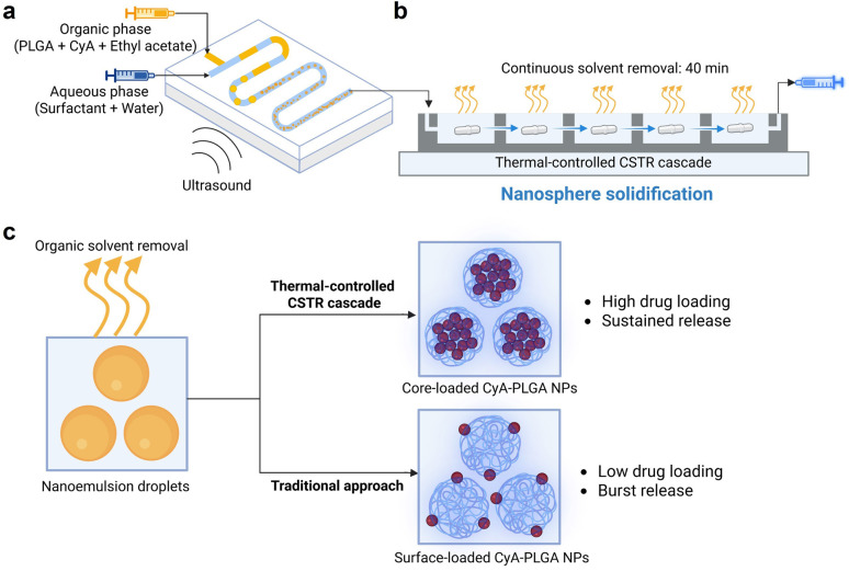 Fig. 1
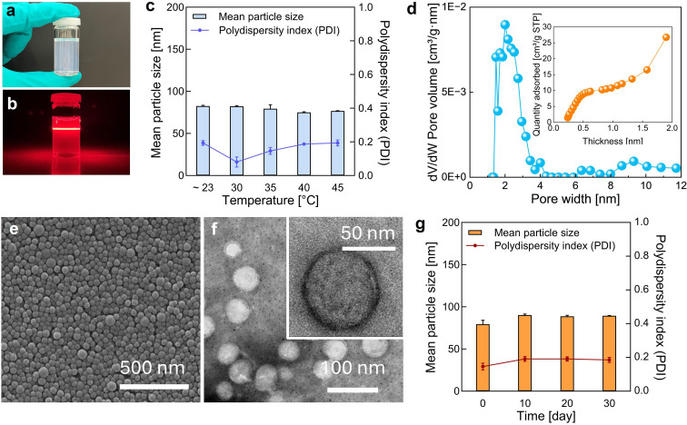 Fig. 2
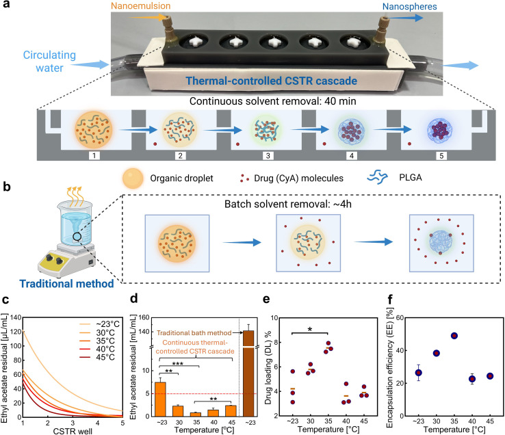 Fig. 3
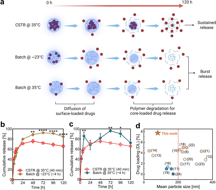 Fig. 4
- —Fonds Wetenschappelijk Onderzoek10.13039/501100003130
- —Flanders10.13039/100020468
- —Spanish Ministry of Science and Innovation10.13039/501100004837
Peer Reviews
No public reviews on file for this paper yet. If you reviewed it on a platform where reviews are public (OpenReview, ICLR, NeurIPS, ICML), you can paste yours below so the community can read it here.
Videos
No videos yet. Explain this paper in a talk, walkthrough, or lecture? Add one.
Taxonomy
TopicsMesoporous Materials and Catalysis · Advanced Drug Delivery Systems · Nanoparticle-Based Drug Delivery
Introduction
Drug-loaded nanospheres represent a powerful strategy for delivering active pharmaceutical ingredients using inert nanoscale carriers. These nanospheres can protect drug molecules from degradation and promote their transport across biological barriers to reach a target site.^1–4^ The inert materials, such as biodegradable polymers, enable sustained drug release, thus allowing for tailored pharmacokinetic properties and reducing the need for repeated dosing.^5,6^ Moreover, increasing the drug load per nanosphere allows higher doses per injection, improving patient compliance and minimizing excipient-related side effects.^7–9^ For many treatments, achieving a high therapeutic mass fraction is a prerequisite due to strict volume limits on injectable formulations (0.1 mL intradermal, 1 mL subcutaneous, and 1–3 mL intramuscular).^10^ These constraints make the development of nanospheres with high drug content critically important.
Engineering high drug-loaded nanospheres is challenging due to inherent limitations arising from their high surface-to-volume ratio. Drugs positioned on the particle surface are susceptible to loss during the formulation and post-formulation processes.^11–13^ Nanospheres are primarily composed of non-therapeutic scaffold polymers, such as poly(lactic-co-glycolic acid) (PLGA). Drug loadings in most reported studies are lower than 4%; some are even substantially below 1%.^11,14–27^ Various strategies have been developed to improve drug loading efficiency through strengthening drug–carrier interactions, such as donor–receptor coordination and covalent conjugation.^28–31^ However, these approaches are limited, as they require both the drug and carrier molecules to have specific structural and chemical properties.
Encapsulating a larger fraction of drug molecules within the nanosphere core, rather than leaving a significant portion on the surface, helps overcome surface-to-volume ratio constraints and enhances overall drug loading. This strategy eliminates the dependence on specific molecular features of the drug and carrier. A core-loaded nanosphere structure not only increases the total drug content but also offers better control over the drug release. Drug release from nanospheres occurs in two phases: an initial burst release caused by the rapid diffusion of surface-located drugs, followed by a more sustained release through the hydrolytic degradation of the PLGA matrix.^32–34^ By increasing the amount of drug loaded in the core, the initial burst release and resulting toxicity risks can be reduced, leading to a more sustained and prolonged release profile. Nanospheres could be formed from nanoemulsion droplets as the organic solvent evaporates (Fig. 1c), leading us to reasonably hypothesize that the solvent removal process would play a critical role in determining the solidification of drug and polymer molecules. By optimizing this process, it may be possible to fabricate core-loaded nanospheres with high drug loading and improved release kinetics.
Synthesis process of cyclosporin A-loaded PLGA nanospheres (CyA-PLGA NPs). The setup consists of (a) an ultrasonic microreactor for emulsion generation and (b) a continuous thermal-controlled CSTR cascade for solvent removal and nanosphere solidification. (c) Solvent removal and solidification processes at the droplet/particle level, along with the resulting nanosphere properties, compared to the traditional batch approach. The illustration was created with Biorender.com.
For continuous nanosphere synthesis, microfluidic nanoprecipitation is used in many studies.^6,35–38^ Although this technique can produce ultra-small nanospheres ranging from 20 to 100 nm, it struggles with a low drug loading capacity (<4%).^14,39–41^ Increasing the particle size can improve drug loading, but it will compromise the ability to cross biological barriers. This hinders particular applications like brain-targeted delivery, which requires particles to be below 100 nm to cross the blood–brain barrier.^42–44^ Furthermore, the small channel sizes required for rapid mixing in microfluidics can lead to issues like low throughput and microchannel clogging.^45–47^
Another commonly used synthesis method is emulsion-solvent evaporation. In 2007, Budhian et al.^20^ attempted to increase drug loading by adjusting the pH during solvent evaporation to reduce drug diffusion into the aqueous phase. However, they achieved a maximum drug loading of 2.5%. In 2016, de Solorzano et al.^48^ used microchannel emulsification for high-throughput synthesis (∼10 g h^−1^) of cyclosporin A-loaded PLGA nanospheres. However, the resulting particles had a mean size far exceeding 200 nm. Recently, Operti et al.^49^ introduced an inline sonicator for continuous emulsification, but the solvent evaporation was performed in a batch mode with dilution and stirring for 1 hour, and the smallest mean particle size achieved was 184.7 nm. To conclude, the synthesis of drug-loaded PLGA nanospheres through emulsion-solvent evaporation has not been fully transitioned to a continuous process. Achieving a mean particle size under 100 nm remains challenging, and the impact of solvent evaporation temperature on drug encapsulation was largely overlooked in previous studies.
In this work, we control the solvent removal process using a continuous stirred-tank reactor (CSTR) cascade (Fig. 1b), which provides thermal regulation, to solidify the drug and polymer molecules. This approach contrasts with previous studies,^48–51^ where solvent removal was performed in batch mode and remained largely unoptimized. This continuous manufacturing strategy eliminates batch-to-batch variations and provides a well-defined scale-up pathway,^52,53^ addressing two major challenges in the industrialization of nanomedicines.^54–58^ To validate our approach, cyclosporin A (CyA) is selected as a model peptide drug due to its poor water solubility and limited bioavailability (class II from the Biopharmaceutical Classification System, BCS). The solvent removal step is systematically optimized, and its impact on particle size distribution, morphology, and drug loading efficiency of cyclosporin A-loaded PLGA nanospheres (CyA-PLGA NPs) is assessed. Furthermore, in vitro drug release studies are conducted to evaluate the sustained release profile. Process efficiency and residual solvent content are also analysed to ensure the robustness and scalability of the developed method.
Results and discussion
Synthesis and characterization of CyA-PLGA NPs
CyA-PLGA NPs were synthesized using the emulsion–solvent evaporation technique. In the first step, an automated ultrasonic microreactor was employed to generate nanoemulsion droplets. The organic phase, consisting of PLGA and cyclosporin A dissolved in ethyl acetate, and the aqueous phase, consisting of the surfactant Poloxamer 407 in Milli-Q water, were introduced into the microreactor. Ultrasonic energy was applied via a piezoelectric transducer plate attached to the bottom of the reactor to facilitate emulsification (Fig. 1a). Following droplet formation, the nanoemulsion was fed into a CSTR cascade for continuous thermal-controlled solvent removal (Fig. 1b). The outlet of the CSTR cascade was connected to a syringe pump, which continuously withdrew the solidified CyA-PLGA NP suspension. The temperature of the CSTR cascade was regulated using a circulating water jacket located beneath the wells (Fig. 3a). The entire process was automated and conducted at a constant flow rate of 250 µL min^−1^, with an overall residence time of ∼40 min. For comparison, a traditional batch method was carried out by collecting the nanoemulsion from the microreactor outlet into a glass vial (Fig. 3b), followed by magnetic stirring for at least 4 hours at room temperature to allow for solvent evaporation.
For effective drug delivery, nanospheres smaller than 200 nm with a polydispersity index (PDI) below 0.2 are desired,^59,60^ while brain-targeted therapies typically demand even smaller sizes—under 100 nm.^42,44,61^ To meet these criteria, the emulsification step in the ultrasonic microreactor was systematically optimized (Table S2) before integrating the thermal-controlled CSTR cascade. After implementing the CSTR cascade for continuous solvent removal, the process temperature was varied from room temperature (∼23 °C) to 45 °C. Across this temperature range, the resulting CyA-PLGA NPs consistently maintained a mean particle size below 82 nm and a PDI below 0.2, fulfilling the size distribution requirements for biomedical applications. The pore size distribution results from the Brunauer–Emmett–Teller (BET) analysis (Fig. 2d) indicate the presence of micropores around 1.5 nm and mesopores in the 2–3 nm range. Larger pores (>5 nm) are likely due to interstitial voids between the nanospheres. The coexistence of micropores and mesopores is further confirmed by the t-plot curve, which exhibits a characteristic profile typical of micro–mesoporous materials.^62^ SEM and TEM images revealed a smooth, spherical morphology with a uniform size distribution. No significant morphological differences were observed between nanoparticles synthesized at ∼23 °C (Fig. S10) and at 35 °C (Fig. 2e and f), indicating that the mild thermal treatment does not compromise particle integrity. Additionally, the CyA-PLGA NPs demonstrated good storage stability, retaining their size distribution for at least 30 days when stored at 2–8 °C (Fig. 2g).
Characterization of CyA-PLGA NPs. (a) Visual appearance of the CyA-PLGA NP suspension. (b) Tyndall effect observed in the suspension using a laser pointer. (c) Mean particle size and polydispersity index (PDI) measured by dynamic light scattering (DLS). (d) Porosity distribution determined by density functional theory (DFT) analysis and t-plot analysis of CyA-PLGA NPs based on Brunauer–Emmett–Teller (BET) isotherms, measured at standard temperature and pressure (STP, 273 K and 101.325 kPa) with N2. Representative (e) scanning electron microscopy (SEM) image, (f) transmission electron microscopy (TEM) images, and (g) stability profile over 30 days (storage 2–8 °C) of CyA-PLGA NPs prepared via the thermal-controlled CSTR cascade at 35 °C. Data are presented as mean ± SD (n = 3).
Thermal-controlled CSTR cascade for enhanced drug loading
The solvent removal time was significantly reduced from over 4 hours (ref. 63) to just 40 min by replacing the conventional batch method (Fig. 3b) with a continuous CSTR cascade (Fig. 3a), which doubled the surface-to-volume ratio (Table S1). At a fixed duration of 40 min and room temperature (∼23 °C), the residual ethyl acetate concentration decreased from 141.3 µL mL^−1^ (batch) to 8.0 µL mL^−1^ (CSTR), demonstrating improved efficiency. However, to meet EMA regulations for Class 3 solvents,^64^ which require residual levels below 5 µL mL^−1^ (5000 ppm), faster solvent removal is necessary. This was addressed by introducing thermal control at elevated temperatures.
*Optimization of the thermal-controlled CSTR cascade and the resulting enhancement in drug loading efficiency. Schematic comparison of the organic solvent removal and nanoparticle formation process using (a) thermal-controlled CSTR cascade and (b) traditional batch method. (c) Residual ethyl acetate concentrations measured in each CSTR well (1–5) during the solvent removal process. (d) Final residual solvent content in the nanoparticle suspension. (e) Drug loading (DL) and (f) encapsulation efficiency (EE) of the resulting CyA-PLGA NPs obtained at varying CSTR cascade temperatures (∼23 °C to 45 °C). In graphic (d), the red dashed line indicates the maximum allowed residual level of ethyl acetate according to EMA guidelines (5 µL mL−1). Data are shown as mean ± SD, n = 3. Statistical significance is indicated as *p < 0.05, **p < 0.01, and **p < 0.001. Only relevant and significant statistical comparisons are highlighted.
The residual ethyl acetate content in each CSTR well during solvent removal (Fig. 3c) and in the final nanosphere suspension (Fig. 3d), along with the drug loading (DL, Fig. 3e) and encapsulation efficiency (EE, Fig. 3f) of the resulting CyA-PLGA NPs, was quantified by HPLC. The results showed that at 30 °C and 35 °C the final residual ethyl acetate was significantly reduced to 2.3 ± 0.3 µL mL^−1^ and 0.9 ± 0.3 µL mL^−1^, respectively, both well below the 5 µL mL^−1^ threshold. Interestingly, a slight increase in residual solvent was observed at 40 °C (1.5 ± 0.4 µL mL^−1^) and 45 °C (2.4 ± 0.1 µL mL^−1^). The drug loading increased from 4.2 ± 1.3% at ∼23 °C to a peak value of 7.6 ± 0.4% at 35 °C, but then declined to 3.6 ± 0.7% and 3.9 ± 0.3% at 40 °C and 45 °C, respectively, even below the value at room temperature (∼23 °C). A similar trend was observed for the encapsulation efficiency, which rose from 26.5 ± 8.5% to 49.0 ± 2.5% at 35 °C and then decreased to 22.7 ± 4.6% and 24.4 ± 2.2% at the higher temperatures.
The initial reduction in residual ethyl acetate at moderately elevated temperatures is attributed to an accelerated solvent removal rate, as further supported by measurements from individual CSTR wells (1–5) during the process (Fig. 3c). The observed improvement in drug loading efficiency results from more efficient solvent removal achieved through the thermal-controlled CSTR cascade. During this process, the organic solvent must first diffuse from the emulsion droplets into the surrounding aqueous phase before it can evaporate.^65,66^ Katou et al.^67^ proposed a mathematical model to describe the kinetics of this solvent transfer from oil droplets to the aqueous phase:
where Cd stands for the solvent concentration, t stands for time, r stands for radial position, and D stands for the solvent diffusion coefficient. According to the Wilke–Chang equation, the solvent diffusion coefficient D is given by
where ∅ is the association parameter, M is the molecular mass of the solvent, T is the temperature, η is the viscosity, and V is the molecular volume. The diffusion coefficient is proportional to the temperature, meaning that at higher temperatures, solvent removal occurs more rapidly. This accelerates droplet solidification and limits the time available for drug diffusion into the external aqueous phase. As a result, a larger amount of the drug is retained within the polymeric matrix of the nanospheres, significantly enhancing drug loading.
However, at further elevated temperatures of 40 °C and 45 °C—surpassing the glass transition temperature of PLGA (Fig. S11)—the polymer does not solidify into a rigid, glassy state but instead transitions into a softer, rubbery phase.^68–70^ In this rubbery state, stronger polymer–solvent interactions hinder solvent diffusion and evaporation, resulting in higher residual ethyl acetate levels despite the elevated temperature. Moreover, this rubbery state leads to two major effects: (i) the diffusion of the organic solvent slows down considerably, giving the drug more time to diffuse out of the polymeric matrix and escape into the aqueous phase, reducing drug retention; (ii) in some cases, the polymer–drug mixture fails to solidify into nanoparticles before reaching the liquid surface during evaporation. Instead, it forms a polymeric film on the liquid surface in the CSTR (as observed during experiments), leading to a loss of the drug material and reduced nanoparticle formation. Consequently, the drug loading efficiency at 40 °C and 45 °C declined, even falling below that achieved at room temperature (∼23 °C).
Therefore, 35 °C was determined to be the optimal solvent removal temperature in the CSTR cascade, as it enables ideal particle size distribution, maximizes the drug loading efficiency, and minimizes the residual ethyl acetate content, while preventing the detrimental effects of PLGA transitioning into its rubbery state above .
To confirm that these improvements were due to the nature of the thermal-controlled CSTR cascade nanotechnology, rather than temperature alone, a batch solvent removal was also performed at 35 °C for comparison (Table S4). When the batch process was run for the same duration as that of the CSTR cascade (40 min), the drug loading efficiency was extremely low at 0.8 ± 0.1%, indicating that almost no drug was encapsulated. As previously discussed, this is attributed to the poor solvent removal efficiency of the batch method, which left a high residual ethyl acetate concentration (91.9 ± 1.6 µL mL^−1^). With the aqueous phase nearing saturation, the diffusion of ethyl acetate from the droplets was restricted, significantly slowing solidification. This prolonged diffusion window allowed drug molecules to escape into the aqueous phase before being entrapped. Even after extending the batch evaporation to 4 hours, the drug loading efficiency only modestly increased to 2.5 ± 0.9%, still far below the value achieved with the CSTR cascade. These results confirm that the significant improvement in drug loading was primarily due to the efficient solvent removal enabled by the thermal-controlled CSTR cascade.
In vitro drug release
The release of a poorly water-soluble drug from PLGA nanospheres typically happens in two phases (Fig. 4a). The first phase is a rapid initial burst release, driven by the diffusion of the drug located near the particle surface. The second phase is more sustained and is attributed to the hydrolysis of the PLGA and erosion of the polymeric matrix, which releases the drug encapsulated in the nanosphere core.^32–34^ If the enhanced solvent removal rate indeed results in increased drug encapsulation in the nanosphere core, CyA-PLGA NPs synthesized using the thermal-controlled CSTR cascade at 35 °C should exhibit less of an initial burst release and more drug release during the second sustained phase.
*In vitro drug release demonstrating the advantage of the core-loaded nanoparticle structure. (a) Schematic illustration of the two-phase drug release mechanism from PLGA nanoparticles. Sustained and burst release profiles resulting from core-loaded and surface-loaded drug distributions, respectively. (b and c) In vitro drug release data of CyA-PLGA NPs prepared via the thermal-controlled CSTR cascade at 35 °C (CSTR @ 35 °C), compared to those prepared using the traditional batch method at room temperature (Batch @ ∼23 °C) and at 35 °C (Batch @ 35 °C). Data are presented as mean ± SD (n = 3). Statistical significance is indicated as follows: *P < 0.05; **P < 0.01; ***P < 0.001; ***P < 0.0001. (d) Positioning our core-loaded CyA-PLGA NPs within the landscape of benchmark studies,11,14–27 highlighting their exceptional ability to simultaneously achieve both a high drug loading efficiency and a small particle size (two critical and often mutually exclusive parameters in nanoparticle synthesis). The red dashed line in panel (d) marks the <200 nm threshold typically considered suitable for drug delivery applications.
To provide a meaningful comparison, we selected the batch solvent evaporation at ∼23 °C (Batch @ ∼23 °C) to represent the slowest evaporation rate. The drug release profile over 120 h, depicted in Fig. 4b, confirms the hypothesis. During the first 24 hours of the initial release phase, there was no statistically significant difference between the two samples. However, by the end of the burst release phase (72 h), 93% of the drug encapsulated in the Batch @ ∼23 °C nanospheres was released, indicating that a major fraction of the CyA molecules was located close to the surface. In contrast, the CSTR @ 35 °C nanospheres only released 76% of their drug content during the initial burst phase (48 h), leaving 24% encapsulated within the nanosphere core for a slower, sustained release. The cumulative drug release gradually declined in the second phase, as the drug continued to be released, but at a slower rate than its degradation, making the net change appear negative. This trend aligns with the CyA stability profile observed in our degradation test (Fig. S9) and is consistent with findings reported in other studies.^71,72^
Next, we conducted an additional in vitro drug release study on CyA-PLGA NPs prepared via batch solvent evaporation at 35 °C (Batch @ 35 °C). The aim was to assess whether the core-loaded drug distribution observed in nanospheres produced by the CSTR cascade resulted primarily from the high solvent removal efficiency of the thermal-controlled CSTR cascade process, or if similar characteristics could be achieved by applying the same elevated temperature in a conventional batch process. As shown in Fig. 4c, the Batch @ 35 °C nanospheres exhibited a sharp burst release, with 86% of the encapsulated drug released within the very first 9 h. This release was even more abrupt than that of the Batch @ ∼23 °C nanospheres, suggesting that the drug was located even closer to the particle surface. By 24 hours, the CyA concentration in the release medium had already begun to decline due to degradation, and the drop was more pronounced because there was little to no sustained release between 9 and 24 hours to replenish the drug. In the end, the sustained release phase began after 48 h, corresponding to the release of the remaining 14% of the drug from the nanosphere core.
Based on the drug release results, we can conclude that the Batch @ 35 °C nanospheres contained a larger fraction of the drug encapsulated in the core (14%) compared to the Batch @ ∼23 °C nanospheres (7%), but still less than the CSTR @ 35 °C nanospheres (24%). This suggests that the elevated temperature enhanced solvent diffusion from the droplets into the aqueous phase, allowing more drug to be retained in the nanoparticle core. However, the batch process is limited by a low surface-to-volume ratio, making the evaporation of solvent from the aqueous phase into air the rate-limiting step. This inefficiency resulted in a high residual solvent concentration in the aqueous phase, which in turn suppressed further diffusion of organic solvent from the droplets. Consequently, solvent accumulated at the droplet/nanoparticle interface, providing more time for drug molecules to diffuse into the aqueous phase and be lost, leading to lower drug loading efficiency (2.5 ± 0.9%) compared to the CSTR @ 35 °C (7.6 ± 0.4%). Moreover, this solvent accumulation forces a larger portion of the drug toward the sphere surface, as evidenced by a rapid burst release of 86% of the drug within the first 9 h, in contrast to the slower burst release observed in Batch @ ∼23 °C nanospheres (93% released over 48 h). Therefore, the improvement was not solely due to the elevated temperature, but was primarily driven by the high solvent removal efficiency achieved through the thermal-controlled CSTR cascade.
Conclusions
In summary, we have developed a thermal-controlled CSTR cascade nanotechnology for the synthesis of CyA-PLGA NPs, achieving a significantly improved drug loading efficiency and a more sustained release profile. The formation of nanospheres with high drug loading is a result of a larger fraction of drug molecules solidifying at the particle core, realized by a more efficient solvent removal in the CSTR cascade at 35 °C. The achieved drug loading is, to our knowledge, the highest reported for sub-100 nm CyA-PLGA NPs, significantly exceeding the previously reported maximum of 4.6%^11,14–27^ (Fig. 4d). The core-loaded structure not only provides a higher drug payload, but also enables gradual release of drug molecules. The sustained release from high drug-loaded nanospheres may contribute to an enhanced therapeutic efficacy and reduced adverse effects for drug delivery applications. Additionally, the robustness and scalability of the thermal-controlled CSTR cascade provide a promising pathway for clinical translation, aligning with FDA guidelines for modern pharmaceutical manufacturing.^73^
Author contributions
Conceptualization: H. C., A. P. U. and V. S. C. Methodology: H. C., A. P. U. and V. S. C. Experiments: H. C., A. P. U., and V. S. C. Writing – original draft: H. C. and A. P. U. Writing – review and editing: C. C., V. S. C., and S. K. Data curation: H. C. and A. P. U. Supervision: C. C., V. S. C., and S. K. Funding acquisition: S. K.
Conflicts of interest
There are no conflicts to declare.
Supplementary Material
NA-OLF-D5NA00897B-s001
The reference list from the paper itself. Each links out to its DOI / PubMed record.
- 1Blanco E. Shen H. Ferrari M. Principles of Nanoparticle Design for Overcoming Biological Barriers to Drug Delivery Nat. Biotechnol.201594195110.1038/nbt.333026348965 PMC 4978509 · doi ↗ · pubmed ↗
- 2Chiang M. R. Hsu C. W. Pan W. C. Tran N. T. Lee Y. S. Chiang W. H. Liu Y. C. Chen Y. W. Chiou S. H. Hu S. H. Reprogramming Dysfunctional Dendritic Cells by a Versatile Catalytic Dual Oxide Antigen-Captured Nanosponge for Remotely Enhancing Lung Metastasis Immunotherapy ACS Nano 20251922117213510.1021/acsnano.4c 0952539739571 PMC 11760334 · doi ↗ · pubmed ↗
- 3Huynh T. M. H. Huang P. X. Wang K. L. Tran N. T. Iao H. M. Pan W. C. Chang Y. H. Lien H. W. Lee A. Y. L. Chou T. C. Chiang W. H. Hu S. H. Reprogramming Immunodeficiency in Lung Metastases via PD-L 1 Si RNA Delivery and Antigen Capture of Nanosponge-Mediated Dendritic Cell Modulation ACS Nano 20251927251342515310.1021/acsnano.5c 0539540616527 PMC 12269356 · doi ↗ · pubmed ↗
- 4Yalamandala B. N. Chen Y. J. Lin Y. H. Huynh T. M. H. Chiang W. H. Chou T. C. Liu H. W. Huang C. C. Lu Y. J. Chiang C. S. Chu L. A. Hu S. H. A Self-Cascade Penetrating Brain Tumor Immunotherapy Mediated by Near-Infrared II Cell Membrane-Disrupting Nanoflakes via Detained Dendritic Cells ACS Nano 20241828187121872810.1021/acsnano.4c 0618338952208 PMC 11256899 · doi ↗ · pubmed ↗
- 5Brigham N. C. Ji R. R. Becker M. L. Degradable Polymeric Vehicles for Postoperative Pain Management Nat. Commun.202112136710.1038/s 41467-021-21438-333649338 PMC 7921139 · doi ↗ · pubmed ↗
- 6Liu D. Zhang H. Fontana F. Hirvonen J. T. Santos H. A. Microfluidic-Assisted Fabrication of Carriers for Controlled Drug Delivery Lab Chip 20171856188310.1039/c 7lc 00242 d 28480462 · doi ↗ · pubmed ↗
- 7Shen S. Wu Y. Liu Y. Wu D. High Drug-Loading Nanomedicines: Progress, Current Status, and Prospects Int. J. Nanomed.20174085410910.2147/IJN.S 132780 PMC 545998228615938 · doi ↗ · pubmed ↗
- 8Della Rocca J. Liu D. Lin W. Are High Drug Loading Nanoparticles the next Step Forward for Chemotherapy?Nanomedicine 201230330510.2217/nnm.11.19122385191 PMC 3777216 · doi ↗ · pubmed ↗