Occurrence and Haematology of Canine Tick‐Borne Protozoa in Dhaka City, Bangladesh
Most. Aklima Khatun, S. M. Abdullah, Md. Kamrul Hassan, Mahfuzul Islam

TL;DR
This study found tick and protozoan infections in stray dogs in Dhaka, Bangladesh, with significant effects on blood parameters.
Contribution
The study reports the prevalence of tick-borne protozoa and their impact on haematology in stray dogs in Dhaka.
Findings
30.62% of dogs were infested with Rhipicephalus sanguineus ticks, mostly on the neck and chest.
23.13% of dogs had protozoan infections, with Babesia canis being the most common at 11.88%.
Infected dogs showed significantly lower RBC, haemoglobin, and PCV, and higher eosinophil counts.
Abstract
Dogs, being companion animals, serve a variety of economic, social and cultural purposes. However, diseases caused by the tick‐borne protozoans are drawing attention globally due to their zoonotic transmission. In this context, the present study aimed to observe the occurrence of tick‐borne protozoan infections as well as some selective haematological parameters of stray dogs in Dhaka city, Bangladesh. A total of 160 dogs from various places were selected randomly and examined for both tick and protozoan infection. Only one species of hard tick, Rhipicephalus sanguineus, was identified, where 49 (30.62%) among the study population were found to be infested with this tick. Ticks ranging from 1–16 were removed from dogs, where most of the ticks were collected from the neck and chest regions (P < 0.001). On the other hand, examinations of blood smears confirmed three protozoan species…
Genes, proteins, chemicals, diseases, species, mutations and cell lines named across the full text — each resolved to its canonical identifier and authoritative record.
Click any figure to enlarge with its caption.
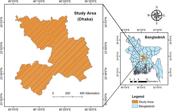 FIGURE 1
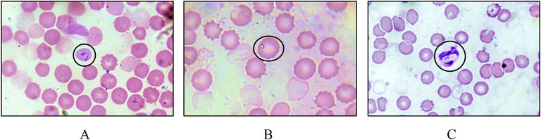 FIGURE 2
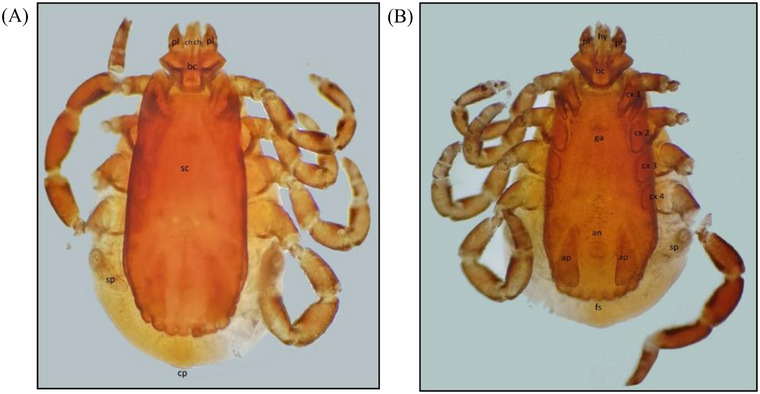 FIGURE 3
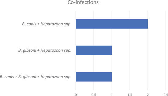 FIGURE 4
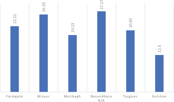 FIGURE 5
| Species | No. of dogs infected (n = 160) | Occurrence % |
|
|---|---|---|---|
|
| 19 | 11.88 | 0.047 |
|
| 7 | 4.38 | |
|
| 16 | 10.00 |
| Types of infection | No. of dogs infected (n = 37) | Occurrence % |
|
|---|---|---|---|
| Single infection | 33 | 89.19 | < 0.001 |
| Multiple infections | |||
| Two species | 3 | 8.11 | |
| More than two species | 1 | 2.70 |
| Group | Variable | No. of examined | No. of infected | Occurrence % |
|
|---|---|---|---|---|---|
| Gender | Male | 47 | 9 | 19.15 | 0.442 |
| Female | 113 | 28 | 24.78 | ||
| Age | < 1 year of age | 24 | 7 | 29.17 | 0.730 |
| 1–2 years of age | 52 | 12 | 23.08 | ||
| > 2 years of age | 84 | 18 | 21.43 |
| Areas | No. of examined | No. of infected | Occurrence% | Collected female ticks | Collected male ticks | Sex ratio |
|---|---|---|---|---|---|---|
| Farmgate | 27 | 8 | 29.63 | 38 | 12 | 3.16 |
| Mirpur | 42 | 12 | 28.57 | 52 | 19 | 2.73 |
| Malibagh | 26 | 8 | 30.77 | 28 | 7 | 4.00 |
| Basundhara R/A | 33 | 14 | 42.42 | 62 | 18 | 3.44 |
| Tejgoan | 24 | 6 | 25.00 | 29 | 8 | 3.62 |
| Gulshan | 8 | 1 | 12.50 | 3 | 2 | 1.50 |
|
|
|
|
|
|
|
|
| Group | Variable | No. of examined | No. of infected | Occurrence % |
|
|---|---|---|---|---|---|
| Gender | Male | 47 | 10 | 21.28 | 0.098 |
| Female | 113 | 39 | 34.51 | ||
| Age | < 1 year of age | 24 | 9 | 37.50 | 0.726 |
| 1–2 years of age | 52 | 15 | 28.85 | ||
| > 2 years of age | 84 | 25 | 29.76 |
| Months | No. of examined | No. of infected | Occurrence % |
| Seasons | Occurrence % |
|
|---|---|---|---|---|---|---|---|
| November | 32 | 9 | 28.13 | 0.787 | Winter | 26.92 | 0.166 |
| December | 21 | 5 | 23.81 | ||||
| January | 31 | 8 | 25.81 | ||||
| February | 20 | 6 | 30.00 | ||||
| March | 37 | 13 | 35.84 | Summer | 37.50 | ||
| April | 19 | 8 | 42.11 |
| Degree of infestation | No. of ticks counted | No. of dogs infected | Occurrence % |
|
|---|---|---|---|---|
| Low (+) | 1–4 | 22 | 44.89 | 0.001 |
| Mild (++) | 5–8 | 16 | 32.65 | |
| Moderate (+++) | 9–12 | 7 | 14.28 | |
| High (++++) | 13–16 | 4 | 8.16 |
| Attachment of ticks | No. of ticks counted | Occurrence % |
|
|---|---|---|---|
| Head with ears | 56 | 20.14 | < 0.001 |
| Neck and chest region | 95 | 34.17 | |
| Back region | 19 | 6.83 | |
| Abdomen | 72 | 25.89 | |
| Legs | 36 | 12.94 |
| Parameters | Non‐infected animals | Infected with protozoa | Infected with ticks | SEM |
| Reference value |
|---|---|---|---|---|---|---|
| RBC (106 cells/µL) | 6.23a | 4.51b | 4.69b | 0.081 | < 0.001 | 5.5–8.5 |
| Hb (g/dL) | 15.48a | 11.44b | 11.18b | 0.265 | < 0.001 | 12–19 |
| PCV (%) | 43.80a | 33.50b | 32.10b | 0.901 | < 0.001 | 37–57 |
| MCV (fL) | 70.34 | 74.78 | 68.56 | 2.140 | 0.143 | 66–77 |
| MCH (pg) | 24.87 | 25.49 | 23.89 | 0.639 | 0.238 | 19.5–24.5 |
| MCHC (%) | 35.43 | 34.24 | 35.06 | 0.857 | 0.662 | 32–36 |
| WBC (103 cells/µL) | 13.66b | 16.90a | 17.30a | 0.295 | < 0.001 | 6–17 |
| Neutrophil (%) | 70.70a | 66.40b | 65.90b | 1.244 | 0.024 | 58–85 |
| Lymphocyte (%) | 14.50 | 13.40 | 11.30 | 0.928 | 0.065 | 8–21 |
| Monocyte (%) | 8.80 | 9.20 | 10.20 | 0.460 | 0.134 | 2–10 |
| Eosinophil (%) | 6.00b | 11.00a | 12.70a | 0.673 | < 0.001 | 0–9 |
- —National Science and Technology Grant, Ministry of Science and Technology, Government of the People's Republic of Bangladesh
Peer Reviews
No public reviews on file for this paper yet. If you reviewed it on a platform where reviews are public (OpenReview, ICLR, NeurIPS, ICML), you can paste yours below so the community can read it here.
Videos
No videos yet. Explain this paper in a talk, walkthrough, or lecture? Add one.
Taxonomy
TopicsVector-borne infectious diseases · Bartonella species infections research · Zoonotic diseases and public health
Introduction
1
One of the most popular pets in the world, dogs serve a variety of economic, social and cultural purposes in society (Swai et al. 2010). Keeping pet animals enhances people's self‐esteem, particularly young people (Robertson et al. 2000; Knoble et al. 2008). However, approximately 75% of dogs across the world are classified as free‐roaming (stray dogs), which has significant implications for public health (Hughes and Macdonald, 2013). Nowadays, many stray, lost, or owner‐surrendered dogs are kept in shelters to provide a temporary home until they can be reclaimed by the owner (Barrera et al. 2010). These stray animals are not even tested for parasites, vaccinated, or treated for diseases. Therefore, they serve as the reservoirs for several significant zoonotic parasites (Dakkak 2010).
These stray dogs are susceptible to different microorganisms, such as bacteria, viruses and protozoa, which can be transmitted by ectoparasites. Among them, tick‐borne protozoa are drawing attention globally for both humans and animals (Fuente et al. 2017). The brown dog tick*, R. sanguineus*, is distributed globally and is the most common in tropical areas, although it is difficult to differentiate this tick from other species having similar morphological characteristics (Walker et al. 2003) and different behaviour and ecology (Walker et al. 2000). Moreover, this tick can cause skin lesions, anaemia and tick paralysis in case of heavy infestations in dogs (Otranto et al. 2012). Furthermore, two protozoal diseases of dogs, namely babesiosis and ehrlichiosis, are transmitted by this tick (Dantas‐Torres et al. 2012). Transmission of these protozoa around the world could be due to the wide distribution of ticks (Jefferies et al. 2007; Yeagley et al. 2009).
In addition to this, the health of dogs is negatively affected by those protozoan diseases, which can result in anaemia and, sometimes, thrombocytopenia and leukopenia (Eiras et al. 2013; Piratae et al. 2017; Rautenbach et al. 2017; Thongsahuan et al. 2020). Despite this, there are very limited comprehensive studies of canine blood protozoa in the study area. Therefore, the objectives of this study were to identify different blood protozoa along with their occurrence, as well as to determine the morphological characteristics of isolated ticks from the dogs in the study areas. The study also aimed to compare the haematological profiles between infected and non‐infected dogs.
Materials and Methods
2
Ethical Approval
2.1
Blood samples were aseptically collected by registered veterinarians through proper restraining of the dogs to avoid any injuries. All the procedures required for the sample collection were fulfilled, based on the ethical guidelines approved by the Animal Welfare Act, 2019. Moreover, permission for sampling was verbally obtained from the Department of Livestock Services.
Study Area
2.2
This research was carried out in Dhaka, the largest city and the capital of Bangladesh (Figure 1). The city has a total area of 118.29 square miles and is situated at 23°42′N 90°22′E. Tropical vegetation covers the region, which has moist soils that are nearly flat and very near sea level. As a result of the excessive rainfall, Dhaka is vulnerable to floods during the monsoon season. The city experiences 2123 millimetres (83.6 inches) of annual rainfall and an average yearly temperature of 26°C (79°F).
Location of study area.
Study Period
2.3
The cross‐sectional study lasted for six months, comprising November 2022 to April 2023, where 160 street dogs from various places were randomly selected and examined. A structured questionnaire was developed, including the tentative age, body weight, gender and so on, to collect data from the study population. During sampling, 47 individuals were male and the remaining 113 were female. Moreover, 24 dogs were of < 1 year, 52 were between 1 and 2 years and the remaining 84 were above two years of age.
Sample Collection
2.4
A common versatile tool, ‘Catchpole’, was used to capture and restrain the street dogs with the help of a group of trained people from the Dhaka North City Corporation (DNCC). A general anaesthesia was performed by Atropine Sulphate (0.2 mg/kg, SC) and Ketamine (2.0 mg/kg, IV) to collect both ticks and blood from the selected dogs. A comprehensive physical examination was carried out to collect ticks using forceps and placed into separate vials containing 70% alcohol with proper labelling for each dog. On the other hand, the superficial and accessible cephalic vein was used as the site for blood sampling, where 2 mL of blood was taken from each animal using a sterile 21G needle (JMI, Bangladesh) and immediately transferred to an EDTA vial (JMI, Bangladesh). Then, both vials containing ticks and blood were transferred to the laboratory, maintaining a cool chain and kept in a refrigerator (4–8°C) for further examination.
Blood Smear Preparation
2.5
At least 2 (two) thin smears per animal were prepared, where the cells were in a monolayer (i.e., not touching one another). A freshly prepared Giemsa working solution was made from a well‐prepared commercial stock (SRL, India) to stain blood protozoa. To conduct proper staining, a Coplin jar was filled with approximately 40 mL of Giemsa working solution (10%) and 2 drops of Triton X‐100 (Sigma‐Aldrich, USA) were added to the solution. Then, the slides were placed into the working Giemsa solution for 30 min. The excess stain was afterwards removed from those slides by dipping them three to four times in Giemsa buffer solution. Finally, the stained slides were dried by keeping them on tissue paper.
Tick Identification
2.6
Firstly, the ectoparasites were cleared by dissolving in 10% KOH (Merck, India) at room temperature overnight, which allowed them to pass light through them. After clearing, the specimens were returned to 50% ethanol, followed by distilled water for 30 min in each to prepare them for staining. Hematoxylin and Eosin (H & E) dye was used to stain the specimens, where the slides were kept in the stain overnight. As the specimens became darker, the excessive stain was removed by keeping them in 3% Acid‐Alcohol. Subsequently, the dehydration process was accomplished to prevent the specimen from spoiling by bacteria. Then, the specimens were cleared by xylene (Sigma‐Aldrich, USA) for a few seconds to remove ethanol and mounted in a fresh slide with Canada balsam (SRL, India). Finally, the ectoparasites (ticks) were examined under a microscope (4X and/or 10X) for morphological identification (Krantz and Walter, 2009).
Haematological Analysis
2.7
For haematological comparison, dogs were categorised into three groups (non‐infected, infected with ticks, and infected with protozoa), where non‐infected animals were defined as those showing the absence of both tick infestation and protozoan infection upon examination. Additionally, to ensure the uniformity among those three groups, randomly 10 blood samples from each group were analysed for different haematological parameters, such as, red blood cells (RBC), Haemoglobin (Hb), packed cell volume (PCV), mean corpuscular volume (MCV), red cell distribution width (RDW), mean corpuscular haemoglobin (MCH), MCH concentration (MCHC), white blood cells (WBC) and so on. Among these parameters, RBC and WBC were calculated in a haemocytometer slide, while Hb was detected by Sahli's method. Moreover, PCV was examined in a Wintrobe tube and recorded using the following formula.
Another three auxiliary indicators (MCV, MCH and MCHC), particularly in the differential diagnosis of anaemia, were recorded using the following formula (Ware 2020).
Statistical Methods
2.8
The obtained data were imported, stored and coded accordingly using Microsoft Excel 2016, where all data analyses were performed by using a statistical software program (SPSS for Windows, Version 19.0, USA). The results of occurrence were expressed as a percentage. Association among various risk factors, namely, sex, age and season, was carried out by Chi‐square (𝜒2‐test). Moreover, the standard error of the mean was also determined for haematological parameters.
Results
3
Morphological Identifications of Blood Protozoa
3.1
Babesia species can be identified by their orientation in RBCs, where B. canis makes an acute angle, while B. gibsoni appears single in most cases. In our study, the shape of B. canis was observed as pyriform, where one end was pointed and the other end was rounded (Figure 2A). On the other hand, B. gibsoni lacked the usual pyriform shapes and had a signet ring form (Figure 2B). Moreover, in the stained blood smear under the microscope, Hepatozoon spp. was easily identified in the cytoplasm of WBCs (mostly in neutrophils) where the gamonts were observed elongated, ellipsoidal and had an eccentrically positioned nucleus (Figure 2C).
Microscopic observation of canine blood protozoa (100X magnifications). (A) Babesia canis; (B) Babesia gibsoni; and (C) Hepatozoon spp. (black circle indicates the organism).
Morphological Identifications of Rhipicephalus sanguineus
3.2
Medium‐sized, yellowish‐brown to reddish‐brown ticks having a dark, inornate brown scutum were measured with a scale, where unfed males and females were found on an average of 3.60 and 4.23 mm, respectively. The capitulum, or anterior portion of the body (Figure 3), was composed of one hexagonal‐shaped basis capitulum, which supports several organs, such as one powerful hypostome for sucking blood, two chelicerae for cutting the skin and two short palps for sensory function. Furthermore, in all the studied specimens in our investigation, setae and sensilla were discovered to be present throughout the body without a distinct pattern of distribution.
Male Rhipicephalus sanguineus (4X); dorsal view (A) & ventral view (B). pl, palp; hy, hypostome; ch, chelicerae; bc, basis capitulam; cx, coxa; sc, scutum; sp, spiracle; an, anus; ap, adanal plate; fs, festoon; ga, genital apron; cp, caudal plate.
Despite intraspecific heterogeneity among the study samples, festoons were located on the posterior margin of the body and were separated into 11 unique rectangular portions (Figure 3). The size of the caudal process varied across the specimens under study in fed males. The two valves and an anal groove that created the anal orifice were articulated with four setae that are positioned symmetrically on each side. Adanal plates and accessory shields were found in male ticks, composing the anogenital region. These structures are highly sclerotised and located on the side of the anus. The adanal plates were long, parallel and had a sharp posterior margin. The accessory shields, varying in form, were located beside the adanal plates. The narrow spiracular plates were located behind the last pair of legs. A circular ostial lip was also present in each spiracular plate. The genital plate was located between the 1^st^ and 2^nd^ pair of coxae and exhibited a round structure in all the specimens in our study.
Occurrence of Blood Protozoa
3.3
Overall Occurrence of Blood Protozoa
3.3.1
Examination of blood smear was performed under a light microscope, where 37 out of 160 samples (23.13%) were infected with any of the three species of blood protozoa, namely Babesia canis, Babesia gibsoni and Hepatozoon spp. In addition to this, the species‐wise occurrence of canine blood protozoa showed statistically significant differences, where B. canis, B. gibsoni and Hepatozoon spp. were detected in 19, 7 and 16 dogs, comprising 11.88%, 4.38% and 10.00%, respectively (Table 1). However, of these 37 positive dogs, 33 (89.19%) were infected with only one species, while infections with more than one protozoan were found only in 4 (10.81%) dogs (Table 2). These mixed infections were observed with two (2) and three (3) species of canine blood protozoa, comprising 8.11% and 2.70%, respectively. These single and mixed infections of protozoa had a statistical significance (P <0.001). Moreover, the occurrence of these co‐infections with canine blood protozoa is exhibited in Figure 4.
The occurrence of co‐infections with canine blood protozoa.
Occurrence of Blood Protozoa in Relation to Different Risk Factors
3.3.2
The proportion of gender was mentioned before, where 70.63% were females and the rest 29.37% were males. In the case of gender, a little difference was observed where females (24.78%) were infected with canine blood protozoa more than males (19.15%). However, all dogs were categorised into three groups (i.e., < 1 year, 1–2 years and > 2 years), where the highest occurrence (29.17%) was seen in the younger groups of age (< 1 year), followed by 23.08% in the age group of 1–2 years and 21.43% in the age group of more than 2 years. These results indicated more canine blood protozoan infections in puppies than in adults. Protozoan infection in relation to gender and age is shown in Table 3.
Occurrence of Canine Blood Protozoa in Different Locations
3.3.3
As mentioned before, a total of 160 stray dogs from 6 different locations in Dhaka city were included in this study. Microscopic examination of the blood revealed the highest incidence in Basundhara R/A (27.27%), followed by Mirpur (26.19%), Farmgate (22.22%), Tejgaon (20.83%), Malibagh (19.23%) and Gulshan (12.50%). This area‐wise infection of blood protozoa is given in Figure 5.
Occurrence of canine blood protozoa in different locations.
Occurrence of Ticks
3.4
Overall Occurrence of Adult Ticks in Dhaka City
3.4.1
A total of 160 stray dogs, varying in age and sex, were selected and examined for ticks after performing general anaesthesia. Upon visual inspection, 49 of those study populations were found to be infested with brown dog tick, Rhipicephalus sanguineus. Area‐wise overall occurrence (30.62%) is shown in Table 4. Concerning area‐wise infestation, the highest infection was encountered in Basundhara R/A (42.42%), followed by Malibagh (30.77%), Farmgate (29.63%), Mirpur (28.57%), Tejgoan (25.00%) and Gulshan (12.50%). The infected dogs produced a total of 278 adult ticks, where the average sex ratio was 3.21, indicating more female ticks on the host.
Occurrence of Ticks in Relation to Different Risk Factors
3.4.2
Among the examined 47 male dogs, 10 (21.28%) were found infested with ticks. On the other hand, 39 dogs out of 113 female dogs, comprising 34.51% of the total population, were found to be infested with ticks during the study. Considerably, females were infected more than the male dogs, where there was no statistical difference. Moreover, the occurrence of tick infestation varied depending on the age of the studied samples. Dogs aged less than 1 year showed the highest infection (37.50%) followed by 1–2 years (28.85%) and more than 2 years (29.76%) age groups. From these results, it was clear that young dogs were affected by ticks more than adults, although there was no statistical significance among them. These variations in relation to different risk factors are given in Table 5.
Seasonal Occurrence of Ticks
3.4.3
The present study was initiated in November 2022, when we collected samples from dogs. Since then, the collection of ticks from dogs has continued till April 2023. Therefore, only two major seasons, namely winter (November–February) and summer (March–April), were included in our study. There were differences in tick burdens in different seasons, with higher infestation levels in summer (37.50%) followed by winter (26.92%). Moreover, the monthly incidence of tick infestation is given in Table 6, where the highest percentage of tick‐infested dogs were examined in March (37.84%) and the lowest percentage was marked in December (23.81%).
Degree of Tick Infestation
3.4.4
In our study, ticks were removed individually from each dog, with an average of 1–16 ticks per dog. To determine the degree of tick infestation, a total of 4 categories were identified: first, low infestation rate comprising 1–4 numbers of ticks, second, mild infestation rate comprising 5–8 numbers of ticks, third, moderate infestation rate comprising 9–12 numbers of ticks and finally, high infestation rate comprising 13–16 numbers of ticks. The degree of tick infestation having a statistical significance is included in Table 7.
Regions of Infestation by Ticks on Dogs’ Bodies
3.4.5
Predominantly, five (5) regions were identified on the dog's body after reviewing the literature and these areas were the head with ears, neck and chest regions, back region, abdomen and legs. As we mentioned before, 278 adult ticks were collected during this study, while 95 ticks, comprising nearly one‐third of the total population, were found around the neck and chest region, which expressed the highest percentage of other parts of the body with a statistical significance (P < 0.001). On the other hand, the back region comprised the lowest percentage of the availability of ticks. The attachment of ticks on the host's body is exhibited in Table 8.
Haematological Parameters of Blood
3.5
Of the 160 samples collected from dogs, 49 were positive for R. sanguineus and 37 were positive for different protozoan infections. The average haematological values obtained from the non‐infected and infected groups are presented in Table 9. The RBC counts, Hb and PCV of all infected dogs were numerically lower compared to the non‐infected group. The average values RBC (6.23, 4.51 and 4.69), Hb (15.48, 11.44 and 11.18) and PCV (43.80, 33.50 and 32.10) were recorded from the non‐infected group, the infected group with protozoa and the infected group with ticks, respectively, which indicated the different degrees of anaemia. The average values of MCV, MCH and MCHC were found in the normal range. On the other hand, the average WBC count was higher in the infected groups, where the average value of WBC for protozoan was 16.90 × 10^3^ cells/µL and the average value of WBC for tick infestation was 17.30 × 10^3^ cells/µL. When compared to the different leukocyte counts, the eosinophils of the infected groups showed higher values (11.00 % and 12.70% for protozoa and ticks, respectively) than the normal range, indicating the parasitic infections.
Discussion
4
A total of 160 street dogs were included in this study, where samples were collected, smeared and stained for microscopic identification following a proper scientific method. Three (3) protozoan species, namely, B. canis, B. gibsoni and Hepatozoon spp., were identified according to the keys and descriptions of various authors (Allison and Little 2013; Saari et al. 2019). On the other hand, only one species of ticks, R. sanguineus, was identified according to various authors (Walker et al. 2000; Guglielmone et al. 2006; Krantz and Walter 2009; Dantas‐Torres et al. 2013; Nava et al. 2015).
Moreover, nearly one‐fourth (23.13%) of the study samples were infected with at least one protozoon, which is very similar to various reports in Southeast Asia where the prevalence of canine blood protozoan infections reached up to 28% (Laummaunwai et al. 2014; Sontigun et al. 2022). However, other authors (Piratae et al. 2017; Juasook et al. 2021) revealed higher prevalence than our study. These higher infection rates may be due to the difference in methodology, where most of them applied molecular techniques that provide more specific genetic and species information (Sainz et al. 2015; Das et al. 2020). Notably, B. canis was encountered in the highest number found during the study, which is similar to the studies in the above‐mentioned areas, including the Indian Sub‐continent (Singh et al. 2014; Jain et al. 2017). Although Piratae et al. 2017 and Thongsahuan et al. 2020 found more infections with Hepatozoon spp. than Babesia. The variation in species‐wise prevalence might be due to the geographic location, distribution of vectors, methods of sampling, etc. (Singla et al. 2016; Rucksaken et al. 2019). Furthermore, the results of mixed infections are supported by Mylonakis et al. (2004), Kumar and Varshney (2007) and Yabsley et al. (2008). In our study, the proportion of gender in the observed population was nearly 2:1, where the females were 70.63% and the rest 29.37% were males. In the case of gender, females were more infected than males and this result is inconsistent with that of Amuta et al. (2010) and Singh et al. (2011). The physiological stress experienced by females during nursing, oestrus and pregnancy may be the cause of this greater incidence. Moreover, the occurrence of blood protozoa was found to be highest in young dogs, due to various risk variables including immunity, habitat, interaction and so on. (Samradhni et al. 2005). Co‐infections with canine blood protozoa are thought to arise because of the research area's warm, humid urban climate, as well as the large number of stray dogs and the frequent encounters between humans and animals. It is probable that the affected dogs were mostly free‐roaming, varied in age and sex and had minimal access to veterinary treatment, which increases their susceptibility to various protozoa (Boonhoh et al. 2023).
During our study, a total of 278 adult ticks were gathered from the infected dogs, where the average sex ratio was 3.21, indicating more female ticks on the host. The sex ratio is in complete disagreement with Dantas‐Torres and Otranto 2011 who reported more male ticks. This variation may be due to the longer attachment time of females than males (Handeland et al. 2013). This study revealed a moderate infestation of ticks in dogs sampled in Dhaka city and the percentage was 30.62%, which was very similar to Zeb et al. (2023), conducted in Pakistan. However, the results of this study showed a lower infection rate than the neighbouring countries, where the prevalence of ticks in India, Pakistan and Indonesia has been reported at 45.0%, 53% and 67.9%, respectively (Bhadesiya et al. 2014; Soundararajan 2016; Grant et al. 2023). On the other hand, Shimada et al. 2003, Ul‐Hasan et al. 2012 and Saleh et al. 2019 reported a much lower prevalence than the present study. This fluctuation in prevalence might be brought on by factors such as climate, geographic distribution, sample size, methods of sample collection, etc (Apanaskevich and Oliver 2014). Considering the gender‐wise occurrence of ticks, females were heavily infected more than the male dogs due to their prolonged sitting habit on the ground and nursing their puppies (James‐Rugu and Jidayi 2004). This study showed young dogs were affected with more ticks than adults, which may be related to gradual immunity development and close proximity to the ground (Abdulkareem et al. 2018). Seasonal variances in tick infestation, higher in summer, could be attributed to the warm and humid environment accelerates their development and reproduction (Bouattour 2002). In addition to this, the neck and chest appeared to be the most favoured preference sites for ticks on dogs, which is supported by Foldvari and Farkas (2005).
Canine babesiosis and hepatozoonosis are important tick‐borne diseases that infect dogs worldwide. The results of this study indicated that the presence of both protozoa and ticks was considered as risk factors, showing significantly lower RBC, Hb and PCV volumes. The results from RBC parameters indicated normocytic normochromic anaemia, which is non‐regenerative due to bone marrow dysfunction (Fleischman 2012). These RBC indices, which were computed from blood samples infected with both protozoa and ticks, were below the accepted reference limits and consistent with previously published findings (Das and Konar 2013; Wongsawang and Jeimthaweeboon 2018; Piratae et al. 2019). In fact, anaemia is a common finding in canine blood protozoan infection, which occasionally can be severe (Das and Konar 2013; Bhadesiya and Raval 2015; Wongsawang and Jeimthaweeboon 2018). Moreover, WBC abnormalities were also found in protozoa and tick‐infected dogs compared to the non‐infected ones, which agreed with Paiz et al. (2016) and Wongsawang and Jeimthaweeboon (2018). Although molecular and serological methods have increased sensitivity and specificity, microscopy persists as one of the most widely used techniques for detecting blood protozoa due to its quick and simple technique. It allows identification of parasites within stained blood smears through direct visualisation and enables the detection of morphological features to distinguish different species (de Waal 2012).
Conclusion
5
Ticks are considered one of the important obligate blood‐sucking arthropods after mosquitoes. Several ecological parameters, including seasonal variations, temperature, relative humidity and vegetation, are linked to the transmission of these ectoparasites. Our study revealed three species of blood protozoa (B. canis, B. gibsoni and Hepatozoon spp.) and only one species of ticks (R. sanguineus) from the study area. Furthermore, haematological abnormalities (low RBC, Hb and PCV) were strongly associated with blood protozoan infections. These findings underscore the necessity of monitoring the haematological parameters for the clinical diagnosis of blood protozoa.
Author Contributions
Most. Aklima Khatun: conceptualisation, methodology, data curation, formal analysis, funding acquisition, writing – original draft, writing – review and editing. S. M. Abdullah: conceptualisation, methodology, data curation, formal analysis, writing – original draft, writing – review and editing. Md. Kamrul Hassan: data curation, supervision, writing – review and editing. Mahfuzul Islam: conceptualisation, data curation, validation, funding acquisition, supervision, writing – review and editing.
Funding
This study was funded by the National Science and Technology (NST) grant from the Ministry of Science and Technology, Government of the People's Republic of Bangladesh (Grant Number: 2022–2023/111).
Conflicts of Interest
The authors declare no conflicts of interest.
The reference list from the paper itself. Each links out to its DOI / PubMed record.
- 1Abdulkareem, B. O. , A. I. Christy , and U. U. Samuel . 2018. “Prevalence of Ectoparasite Infestations in Owned Dogs in Kwara State, Nigeria.” Parasite Epidemiology and Control 3: e 00079. 10.1016/j.parepi.2018.e 00079.PMC 632401330662964 · doi ↗ · pubmed ↗
- 2Allison, R. W. , and S. E. Little . 2013. “Diagnosis of Rickettsial Diseases in Dogs and Cats.” Veterinary Clinical Pathology 42: 127–144. 10.1111/vcp.12040.23647393 · doi ↗ · pubmed ↗
- 3Amuta, E. U. , B. O. Atu , R. S. Houmsou , and J. G. Ayashar . 2010. “ Rhipicephalus sanguineus Infestation and Babesia canis Infection Among Domestic Dogs in Makurdi, Benue State‐Nigeria.” International Journal of Academic Research 2, no. 3: 170–172. 10.1016/j.parepi.2020.e 00186. · doi ↗
- 4Apanaskevich, D. A. , and J. H. Oliver . 2014. “Life Cycles and Natural History of Ticks.” In Biology of Ticks. 2nd ed. edited by D. E. Sonenshine and R. M. Roe , 59–73. Oxford University Press.
- 5Barrera, G. , A. Jakovcevic , and A. M. Elgier . 2010. “Responses of Shelter and Pet Dogs to an Unknown Human.” Journal of Veterinary Behavior 5: 339–344. 10.1016/j.jveb.2010.08.012. · doi ↗
- 6Bhadesiya, C. M. , and S. K. Raval . 2015. “Hematobiochemical Changes in Ehrlichiosis in Dogs of Anand Region, Gujarat.” Veterinary World 8, no. 6: 713–717. 10.14202/vetworld.2015.713-717.27065635 PMC 4825270 · doi ↗ · pubmed ↗
- 7Bhadesiya, C. M. , S. K. Raval , J. J. Hasnani , P. V. Patel , and D. M. Patel . 2014. “Prevalence and Epidemiological Aspects of Rhipicephalus sanguineus in and around Areas of Anand, Gujarat.” International Journal of Interdisciplinary Research and Innovations 2, no. 4: 164–168.
- 8Bouattour, A. 2002. “Clé Dichotomique et Identification Des Tiques (Acari: Ixodidae) Parasites Du Bétail au Maghreb.” Archives De L'institut Pasteur De Tunis Journal 79: 43–50.15072244 · pubmed ↗