Incidental Detection of Microfilariae in Saguinus bicolor and Saguinus midas From Central Amazon
Christiano T. Figueiredo, David F. Conga, Larissa Q. Pereira, Alessandra Ferreira Dales Nava, Marcelo Gordo

TL;DR
Researchers found microfilariae in two species of callitrichid primates in the Central Amazon, suggesting potential parasitic infections in these animals.
Contribution
This study reports the incidental detection of microfilariae in Saguinus bicolor and Saguinus midas, contributing new data on parasitic prevalence in these primate species.
Findings
56.5% of Saguinus bicolor individuals had Dipetalonema or Mansonella microfilariae.
13.3% of Saguinus midas individuals showed presence of these microfilariae.
Abstract
Callitrichid primates Saguinus bicolor and Saguinus midas from urban accidents in peri‐urban forests from Central Amazon were necropsied. Analysis of thoracic and peritoneal fluid showed that 56.5% (13/23) of S. bicolor individuals and 13.3% (4/30) of S. midas individuals had the presence of Dipetalonema and Mansonella (Tetrapetalonema) microfilariae.
Genes, proteins, chemicals, diseases, species, mutations and cell lines named across the full text — each resolved to its canonical identifier and authoritative record.
Click any figure to enlarge with its caption.
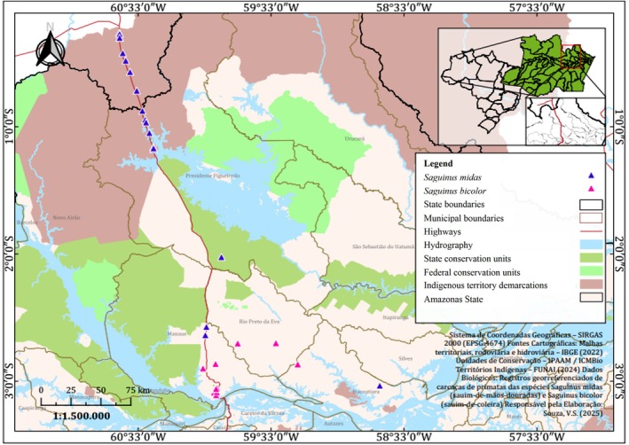 FIGURE 1
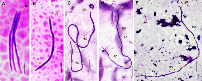 FIGURE 2
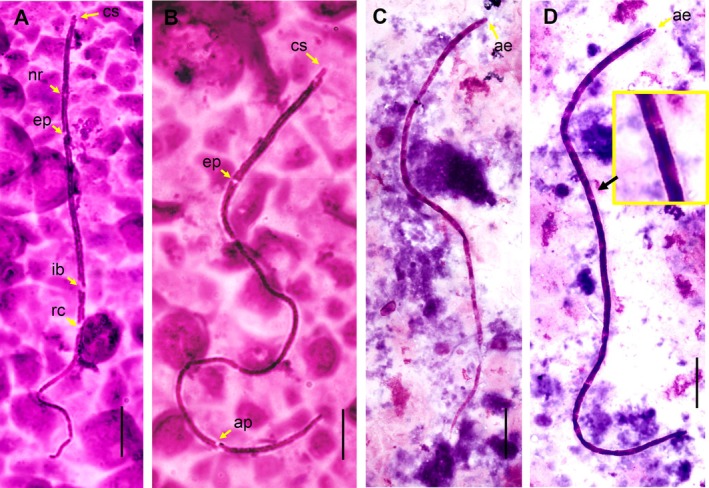 FIGURE 3
- —Universidade Federal do Amazonas 10.13039/100019301
Peer Reviews
No public reviews on file for this paper yet. If you reviewed it on a platform where reviews are public (OpenReview, ICLR, NeurIPS, ICML), you can paste yours below so the community can read it here.
Videos
No videos yet. Explain this paper in a talk, walkthrough, or lecture? Add one.
Taxonomy
TopicsParasitic Diseases Research and Treatment · Insects and Parasite Interactions · Dermatological diseases and infestations
Introduction
1
The filarial worms that naturally infect Primates Platyrrhini (PP) are restricted to the genus Dipetalonema, parasitizing the thoracic and abdominal cavities, and the genus Mansonella (Tetrapetalonema), parasitizing subcutaneous tissue [1, 2]. The larval forms of these nematodes, called microfilariae, are found in the bloodstream of their hosts and exhibit morphometric and morphological characteristics used as taxonomic keys, such as the presence of a sheath, cephalic space, body size, and tail shape [3, 4]. In human filariasis, these characteristics of microfilariae are widely described and facilitate the diagnosis of diseases related to each species [5]. In veterinary medicine, there is a lack of detailed information on the morphology and morphometry of microfilariae in PP, which is useful for rapid diagnosis of filarial infection in both captive animals and environmental monitoring in wild animals.
Materials and Methods
2
Necropsies were performed on 53 adult callitrichids (30 Saguinus midas and 23 S. bicolor ) from accidents (electroshock and roadkill) from Central Amazon: Waimiri Atroari Indigenous Territory and the municipalities of Presidente Figueiredo (BR174 and AM 240), Manaus (Universidad Federal de Amazonas, Complejo Acariquara, Distrito Industrial, Reserva Adolpho Ducke), Rio Preto da Eva (AM 010) and Itacoatiara (Figure 1) and kept frozen in the collection of the Projeto Sauim‐de‐Coleira (2010–2022). During the necropsies, smears were made on slides of fluid samples from the thoracic and abdominal cavities and stained with Panótico Rápido. Morphological and morphometric characteristics of microfilariae were analyzed and taxonomically classified using the microfilariae identification keys in PP [3], all measurements are given in micrometers and average values followed by standard deviation. Adult nematodes recovered from the abdominal cavity were stored in 70°GL ethanol and clarified in Amann's lactophenol for morphological studies.
Map showing sample collection of Saguinus midas and Saguinus bicolor in the Waimiri Atroari Indigenous Territory and the municipalities of Presidente Figueiredo, Manaus, Rio Preto da Eva, and Itacoatiara (Amazonas State, Brazil).
Results
3
In total, 56.5% (13/23) individuals of S. bicolor and 13.3% (4/30) of S. midas examined were observed to have microfilariae. Based on morphology and morphometry, the microfilariae analyzed were classified into four morphotypes: The first morphotype (36 microfilariae analyzed), observed only in S. midas , had a short and robust body, reduced cephalic space, short conic tail, and sheath retracted to the body, a total length of 117.9 (SD 5.6) × 3.3 (SD 0.46) in width at mid‐body length (Figure 2A,B), and was classified as microfilariae of Dipetalonema gracile. The second morphotype (129 microfilariae analyzed) observed in S. bicolor and S. midas had a slender and elongated body with somatic nuclei arranged in two parallel rows along the body, narrowing finely towards the end of the tail, giving a vertebrate appearance with nuclei at the tip of the tail. The total length was 316.1 (SD 14.6) × 2.2 (SD 0.3) in width. In this microfilaria, it was possible to identify structures (mean to 18 specimens) such as the nerve ring (72.3, SD 9.2), excretory pore (102.8, SD 11.5), excretory cell (114.7, SD 10.7), inner body (173.9, SD 17.4), rectal cell (216.9, SD 16.4) and anal pore (263.0, SD 15.7), measurement from anterior end. This microfilariae was classified as Mansonella (Tetrapetalonema) mariae (Figure 2C–E).
(A, B). Microfilaria of D. gracile, showed short and robust body with short cephalic space (cs) and sheath (s). bar: 20 μm. (C, D and E). Microfilaria of Mansonella (T.) mariae, body of the microfilariae with compact column of the nuclei purple in color, with exception of areas without nuclei (yellow arrow), cephalic space (cs), nerve ring (nr), excretore pore (ep), excretore cells (ec), inner body (ib), rectal cells (rc), anal pore (ap), bar: 25 μm.
The third morphotype (40 microfilariae analyzed) observed in S. bicolor and S. midas had a rounded anterior end, somatic nuclei arranged in two parallel rows along the body and narrowing at the tip of the tail, with nuclei at the tip of the tail, sheath absent and no striations were observed on the cuticle. The total length was 256.2 (SD 30.7) × 2.2 (SD 0.3) in width. In this microfilaria, it was possible to identify structures (mean to three specimens) such as the nerve ring (57.8, SD 4.1), excretory pore (82.1, SD 1.4), excretory cell (93.3, SD 3.4), inner body (148.0, SD 5.4), rectal cell (171.5, SD 12.3) and anal pore (217.3, SD 9.4), measurement from anterior end (Figure 3A,B). This microfilariae was classified as Mansonella (Tetrapetalonema) sp. The fourth morphotype (39 microfilariae analyzed) observed in S. bicolor and S. midas had a slender cuticle with striated markings along the body and rounded anterior end. The total length was 241.4 (SD 22.8) × 2.5 (SD 0.3) in width and it was classified within the genus Mansonella (Tetrapetalonema) sp. (Figure 3C,D). Additionally, adult filariae were recovered from the peritoneal cavity (7 females and 4 males) of D. gracile in an S. midas individual. In the S. bicolor individuals, no adult filariae were observed in the peritoneal‐thoracic cavities or subcutaneous tissue examined.
(A, B). Microfilaria of Mansonella (Tetrapetalonema). Morphotype 3, showed compact double column of the nuclei purple in color, with exception of areas without nuclei (yellow arrow), cephalic space (cs), nerve ring (nr), excretore pore (ep), inner body (ib), rectal cells (rc), anal pore (ap), bar: 25 μm. (C, D) Morphotype 4, showed rounded anterior end (ae) and body striated (black arrow).
Discussion
4
Microfilariae of the genus Dipetalonema are characterized by the presence of a sheath, a structure observed to be highly retracted to the body of D. gracile microfilariae of this study, possibly due to post‐mortem changes and thawing of the host. The classification of this microfilaria is confirmed by the presence of adult specimens of D. gracile in the peritoneal cavity of S. midas . The microfilaria of M. (T.) mariae is one of the longest and thinnest of the genus Mansonella. Infection by this species was recorded in S. bicolor in Manaus city using molecular analysis [6]; however, details of larval morphology were not provided. This study complements this important information for the conservation of this primate, classified as critically endangered (CR) according to the IUCN [7]. For S. midas , infection by M. (T.) mariae is recorded for the first time in this study. On the other hand, the third morphotype is morphologically compatible, both in length and width, and in the presence of evident cuticular striations along the body, with the species Mansonella (T.) marmosetae. The fourth morphotype, classified as Mansonella (Tetrapetalonema), is morphologically compatible with the species M. (T.) mystaxi, both in size and width, and in the presence of caudal nuclei. However, as a limitation of this study, the presence of these two species should be confirmed by molecular analyzing and adult specimens in future research integrative on these callitrichids.
Observing adult specimens of the genus Mansonella at infection organ is challenging due to their minute size and the large search area in subcutaneous tissue. Microfilariae that infect humans or domestic animals were not observed in this study. However, within a One Health perspective, we highlight the relevance of monitoring filarial infections in primates inhabiting anthropized landscapes to detect the presence of Dirofilaria immitis, Mansonella ozzardi, and Mansonella perstans [8, 9], the latter previously reported in captive Saguinus leocopus [10]. On the other hand, apparently the filariae Dipetalonema and Mansonella do not have host specificity and are highly likely to be found in other platyrrhine hosts and in multispecies infections. Hyperinfections caused by adult filariae promote inflammatory reactions such as pleuritis and fibrinopurulent and fibrinous peritonitis, while microfilariae can invade various vital organs, compromising the lives of primates [6, 11, 12]. A rapid diagnosis of microfilariae using only a simple blood smear would aid in medical treatment and preventive actions, especially in captive animals and wildlife rehabilitation programs.
Ethics Statement
This study also has a license with the Sistema de Autorização e Informação em Biodiversidade (SISBio), provided by the Instituto Chico Mendes de Conservação da Biodiversidade (ICMBio) to Marcelo Gordo (code: 10286‐7).
Conflicts of Interest
The authors declare no conflicts of interest.
The reference list from the paper itself. Each links out to its DOI / PubMed record.
- 1O. Bain , G. Petit , and L. Rosales‐Loesener , “Filaires de Singes sud‐américains,” Bulletin du Muséum National D'histoire Naturelle 8, no. 3 (1986): 513–542, 10.5962/p.326708. · doi ↗
- 2M. L. Eberhard and T. C. Orihel , “The Genus Mansonella (Syn. Tetrapetalonema): A New Classification,” Annales de Parasitologie Humaine et Comparée 59, no. 5 (1984): 483–496, 10.1051/parasite/1984595483.6508144 · doi ↗ · pubmed ↗
- 3H. N. Fraiha and J. A. P. C. Muniz , “Chave preliminar para identificação de microfilárias do sangue de primatas não humanos da região neotropical,” Boletim Do Museu Paraense Emílio Goeldi. Série Zoologia 9, no. 2 (1993): 195–201.
- 4J. Notarnicola , C. M. Pinto , and G. T. Navone , “Host Occurrence and Geographical Distribution of Dipetalonema spp. (Nematoda: Onchocercidae) in Neotropical Monkeys and the First Record of Dipetalonema gracile in Ecuador,” Comparative Parasitology 75, no. 1 (2008): 61–68, 10.1654/4284.1. · doi ↗
- 5B. A. Mathison , M. R. Couturier , and B. S. Pritt , “Diagnostic Identification and Differentiation of Microfilariae,” Journal of Clinical Microbiology 57, no. 10 (2019): e 00706–19, 10.1128/JCM.00706-19.31340993 PMC 6760958 · doi ↗ · pubmed ↗
- 6C. A. Dias , T. R. Silva , M. Gordo , et al., “First Report of Mansonella sp. and Dipetalonema Gracile in the Amazonian City‐Dwelling Threatened Primate, Saguinus bicolor ,” Frontiers in Tropical Diseases 4 (2023): 1080218, 10.3389/fitd.2023.1080218. · doi ↗
- 7M. Gordo , F. Röhe , and M. D. Vidal , Saguinus bicolor (Amended Version of 2019 Assessment). The IUCN Red List of Threatened Species (2021), 10.2305/IUCN.UK.2021-1.RLTS.T 40644 A 192551696.en. · doi ↗
- 8S. A. Basano , J. S. A. Camargo , L. J. S. Vera , et al., “Investigação da ocorrência da Mansonella ozzardi no Estado de Rondônia, Amazônia Ocidental,” Revista da Sociedade Brasileira de Medicina Tropical 44, no. 5 (2011): 600–603, 10.1590/S 0037-86822011005000055.21877064 · doi ↗ · pubmed ↗