Endoscopic management of a giant ampullary cyst causing acute pancreatitis
Arvind Bussetty, Michael Ma, Arvind Julius Trindade, Ankoor Patel, Petros Benias

Abstract
Genes, proteins, chemicals, diseases, species, mutations and cell lines named across the full text — each resolved to its canonical identifier and authoritative record.
Click any figure to enlarge with its caption.
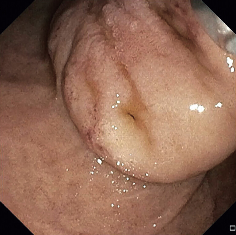 Fig. 1
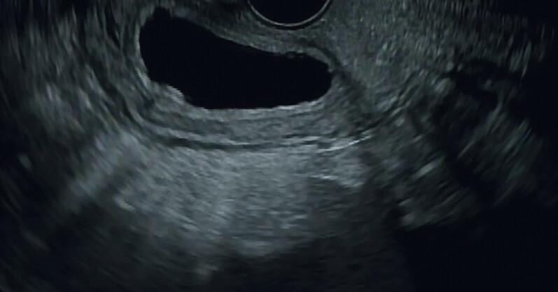 Fig. 2
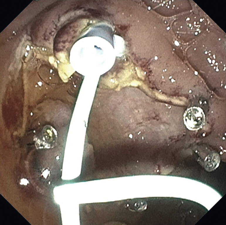 Fig. 3
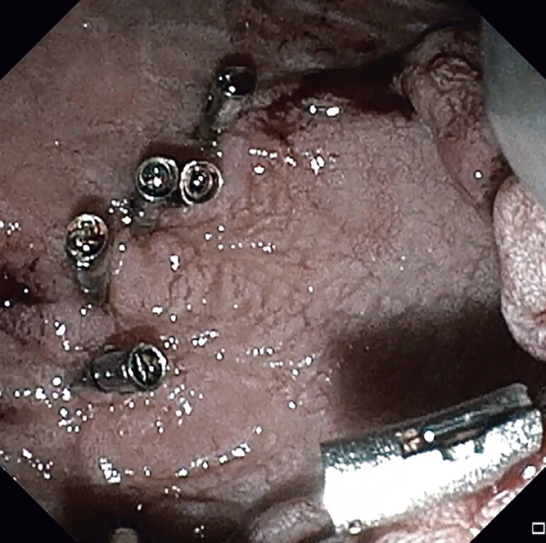 Fig. 4
Peer Reviews
No public reviews on file for this paper yet. If you reviewed it on a platform where reviews are public (OpenReview, ICLR, NeurIPS, ICML), you can paste yours below so the community can read it here.
Videos
No videos yet. Explain this paper in a talk, walkthrough, or lecture? Add one.
Taxonomy
TopicsPancreatitis Pathology and Treatment · Pancreatic and Hepatic Oncology Research · Gastrointestinal disorders and treatments
Ampullary cysts (ACs) are congenital abnormalities of the gastrointestinal tract and are uncommon forms of duodenal duplication cysts 1 . They are anechoic on endoscopic ultrasound (EUS) arising from any of the three to five enteric layers with a wall of intestinal origin 2 . ACs can present with many symptoms such as duodenal/biliary obstruction, abdominal pain, infection, and pancreatitis.
We present a 17-year-old female with right upper quadrant abdominal pain, nausea, and poor oral intake found to have acute pancreatitis ( Video 1 ). On MRI, she was found to have a large cyst. Upper endoscopy with endoscopic ultrasound was notable for visualization of a large ampullary duplication cyst, not directly involving in the biliary duct ( Fig. 1 and Fig. 2 ). An attempt at cannulating the bile duct was performed which identified a false tract within the AC, likely leading to obstruction and the resultant pancreatitis. The medial aspect was cut with sphincterotome and the remainder of the AC was unroofed with snare resection ( Fig. 3 ). The ampulla was identified at the apex after resection. The bile duct and pancreatic duct were stented ( Fig. 3 ). Endoclips were placed around the edge of the dissected AC for bleeding prophylaxis. Follow-up endoscopy showed well-healed resection edges ( Fig. 4 ). The patient tolerated both procedures well. She was followed up as an outpatient and did not experience any further episodes of recurrent pancreatitis.
Endoscopic intervention of a giant ampullary cyst involving mucosectomy and stenting of the pancreatic and biliary ducts to resolve pancreatitis from biliary obstruction.Video 1
Giant cyst located at the ampulla during endoscopic intervention.
Endoscopic ultrasound demonstrating cyst with simple fluid content.
Mucosectomy of ampullary cyst completed and plastic stents placed in the biliary and pancreatic ducts.
Follow-up endoscopy of mucosectomy site at ampulla, which shows well-healed edges.
Symptomatic ACs may occasionally require surgical resection 3 4 . The cyst’s location and proximity to the biliary tree rendered surgery high-risk with potential need for biliary reconstruction. This endoscopic approach avoided the morbidity of surgical intervention and resolved pancreatitis without extensive intervention.
Endoscopy_UCTN_Code_CCL_1AZ_2AK
The reference list from the paper itself. Each links out to its DOI / PubMed record.
- 1Trobs RB Hemminghaus M Cernaianu G Stone-containing periampullary duodenal duplication cyst with aberrant pancreatic duct J Pediatr Surg 200944 e 33e 3510.1016/j.jpedsurg.2008.10.10619159708 · doi ↗ · pubmed ↗
- 2Narlawar RS Rao JR Karmarkar SJ Sonographic findings in a duodenal duplication cyst J Clin Ultrasound 20023056656810.1002/jcu.1011712404525 · doi ↗ · pubmed ↗
- 3Chen JJ Lee HC Yeung CY Meta-analysis: the clinical features of the duodenal duplication cyst J Pediatr Surg 2010451598160610.1016/j.jpedsurg.2010.01.01020713206 · doi ↗ · pubmed ↗
- 4Kurita S Kitagawa K Toya N Endoscopic resection of a duodenal duplication cyst: A case report DEN Open 20222 e 8810.1002/deo 2.8835310702 PMC 8828194 · doi ↗ · pubmed ↗