Endoscopic ultrasound-guided radiofrequency ablation followed by ethanol ablation of a solid pseudopapillary neoplasm of the pancreas
Marcin Polkowski, Mateusz Szmit, Krzysztof Skoczylas, Jakub Krzyżkowiak, Andrzej Mróz, Jakub Pałucki, Jarosław Reguła

Abstract
Genes, proteins, chemicals, diseases, species, mutations and cell lines named across the full text — each resolved to its canonical identifier and authoritative record.
Click any figure to enlarge with its caption.
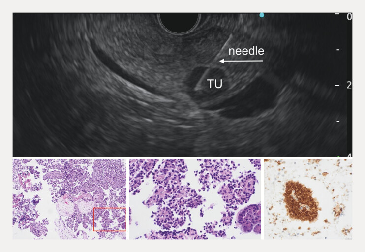 Fig. 1
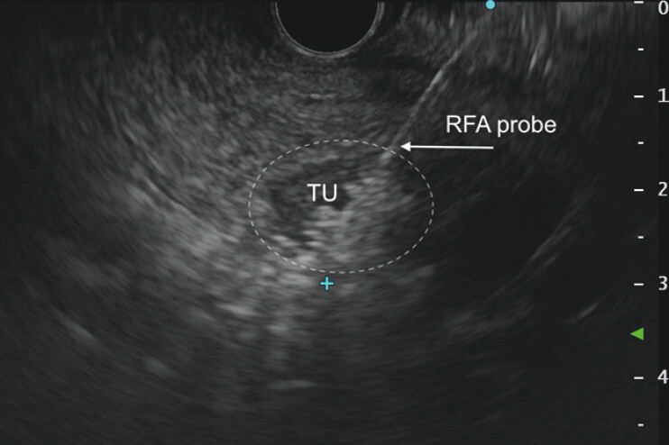 Fig. 2
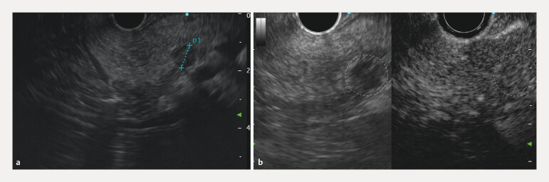 Fig. 3
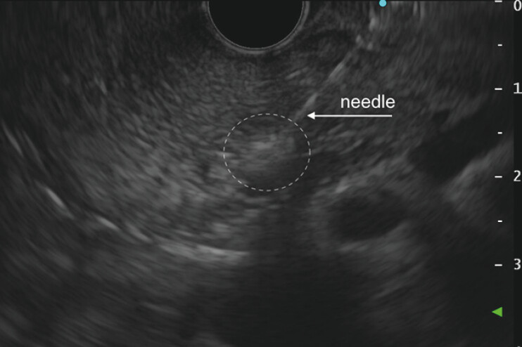 Fig. 4
Peer Reviews
No public reviews on file for this paper yet. If you reviewed it on a platform where reviews are public (OpenReview, ICLR, NeurIPS, ICML), you can paste yours below so the community can read it here.
Videos
No videos yet. Explain this paper in a talk, walkthrough, or lecture? Add one.
Taxonomy
TopicsPancreatic and Hepatic Oncology Research · Gallbladder and Bile Duct Disorders · Hepatocellular Carcinoma Treatment and Prognosis
A solid pseudopapillary neoplasm (SPPN) is a rare pancreatic tumor that predominantly affects young females and typically presents as a large mass with a cystic component. With the increasing use of diagnostic imaging, small SPPNs (≤2 cm) are now detected more frequently. Although their natural history remains unclear and malignant potential is considered low, surgical resection is often recommended 1 . Recently, several cases of small SPPNs successfully treated with endoscopic ultrasound (EUS)–guided radiofrequency ablation (RFA) have been reported 2 3 . We describe an additional case managed with EUS-RFA followed by ethanol ablation (EA) for a residual tumor post-RFA.
A 38-year-old woman had a 13-mm pancreatic head nodule incidentally detected on transabdominal ultrasound and subsequently confirmed by magnetic resonance imaging. EUS-guided biopsy diagnosed SPPN ( Fig. 1 ). The patient declined surgical resection and opted for EUS-guided RFA.
Fine-needle biopsy of the tumor (TU) with a 25G needle (upper panel). Histopathological images (lower panel, from left to right) show the hematoxylin and eosin (H&E) low-power view demonstrating pseudopapillary structures composed of uniform tumor cells; H&E high-power view; immunostaining for β-catenin demonstrating strong nuclear and membranous expression in tumor cells.
After EUS evaluation, a RFA probe (EUSRA, 19G, 10-mm active tip; TaeWoong Medical) was advanced under EUS guidance into the tumor. The radiofrequency energy (50 W) was delivered to three regions of the tumor in applications lasting for 8, 7, and 7 seconds, guided by EUS imaging and the rise in impedance measured using a RFA generator ( Fig. 2 , Video 1 ).
An EUS image of the tumor (TU, outlined with a dashed line) during radiofrequency ablation (RFA). The hyperechoic blush around the RFA probe in the lower right portion of the tumor represents gas microbubbles formed by tissue heating, indicating effective ablation. EUS, endoscopic ultrasound.
Successful EUS-guided ablation of a small (13 × 11mm) solid pseudopapillary neoplasm of the pancreatic head using radiofrequency ablation (RFA) followed by ethanol ablation of a residual post-RFA lesion. EUS, endoscopic ultrasound.Video 1
Follow-up EUS performed 6 months after EUS-RFA revealed an 8 × 7mm residual lesion at the treatment site ( Fig. 3 a ). Contrast-enhanced EUS demonstrated enhancement, suggesting viable tumor tissue ( Fig. 3 b ). Given the small volume of the residual lesion, EA was performed to achieve complete ablation. A total of 0.4 mL of 96% ethanol (Ethanol Sterop) was injected through a 25G EUS needle (EZ Shot 3 Plus, Olympus) in three fractions ( Fig. 4 , Video 1 ). For both RFA and EA, rectal diclofenac (100 mg) and intravenous antibiotics were administered; the post procedure course was uneventful.
a An EUS image showing a residual hypoechoic lesion (between crosses), 8 × 7 mm in size, at the site of the 13 × 11 mm tumor treated with radiofrequency ablation 6 months earlier. b Contrast enhancement of the residual lesion indicates the viable tumor tissue. The locations of the residual lesion in both the B-mode image and the CHE image are outlined with a dashed line. EUS, endoscopic ultrasound.
A residual post-RFA lesion (outlined with a dashed line) during ethanol injection with a 25G needle. A hyperechoic cloud around the needle tip represents ethanol spread in the tissue. RFA, radiofrequency ablation.
Follow-up EUS and computed tomography 6 months post-EA (12 months post-RFA) showed no residual or recurrent lesion. This case adds to previous reports of successful EUS-guided ablation of SPPNs 3 .
Endoscopy_UCTN_Code_TTT_1AS_2AI
The reference list from the paper itself. Each links out to its DOI / PubMed record.
- 1Khoury T Farraj M Sbeit W Solid pseudopapillary neoplasm of the pancreas: A comprehensive review focusing on the role of endoscopic ultrasound-guided radiofrequency ablation as an alternative treatment Cancers (Basel)202517224010.3390/cancers 1713224040647537 PMC 12249432 · doi ↗ · pubmed ↗
- 2Coupier A Khoury T Gincul R Endoscopic ultrasound-guided radiofrequency ablation for solid pseudopapillary neoplasm of the pancreas Endoscopy 202355 e 951e 95210.1055/a-2127-489037604456 PMC 10442209 · doi ↗ · pubmed ↗
- 3Choi JH Seo DW Song TJ Endoscopic ultrasound-guided radiofrequency ablation for management of benign solid pancreatic tumors Endoscopy 2018501099110410.1055/a-0583-838729727904 · doi ↗ · pubmed ↗