Immature Caenorhabditis elegans motor neurons control early embryo behavior via both synaptic and nonsynaptic GABA release
James Marvel-Coen, Evan Ardiel, Jian Zhao, Stephen Nurrish, Joshua M. Kaplan

TL;DR
Immature neurons in C. elegans embryos use nonsynaptic GABA release to control early movement before synapses form.
Contribution
The study reveals that nonsynaptic GABA release from immature motor neurons regulates embryo behavior before synapse formation.
Findings
Early embryo motion inhibition in C. elegans is caused by immature GABAergic motor neurons.
Nonsynaptic GABA mechanisms predominate over synaptic ones in controlling early embryo behavior.
Motion inhibition requires GABA synthesis in DD neurons and GABAA receptors in muscles.
Abstract
Little is known about how prenatal circuits control embryo behavior. We show that the motion of early Caenorhabditis elegans embryos is transiently inhibited by immature GABAergic motor neurons that have not yet completed neurite outgrowth. GABA’s inhibitory effect on early embryo behavior occurs before most synapses have formed in the worm’s central neuropil. For this early behavior, we find that synaptic GABA release plays a minor role while nonsynaptic mechanisms predominate. Our results suggest that nonsynaptic forms of GABA transmission play a significant role in prenatal circuits. Prenatal brain activity has long lasting effects on subsequent neurodevelopment. It is unclear if early brain activity is dominated by cell intrinsic, synaptic, or nonsynaptic mechanisms. We address this question by analyzing Caenorhabditis elegans embryo behavior in snf-11 mutants, which lack a plasma…
Genes, proteins, chemicals, diseases, species, mutations and cell lines named across the full text — each resolved to its canonical identifier and authoritative record.
Click any figure to enlarge with its caption.
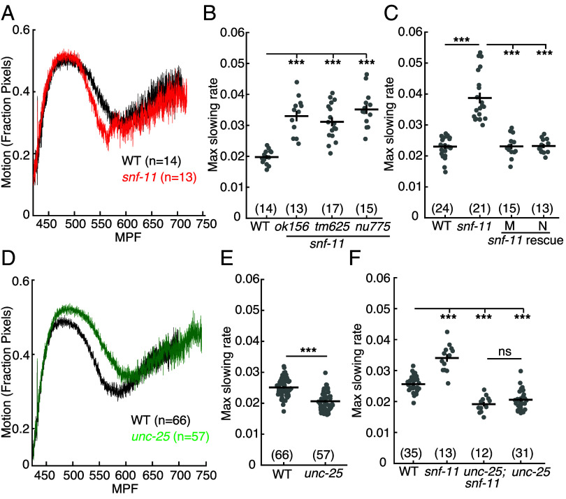 Fig. 2
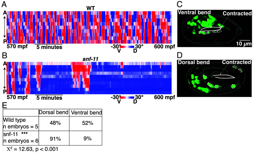 Fig. 3
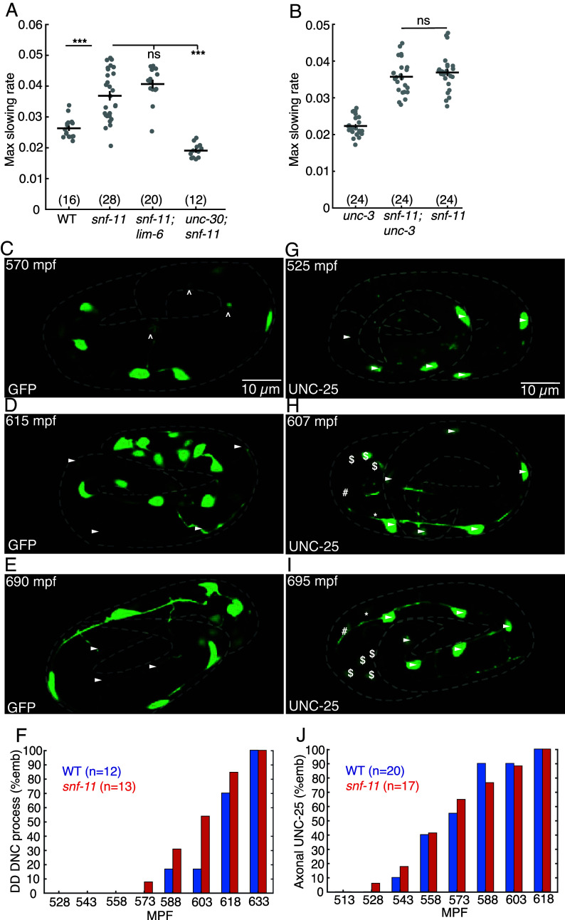 Fig. 4
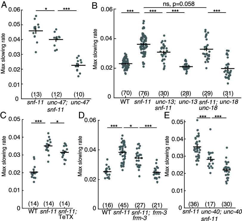 Fig. 5
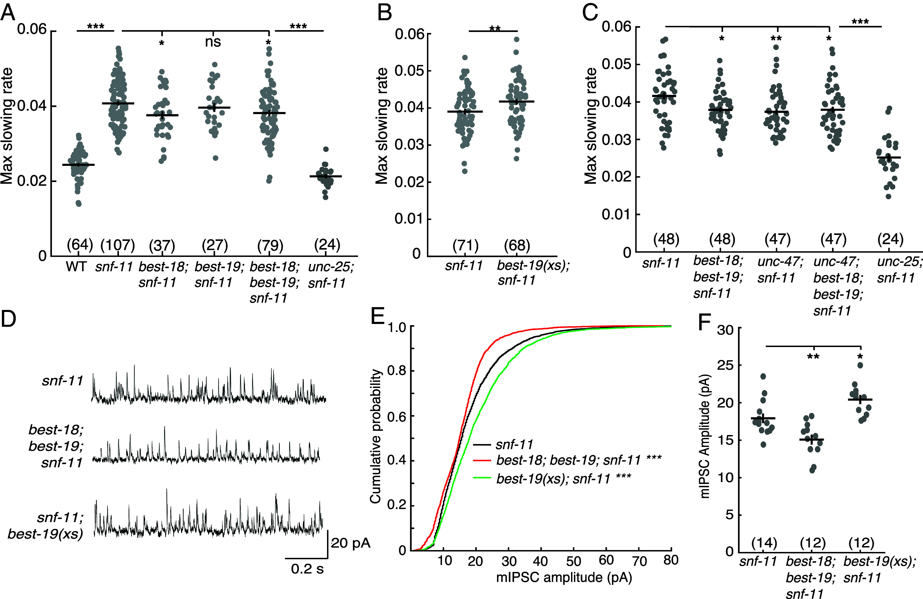 Fig. 6
- —HHS | NIH | National Institute of Neurological Disorders and Stroke (NINDS)100000065
Peer Reviews
No public reviews on file for this paper yet. If you reviewed it on a platform where reviews are public (OpenReview, ICLR, NeurIPS, ICML), you can paste yours below so the community can read it here.
Videos
No videos yet. Explain this paper in a talk, walkthrough, or lecture? Add one.
Taxonomy
TopicsGenetics, Aging, and Longevity in Model Organisms · Reproductive Biology and Fertility · Neurobiology and Insect Physiology Research
Many results suggest that early events in brain development play a pivotal role in postnatal developmental outcomes. Development of some circuits is restricted to particular times early in development (i.e., critical periods) (1?–3). Maternal infection, maternal drug exposure, and prematurity are all associated with increased risk for neurodevelopmental disorders (4?–6). These and other results suggest that early brain circuits may be particularly vulnerable to genetic and environmental perturbation. Although behavior and circuit function have been intensively investigated, the vast majority of these studies have used postnatal samples, presumably due to the inaccessibility of prenatal tissues. Consequently, very little is known about prenatal behaviors and the circuit mechanisms producing them. Several important questions about early brain development remain to be answered. What are the first behaviors exhibited by embryos? When do neurons first assume control of embryo behavior? Do neurons control behavior prior to completing their developmental maturation? Do immature neurons encode behavior by mechanisms distinct from those used in postnatal circuits?
In many systems, embryos begin exhibiting behaviors before birth. C. elegans embryos begin moving shortly before the twofold stage; Drosophila embryos at stage 16, 800 to 900 mpf; chick embryos at stage 21 (~3.5 d), and human embryos at 8 wk (7?–9). Embryonic movement is necessary for circuit formation and even survival in invertebrate model systems including Drosophila and Caenorhabditis elegans (8, 10?–12).
Although prenatal behavior is widely observed, when synapses form and assume control of embryo behavior has not been determined in most systems. In vitro, developing neurons can release neurotransmitters from growth cones (13, 14). In mice, motor axons reach muscles by E12.5 but clustered acetylcholine receptors (AChR) are not observed until E14.5 (15). In vivo recordings of pyramidal neurons in fetal mice indicate synaptic activity commences by E14.5 (16). In contrast, perinatal rat (day E18-P1) hippocampal slices exhibited both evoked and tonic GABA activated currents, which were not blocked by botulinum toxin nor by a general voltage-gated calcium channel antagonist (CdCl_2_), suggesting that these were mediated by nonsynaptic mechanisms (17).
The timing of embryonic neurodevelopment has also been extensively analyzed in C. elegans (Fig. 1A). C. elegans embryos develop inside an eggshell before hatching as a first stage larva, at ~800 min postfertilization (mpf). Neurodevelopmental landmarks occur at stereotyped times of C. elegans embryonic development. Neuron births occur in two waves (230 to 290 and 400 mpf) in the cell lineage (7). Axon outgrowth in the nerve ring (the major neuropil in the worm’s head) is completed at 460 mpf (18). Several presynaptic proteins are localized to the nerve ring at ~400 mpf (19). At 550 mpf, small neuromuscular junctions (NMJs) are observed in electron micrographs of the dorsal nerve cord whereas ventral NMJs are not yet seen at this time (20).
In parallel to this developmental timeline, C. elegans embryos also exhibit a stereotyped pattern of behavioral maturation (Fig. 1A). Spontaneous apparently uncoordinated movement (termed twitches) appear at 430 mpf (7, 12). At 530 to 570 mpf, embryos exhibit an alternating pattern of all ventral and all dorsal body bends, hereafter designated flipping behavior (21). Flipping is not significantly disrupted in unc-13 mutants (21), which nearly completely lack SV exocytosis (22), suggesting that flipping is not mediated by a synaptic circuit. Finally, at 650 to 700 mpf, a mature pattern of sinusoidal motion and rhythmic quiescent bouts emerge, both of which are profoundly inhibited in unc-13 mutants (indicating that they are driven by synaptic circuits) (21).
To further address the mechanisms controlling early brain activity, we analyzed the impact of the neurotransmitter γ-aminobutyric acid (GABA) on C. elegans embryos. For several reasons, GABA is a particularly interesting case to consider. In mice, GABA plays a crucial role in early circuit formation (23?–25). Inhibitory signaling from GABAergic neurons is required for the onset of plasticity during the critical period in mouse visual cortex development, and mutations in MeCP2, associated with Rett syndrome, alter visual cortex development by increasing GABA transmission (2, 11, 26). The GAT1 GABA reuptake pump and GABRB3 GABA_A_ receptors are risk genes for autism spectrum disorder (ASD) (27?–29), which is thought to arise from changes in fetal brain development (30?–32). For these reasons, there is significant interest in understanding how and when GABA release occurs in early brain development.
Here, we further investigate when synaptic circuits first form in C. elegans embryos. We find that many nerve ring synapses form after 600 mpf, and that an earlier embryo behavior (flipping) is controlled by both synaptic and nonsynaptic GABA transmission. These results suggest nonsynaptic GABA release plays an important role in early brain activity.
Results
Synaptic Ion Channels Localize to the Nerve Ring at Approximately 600 mpf.
Prior studies provide conflicting conclusions about when functional synaptic circuits first form in C. elegans embryos. Depending on how synapses were assayed, estimates for when synapses form ranged from 400 mpf (19), to 550 mpf (20), to 650 mpf (21).
To further address this question, we asked when several synaptic ion channels are localized to the nerve ring (a large circumferential axon bundle in the worm’s head that contains many synaptic connections). Trafficking of large multimeric ion channels (e.g., ionotropic glutamate receptors) to the cell surface takes many hours due to their slow protein folding and subunit assembly in the endoplasmic reticulum (33, 34). Consequently, we reasoned that ion channels likely represent the last components to be localized to synapses. We analyzed the localization of endogenously expressed UNC-2 Ca_V_2 (a voltage-activated calcium channel), UNC-49 GABA_A_R (a GABA activated chloride channel), and ACR-16 CHRNA7 (a nicotinic acetylcholine receptor) in the nerve ring of 540 to 660 mpf embryos, using mNeonGreen (mNG)-tagged CRISPR alleles of each gene (Fig. 1 B–J). Electrophysiological recordings show that the mNG tags did not impair the function of UNC-2 (35), UNC-49, and ACR-16 channels (SI Appendix, Fig. S1 A–D). All three channels arrived in the nerve ring at a similar time, starting at ~600 mpf (Fig. 1 B–J), which was ~1 to 3 h after mRNA expression onset (defined as the time when expression reached 20% of the peak value) (Fig. 1 H–J) (36?–38). By contrast, mNG tagged NRX-1 and NLG-1 (pre- and postsynaptic adhesion molecules respectively) arrived in the nerve ring significantly earlier than these ion channels (SI Appendix, Fig. S2), consistent with the earlier arrival times reported for other synaptic proteins (19). These results imply that some essential synaptic proteins do not appear in the nerve ring until 600 mpf or later, which coincides with when motion defects are first observed in unc-13 mutants (21).
GABA Signaling Decreases Early Embryo Motion.
To assess GABA function in early embryos, we analyzed the behavior of snf-11 mutants. SNF-11 is a plasma membrane GABA reuptake pump (orthologous to GAT1) (39, 40). Thus, snf-11 GAT1 mutants are predicted to have exaggerated GABA signaling. Embryo motion was assessed by frame subtraction of bright field images (21). Using this assay, we found that snf-11 mutant embryos exhibit a unique behavioral defect whereby motion is dramatically decreased at a consistent developmental time point (~510 to 570 mpf) before abruptly returning to a wild type pattern thereafter (Fig. 2A). Motion inhibition was quantified by measuring the maximal decrease in motion rate in individual 480 to 620 mpf embryos (as detailed in Experimental Methods). This phenotype was observed in mutants containing three independent snf-11 GAT1 loss of function alleles (Fig. 2B). This snf-11 GAT1 mutant motion defect was rescued by transgenes expressing SNF-11 in either muscles or neurons (Fig. 2C), both of which express the endogenous snf-11 gene (36). These results confirm that decreased embryo motion is a consequence of snf-11 GAT1 inactivation. Because snf-11 encodes a GABA reuptake pump, we next asked if GABA synthesis is required for the reduced motion of snf-11 embryos. Consistent with this idea, a mutation inactivating unc-25, which encodes the GABA biosynthetic enzyme glutamic acid decarboxylase (GAD), restores wild type motion in snf-11 embryos (Fig. 2F). By contrast, unc-25 GAD single mutants exhibited a significant increase in motion at ~530 to 570 mpf relative to wild type (Fig. 2 D and E). Collectively, these results suggest that decreased motion of snf-11 GAT1 mutant embryos results from exaggerated GABA signaling.
*GABA transiently inhibits embryo motion. Motion of snf-11 GAT1 (A–C) and unc-25 GAD (D–F) mutant embryos is analyzed. Average motion traces (A and D) and maximal slowing rate of 480 to 620 mpf embryos (B, C, E, and F) are shown for the indicated genotypes. (A) Embryo motion was transiently inhibited in snf-11(ok156) GAT1 embryos (red) compared to WT controls (black). (B) Three independent snf-11 GAT1 loss of function alleles caused similar decreases in motion rate. (C) The decreased motion defect in snf-11(ok156) mutants was rescued by transgenes restoring SNF-11 expression in muscles (M) or in neurons (N). (D and E) Increased embryo motion was observed in unc-25(nu836) GAD (green) embryos compared to WT controls (black). (F) The snf-11 GAT1 decreased motion defect was eliminated in unc-25; snf-11 double mutants. Sample sizes for each genotype are indicated in all figure panels. Values that differ significantly are indicated (ns, not significant; **P < 0.001). Error bars indicate SEM.
GABA Inhibits Embryo Flipping Behavior.
The brief inhibition of motion observed in snf-11 GAT1 mutants occurs when behavior is dominated by embryo flipping (21). To confirm that snf-11 GAT1 mutations disrupt flipping, we used lightsheet microscopy to analyze snf-11 GAT1 embryo postures, as described (21). Embryo postures were calculated by tracking the location of paired hypodermal skin cells that run along the left and right sides of the body. As previously described, ~570 mpf wild type embryos regularly flip between full dorsal and full ventral coils (Fig. 3A) (21). By contrast, snf-11 mutant embryos (at 570 mpf) adopted a single prolonged coiled posture (Fig. 3B). Taken together, these results suggest that exaggerated GABA signaling in snf-11 GAT1 mutants dramatically (albeit transiently) suppresses early embryo flipping behavior. Hereafter, we refer to the snf-11 mutant motion defects as inhibition of embryo flipping.
Exaggerated GABA signaling in snf-11 mutants inhibits flipping behavior. (A–C) Posture analysis is shown for a WT embryo (A) and a snf-11(nu775) (B) embryo from 570 to 600 mpf. Each column is one frame (0.33 s) and each row represents dorsoventral bending for each left–right seam cell pair along the anterior–posterior axis. Colors indicate the bend angle (dorsal or ventral) for each seam cell pair. An entirely red column indicates a full body bend in one direction; a full blue column indicates a full body bend in the opposite direction. At this time, WT embryos (A) rapidly alternate between full dorsal and full ventral body coils (i.e., flipping behavior) whereas snf-11 GAT1 mutants (B) exhibit a prolonged coil in one direction. The WT embryo data (A) was taken from a prior publication (21, 77). (C–E) Comparison of dorsoventral bending in WT and snf-11 GAT1 mutants. Dorsoventral bending was profiled in embryos expressing soluble GFP in cholinergic neurons (using the unc-17 promoter). The cholinergic DA/DB motor neuron cell bodies (dashed circles) identify the embryo’s ventral surface. The contracted embryo surface is indicated (solid white outline). Representative images of a ventrally coiled embryo (C), a dorsally coiled embryo (D), and summary data (E) are shown. snf-11 GAT1 embryos were significantly more likely to exhibit a dorsally coiled posture than WT controls (P < 0.001; Χ2 = 12.63).
The embryo posture analysis does not distinguish between dorsal and ventral body bends. Consequently, these data do not indicate if snf-11 embryos exhibit a dorsal or ventral coiling bias. To address this question, we profiled body bend direction in wild type and snf-11 embryos at ~540 to 600 mpf by confocal microscopy. For this analysis, we used cholinergic (DA/DB) motor neuron cell bodies (expressing GFP) to identify the embryo’s ventral surface. In WT controls, DA/DB cell bodies were equally likely to be on the contracted and relaxed sides of embryo postures (Fig. 3 E–G), consistent with the rapid flipping between dorsal and ventral bends that occurs at this time (21). By contrast, DA/DB cell bodies were significantly more likely to be on the relaxed side of snf-11 GAT1 mutant embryos (Fig. 3G). We conclude that excess GABA signaling causes embryos to adopt prolonged dorsal body bends. Because snf-11 mutants exhibited such a strong phenotype, we used them to identify other components required for GABA signaling in early embryos.
GABA Released from DD Motor Neurons Inhibits Embryo Flipping.
We next asked which cells produce the GABA that inhibits embryo motion in snf-11 GAT1 mutants. C. elegans has several classes of GABAergic neurons (41). Different transcription factors specify the differentiation of each class of GABA neurons (42). Embryo motion was restored to wild type levels in snf-11 double mutants containing an unc-30 PITX2 mutation, which blocks differentiation of the D-type GABA motor neurons (Fig. 4A) (43). By contrast, a lim-6 LMX1B mutation (which prevents RIS and AVL differentiation) (44) had no effect on the motion of snf-11 mutants (Fig. 4A). In addition to D-type GABA neurons, unc-30 PITX2 is also expressed in DA/DB cholinergic motor neurons in embryos (36). An unc-3 COE mutation (which blocks DA/DB differentiation) (45) had no effect on the motion of snf-11 mutant embryos (Fig. 4B), suggesting that DA/DB defects are unlikely to account for the increased embryo motion seen in unc-30;snf-11 double mutants. These results suggest that GABA produced by the DD motor neurons (the only D neuron class present in embryos) controls embryo flipping.
GABA release from immature DD motor neurons inhibits embryo motion. (A and B) Maximal slowing rate of 480 to 620 mpf embryos are shown for the indicated genotypes. The decreased embryo motion seen in snf-11(ok156) GAT1 embryos was unaffected in double mutants lacking lim-6 Lmx1b (which is required for AVL, RIS, and DVB cell fates) (A), was eliminated in double mutants lacking unc-30 PITX2 (which is required for the DD cell fate) (A), and was unaffected in double mutants lacking unc-3 COE (which is required for cholinergic DA/DB motor neuron cell fates) (B). (C–E) Representative images of soluble GFP expressed in DD neurons (using the unc-47 promoter) are shown at the indicated developmental times. At 570 mpf, DD neurites in the ventral cord are complete but dorsally extending neurites have growth cones and have not yet reached the dorsal cord. At 615 mpf, DD ventral and dorsal neurites, and DD commissures are mostly complete, and little change is observed from 615 to 690 mpf. Growth cones (carats) and completed commissures (arrowheads) are indicated. (F) Summary data for DD dorsal nerve cord neurite presence over time for wild type (blue, n = 12) and snf-11 (red, n = 13) embryos. No significant differences were observed (chi-squared test; P > 0.05). (G–I) Expression of UNC-25(nu808 mNG) is shown at the indicated developmental times. At 525 mpf (G), UNC-25 is observed in six DD cell bodies (arrowheads) but fluorescence is not observed in the DD ventral cord processes. At 607 mpf (H), UNC-25 is observed in DD cell bodies, DD ventral processes, and in some DD dorsal neurites and commissures. At this time, UNC-25 is also seen in six head neurons, including RIS (), AVL (#), and four RME ($) neurons. At 695 mpf (I), UNC-25 is observed in DD cell bodies, DD commissures, and throughout DD ventral and dorsal cord neurites, as well as in the head neurons. (J) Summary data for UNC-25 (nu808 mNG) presence in the ventral process over time for wild type (blue, n = 20) and snf-11 (red, n = 17) embryos. No significant differences were observed (chi-squared test; P > 0.05).*
Next, we asked if DD neurons are developmentally mature during embryo slowing in snf-11 GAT1 mutants. DD neurite morphology was examined by expressing GFP in DD neurons (using the unc-47 VGAT promoter) (Fig. 4 C–F). DD ventral nerve cord processes were complete at 540-570 mpf (Fig. 5B), while DD commissures and dorsal nerve cord processes were completed after 600 mpf (Fig. 5 D–F). At 570 mpf, DD neurons typically had incomplete commissures with dorsally directed growth cones (Fig. 5B). We assessed UNC-25 GAD expression using a CRISPR allele that contains mNG inserted into the endogenous unc-25 locus, just after the ATG codon (Fig. 5 G–J). At 510 to 540 mpf, UNC-25 (nu808 mNG) appears primarily in the DD motor neurons (Fig. 5G), whereas expression in other GABA neurons (RIS, AVL, and RME) begins significantly later (after 600 mpf) (Fig. 4 H and I). The onset of UNC-25 (nu808 mNG) expression in DD neurons coincides with the timing of motion inhibition in snf-11 GAT1 mutants (Fig. 3A) and the timing of unc-25 gene transcription in DD neurons (36). Interestingly, UNC-25 (nu808 mNG) was primarily found in DD cell bodies at 510 to 550 mpf (when motion inhibition occurs in snf-11 mutants). Very little UNC-25 (nu808 mNG) was detected in DD neuron commissures, ventral cord, and dorsal cord processes until after 560 mpf (Fig. 4J). Neither the timing of DD neurite outgrowth (Fig. 4F) nor the appearance of UNC-25(nu808 mNG) in DD neurites (Fig. 4J) was significantly altered in snf-11 mutants, indicating that DD maturation was not dramatically altered. These results suggest that immature DD neurons inhibit embryo flipping. These results also suggest that nonsynaptic GABA release from DD cell bodies could contribute to flipping inhibition, since UNC-25 GAD protein is restricted to DD cell bodies and DD NMJs are not observed in the ventral cord at this time in development (20).
*Synaptic and nonsynaptic GABA release both contribute to motion inhibition in snf-11 GAT1 mutants. (A–E) Maximal slowing rates of 480 to 620 mpf embryos are shown for the indicated genotypes. (A) Inactivating unc-47 VGAT, which is required to transport GABA into SVs (51), significantly reduces motion inhibition in snf-11 GAT1 embryos but does not eliminate it. (B) Loss of the SV priming factor unc-13 MUNC13 partially decreased motion inhibition in snf-11 GAT1 embryos, while loss of the priming factor unc-18 MUNC18 did not. (C) GABA neuron expression of tetanus toxin (TeTX), which cleaves the SV SNARE synaptobrevin (53), decreased motion inhibition in snf-11 GAT1 embryos. (D and E) Two mutations preventing UNC-49 clustering at postsynapses, frm-3 FARP (D) and unc-40 DCC (E) decreased motion inhibition in snf-11(ok156) (D) and snf-11(nu775) (E) mutants. Sample sizes for each genotype are indicated in each figure panel. Values that differ significantly are indicated (ns, not significant; *P < 0.05; **P < 0.001). Error bars indicate SEM.
Muscle UNC-49/GABAA Receptors Are Required for Inhibition of Flipping.
Because DD motor neurons provide synaptic input to body muscles, we next asked if muscle GABA receptors control embryo flipping. Consistent with this idea, mutations inactivating UNC-49 GABA_A_ receptors increased embryo motion in snf-11 double mutants and this effect was reversed by a transgene that restores UNC-49 expression in body muscles (SI Appendix, Fig. S3A). By contrast, inactivating subunits that form the GABA activated G protein–coupled receptor (GBB-1 and GBB-2) had no effect on snf-11 embryo motion (SI Appendix, Fig. S3B). Collectively, these results suggest that inhibition of embryo flipping in snf-11 GAT1 mutants is mediated by GABA released by the DD motor neurons, which activates UNC-49 GABA_A_ receptors in body muscles.
GABA Inhibits Embryo Flipping by Hyperpolarizing Body Muscles.
In mammals, GABA often has excitatory effects on immature neurons and only becomes inhibitory as neurons mature (46, 47). This pattern of early excitatory GABA effects is also thought to occur in C. elegans (48). Prompted by these results, we next asked if GABA’s effects on embryo motion are mediated by depolarizing or hyperpolarizing body muscles. Because UNC-49 GABA_A_ subunits form GABA-activated chloride channels, GABA’s impact on muscle activity depends on the transmembrane chloride gradient. UNC-49 activation hyperpolarizes muscles when extracellular chloride exceeds cytoplasmic levels, while UNC-49 depolarizes muscles in the converse conditions. The transmembrane chloride gradient is controlled by the activity of two chloride ion extruders (ABTS-1 and KCC-2) and one chloride ion importer (NKCC-1) (48, 49). Loss of the chloride importer NKCC-1 further inhibits motion in snf-11 double mutants, loss of the chloride extruder KCC-2 increased motion in snf-11 double mutants, while loss of the ABTS-1 chloride extruder had no effect (SI Appendix, Fig. S3 C–E). Taken together, these results suggest that GABA prolongs dorsally coiled postures by hyperpolarizing ventral body muscles. GABA’s preferential effect on ventral body muscles most likely results from the fact that UNC-49 receptors are found in ventral but not dorsal body muscles in both embryos (SI Appendix, Fig. S3F) and newly hatched first stage larvae (50).
SV Exocytosis Modestly Contributes to GABA Mediated Inhibition of Embryo Motion.
We next asked if SNF-11’s impact on embryo motion is mediated by synaptic GABA release. Synaptic GABA release is mediated by exocytosis of SVs at presynaptic active zones. To test its role in controlling early embryo motion, we constructed snf-11 GAT1 double mutants containing mutations that prevent synaptic GABA release. Embryo motion in snf-11 GAT1 mutants was modestly increased by mutations that prevent SV loading with GABA (unc-47 VGAT) (51) (Fig. 5A) or SV docking (unc-13 MUNC13) (52) (Fig. 5B); and by expression of tetanus toxin in DD neurons, which cleaves the vesicle SNARE protein synaptobrevin (53) (Fig. 5C). In all cases, snf-11 GAT1 double mutants lacking synaptic GABA release had significantly less motion than that observed in unc-25 GAD; snf-11 GAT1 double mutants (Fig. 2F), suggesting that DD neurons exhibit significant residual GAIBA release even when synaptic release was blocked. To further address the role of vesicular GABA release, we also analyzed unc-18 mutants (which reduce SV exocytosis) (54) and cat-1 VMAT mutants, because VMAT2 promotes GABA transport into SVs in mouse dopaminergic neurons (55). Embryo motion in snf-11 GAT1 mutants was not affected by cat-1 VMAT mutations (SI Appendix, Fig. S4A). There was a trend for increased motion in snf-11; unc-18 double mutants, which was not statistically significant (P = 0.058, Fig. 5B). Finally, it is possible that synaptic GABA release inhibits embryo motion only in snf-11 GAT1 mutants, where GABA signaling is exaggerated. Contrary to this idea, several mutations that impair synaptic GABA release significantly increased embryo motion in single mutants (SI Appendix, Fig. S4B). Taken together, these results suggest that synaptic release contributes to GABA mediated inhibition of embryo flipping.
Synaptic UNC-49/GABAA Receptors Contribute to GABA Mediated Inhibition of Embryo Flipping.
Thus far, our results suggest that SV exocytosis contributes to GABA release from DD neurons in early embryos (510 to 570 mpf). In mature neurons, SV exocytosis is restricted to presynaptic active zones; however, SV exocytosis could occur at nonsynaptic sites in immature neurons. To further address whether GABA is released at synapses, we asked if clustered postsynaptic GABA receptors are required for inhibiting embryo motion. To address this possibility, we analyzed mutants lacking two scaffolding proteins (FRM-3/FARP and UNC-40/DCC) that immobilize UNC-49 GABA_A_ receptors at postsynaptic elements (56, 57). We found that embryo motion was modestly increased in both frm-3 FARP; snf-11 GAT1 and unc-40 DCC; snf-11 GAT1 double mutants compared to snf-11 single mutant controls (Fig. 5 D and E). The magnitude of these increases in embryo motion are similar to those in snf-11 GAT1 double mutants lacking UNC-47 VGAT or UNC-13. These results suggest that synaptic GABA release and activation of clustered synaptic UNC-49 GABA_A_ receptors both contribute to the inhibited embryo motion seen in snf-11 GAT1 mutants.
Bestrophin Channels Promote GABA Mediated Inhibition of Embryo Motion.
Because mutations that eliminate synaptic GABA transmission only partially block inhibition of embryo flipping in snf-11 GAT1 mutants, we next asked if immature DD neurons also release GABA by a nonsynaptic mechanism. In tonic transmission, GABA is released by plasma membrane channels rather than by SV exocytosis (58). In mammals, two classes of channels are implicated in tonic GABA release: bestrophin channels and SWELL1 (LRC88A) osmolyte channels (58). The C. elegans genome encodes 26 bestrophin paralogs but lacks an obvious SWELL1 equivalent. Two bestrophin genes (best-18 and best-19) are weakly expressed in embryonic DD motor neurons (36). We therefore asked if best-18 or best-19 are required for GABA to inhibit motion in snf-11 GAT1 mutant embryos. Embryo motion was significantly increased in best-18; best-19; snf-11 triple mutants as well as in best-18; snf-11 double mutants (compared to snf-11 single mutants) (Fig. 6A). The motion of best-18; best-19; snf-11 triple mutant embryos was significantly decreased by transgenes expressing best-19 in DD neurons using either of two promoters (unc-30 or cpg-8) (SI Appendix, Fig. S5A), suggesting that BEST-19 functions in DD neurons to promote GABA signaling. The unc-25 GAD mutant had significantly more embryo motion than in best-18; best-19; snf-11 triple mutants (Fig. 6A), suggesting that DD neurons had significant residual GABA release in the triple mutants.
*Bestrophins promote synaptic GABA transmission. (A–C) Maximal slowing rate of 480 to 620 mpf embryos is plotted for the indicated genotypes. (A) Inhibition of embryo motion in snf-11 GAT1 mutants is modestly decreased in mutants lacking BEST-18, and in double mutants lacking both BEST-18 and BEST-19 but is not significantly altered in mutants lacking just BEST-19. (B) BEST-19 overexpression in GABA neurons further inhibits embryo motion in snf-11 mutants. (C) Inactivating unc-47 VGAT and bestrophins do not have additive effects on embryo motion in snf-11 mutants, implying that UNC-47 and Bestrophins act together to inhibit embryo motion. (D–F) Bestrophin inactivation and over expression alter synaptic transmission at adult DD NMJs. Representative mIPSC traces from adult muscles (D), cumulative probability distributions for mIPSC amplitude (E), and mean mIPSC amplitudes (F) are shown for the indicated genotypes. mIPSC amplitudes are significantly increased in snf-11; best-19(xs) and decreased in best-18; best-19; snf-11 triple mutants. Sample sizes for each genotype are indicated in each figure panel. Values that differ significantly are indicated (ns, not significant; *P < 0.05; **P < 0.01; **P < 0.001). Error bars indicate SEM.
Bestrophin mediated GABA release has been documented in glia in both C. elegans and mammals (59, 60) but has not been described in neurons. To further test their role in GABA release from neurons, we asked if increased Bestrophin gene expression could further reduce flipping in snf-11 embryos. Consistent with this idea, overexpressing BEST-19 in GABA neurons (using the unc-25 GAD promoter) inhibits motion in snf-11 GAT1 mutant embryos (Fig. 6B). Thus, inactivating BEST-18 and BEST-19 increased embryo motion while overexpressing BEST-19 in GABA neurons decreased motion. By contrast, loss and gain of bestrophin function had no effect in unc-25; snf-11 embryos, indicating bestrophins likely act via GABA signaling (SI Appendix, Fig. S5B).
Bestrophin Channels Promote Synaptic GABA Transmission.
If bestrophin channels mediate nonsynaptic GABA release, bestrophin mutations and mutations inactivating synaptic GABA release should have additive effects on embryo behavior. However, we found no significant differences in motion inhibition between unc-47; snf-11 double mutants (which lack GABA SV loading), best-18; best-19; snf-11 triple mutants, and best-18; unc-47; best-19; snf-11 quadruple mutants (Fig. 6C). We further find that bestrophins and unc-13 (which lacks SV exocytosis) also do not have additive effects on embryo behavior; in fact, unc-13; best-18; best-19; snf-11 embryos have significantly more motion inhibition than unc-13; snf-11 embryos (SI Appendix, Fig. S5C). By contrast, embryo motion of unc-13; snf-11 double mutants and unc-13; unc-47; snf-11 triple mutants were not significantly different (SI Appendix, Fig. S5D), consistent with UNC-13 and UNC-47 acting together to promote synaptic GABA release. Lack of additivity with unc-47 VGAT and unc-13 mutations suggests that BEST-18 and BEST-19 promote synaptic GABA release.
One possible explanation for these results is that bestrophins are required for UNC-47/VGAT mediated GABA transport into synaptic vesicles. To test this idea, we recorded miniature inhibitory postsynaptic currents (mIPSCs), which corresponds to the synaptic current evoked by fusion of a single SV. Thus, mIPSC amplitude scales with the amount of GABA packaged into SVs. In adult body muscles, mIPSC amplitudes were significantly decreased in double mutants lacking BEST-18 and BEST-19 and were significantly increased in transgenic animals overexpressing BEST-19 in GABA neurons (Fig. 6 D–F). These results provide further support for the idea that bestrophins promote synaptic GABA signaling.
How do bestrophins alter mIPSC amplitude? A chloride channel (CLC-3) promotes acidification of SVs, thereby promoting VGAT mediated GABA transport into SVs (61). To determine if bestrophins (which are also permeable to chloride ions) (62) promote SV acidification, we analyzed synaptopHluorin (SpH) fluorescence at DD synapses (SI Appendix, Fig. S6 A–C). SpH contains a pH-sensitive GFP variant fused to the lumenal domain of SNB-1/synaptobrevin (63, 64). We expressed SpH carrying an N-terminal scarlet tag in GABA neurons (using the unc-25 promoter) (SI Appendix, Fig. S6 A and B). SpH was restricted to the intracellular SV pool by analyzing Scarlet-SpH fluorescence in unc-13 mutants, which lack a surface SpH pool (65). To assess SV acidification, we measured the Green/Red fluorescence ratio (G/R) produced by Scarlet-SpH. Scarlet-SpH G/R ratios were not significantly altered in best-18; best-19 double mutants nor in transgenic animals overexpressing BEST-19 in GABA neurons (SI Appendix, Fig. S6C), suggesting that the lumenal pH of SVs had not been significantly altered.
An alternative explanation for changes in mIPSC amplitude is that bestrophins alter UNC-49/GABA_A_ abundance at postsynaptic elements. Contrary to this idea, endogenous UNC-49 levels at adult GABAergic NMJs were not detectably altered in best-18; best-19; snf-11 triple mutants nor in adults overexpressing BEST-19 in GABA neurons but were significantly reduced in frm-3 FARP mutants (SI Appendix, Fig. S6 D and E). These results suggest that changes in mIPSC amplitudes are unlikely to result from a change in synaptic UNC-49 levels. Taken together, these results suggest that bestrophin channels promote synaptic GABA release from DD motor neurons, most likely by enhancing GABA loading into SVs and not by acting as plasma membrane GABA channels, as in glia.
Discussion
Our results lead to five principal conclusions. First, most nerve ring synapses form after 600 mpf, which coincides with the onset of UNC-13*-*dependent locomotion behavior. Second, embryo flipping behavior is inhibited by GABA release from immature DD motor neurons. Third, GABA inhibits embryo flipping by hyperpolarizing ventral body muscles. Fourth, DD neurons inhibit embryo flipping via both synaptic and nonsynaptic (i.e., tonic) GABA release. Fifth, bestrophins function in DD neurons to promote synaptic GABA transmission. Below we discuss the significance of these findings.
Most Nerve Ring Synapses Form After 600 mpf.
When do synapses first form and exert control over embryo behavior? Prior studies gave conflicting answers to this question. Several active zone proteins (including SYD-2/liprin-α, UNC-10/RIM, ELKS-1, CLA-1/Clarinet) were reported to be present in nerve ring axons at ~420 mpf (19). Small NMJs were seen in electron micrographs of the dorsal nerve cord of 550 mpf embryos, whereas ventral NMJs were not seen at this time (20). Mutations inactivating UNC-13 strongly disrupt sinusoidal locomotion and rhythmic quiescent bouts at 650 mpf, but did not alter motion prior to 600 mpf (21). Thus, analysis of synapse structure (using active zone proteins or electron micrographs) and function (using behavior) provide different estimates for when synapses form during embryogenesis. To reconcile the differences between these prior studies, we analyzed localization of three synaptic ion channels (UNC-2/CaV2, UNC-49/GABA_A_, and ACR-16/CHRNA7) and found that all three appear in the nerve ring after 600 mpf, which was ~1 to 3 h after the onset of their mRNA expression. These channels are essential for synaptic function; consequently, their nerve ring localization provides a good estimate for when these synapses begin to function. It is noteworthy that the nerve ring localization of these ion channels coincides with the timing of UNC-13-dependent behaviors (21). These results suggest that most nerve ring synapses form after 600 mpf and that earlier arriving active zone proteins represent intermediates in synapse assembly. These results further suggest that nonsynaptic signaling mechanisms are likely to be involved in behaviors occurring before 600 mpf. These results do not exclude the possibility that these ion channels are present at low levels at some nerve ring synapses prior to 600 mpf, since low levels of mNG fluorescence may not be detected. Finally, these results highlight the importance of combining structural and functional studies to analyze synapse development.
GABA Release from Immature DD Neurons Inhibits Embryo Flipping.
Using embryo motion to assess nervous system function, we identify an early role for GABA release inhibiting alternation of full body bends (flipping). Flipping is strongly inhibited in embryos with exaggerated GABA signaling (i.e., in snf-11 GAT1 mutants), and inhibition of flipping is eliminated by mutations inactivating the GABA biosynthetic enzyme UNC-25 GAD, UNC-49 GABA_A_ receptor expression in body muscles, and UNC-30 PITX2 (which is required for DD neuron differentiation). Flipping inhibition appears at approximately 510 to 570 mpf, before DD neuron commissures and dorsal cord neurites are completed, before UNC-25 GAD is observed in DD ventral cord processes, and before DD NMJs are observed in the ventral cord (20). Thus, immature DD neurons inhibit embryo flipping and this represents a very early neuronally controlled behavior, prior to formation of most nerve ring synapses.
Components of the SV Release Machinery Contribute to but Are Not Essential for GABA Release in Early Embryos.
Synaptic neurotransmitter release is crucial for adult C. elegans movement and viability (66, 67). In late embryos (650 mpf), a strong unc-13 loss of function mutation disrupts sinusoidal motion and rhythmic behavioral quiescence (21). In early embryos, we find that loss of GABA synthesis eliminates the inhibition of embryo motion in snf-11 GAT1 mutants. However, mutations preventing GABA loading into SVs (unc-47 VGAT), those preventing SV fusion (unc-13 and tetanus toxin expression), and those preventing postsynaptic clustering of UNC-49 GABA_A_ (frm-3 FARP and unc-40 DCC) only modestly (but significantly) increased embryo motion in snf-11 GAT1 mutants. These results suggest that synaptic GABA transmission commences by 510 to 570 mpf, before DD neurite outgrowth is completed, and before DD neuron NMJs are observed in the ventral cord (20). These results also suggest that nonsynaptic GABA release from DD neurons plays a significant role in inhibition of embryo flipping behavior. Further experiments will be required to determine the mechanism mediating nonsynaptic GABA release.
Bestrophin Channels Promote Synaptic GABA Release.
Bestrophin channels (plasma membrane channels that flux chloride, GABA, and glutamate) mediate nonsynaptic tonic GABA release from glia (68). Surprisingly, we find that bestrophin channels contribute to synaptic GABA release from DD motor neurons. Several results support this conclusion. First, BEST-19 overexpression in GABA neurons significantly enhanced embryo slowing in snf-11 mutants. Second, expression of BEST-19 in DD neurons rescued the embryo slowing defect in best-18; best-19; snf-11 triple mutants. Third, best-18 and best-19 mutations and mutations that prevent synaptic GABA release (unc-47 VGAT and unc-13 MUNC13) did not have additive effects on GABA mediated inhibition of embryo motion, suggesting that bestrophins and UNC-47 function together to promote synaptic GABA transmission. Fourth, decreased and increased bestrophin function in GABA neurons had reciprocal effects on adult mIPSC amplitudes, indicating that bestrophins promote synaptic GABA transmission. Taken together, these results strongly support the idea that BEST-18 and BEST-19 act in DD neurons to promote synaptic GABA signaling.
How do bestrophins promote synaptic GABA transmission? We can imagine three potential mechanisms. First, if BEST-18 and BEST-19 bind GAD (as previously reported for mammalian BEST1) (69), they could function as scaffold proteins that recruit UNC-25/GAD to presynaptic elements, thereby promoting GABA loading into SVs. Second, BEST-18 and BEST-19 channels (which like all known bestrophins are likely permeable to chloride ions) (62) could promote SV acidification, as previously shown for mammalian CLC-3 chloride channels (61). Contrary to this hypothesis, bestrophin mutations and overexpression did not alter SV acidification. Third, BEST-18 and BEST-19 could increase UNC-49 levels at DD NMJs; however, bestrophin mutations and overexpression did not alter postsynaptic UNC-49 levels in the dorsal nerve cord. Our results do not exclude any of these hypotheses because we could have missed small changes in SV pH or in synaptic UNC-49 levels. Thus, further experiments will be required to determine how BEST-18 and BEST-19 promote synaptic GABA transmission. Because BEST1 is expressed in mouse cortical neurons (70), bestrophins could also promote synaptic GABA transmission in vertebrates.
Loss of bestrophins caused a minor reduction in mIPSC amplitude, while loss of unc-47 VGAT nearly abolishes mIPSCs (71). Yet, loss of bestrophins and loss of unc-47 VGAT had similar effects on behavior in snf-11 mutant embryos. It is possible that bestrophins have a more significant impact on GABA vesicle loading in early embryos, when GAD expression has only just begun, GAD is primarily retained in DD cell bodies, and DD synapse formation is incomplete.
ASD Risk Genes Function in GABA Signaling Pathways Active in Early Embryonic Development, a Critical Period for ASD.
Multiple genes involved in GABA transmission are linked to neurodevelopmental disorders such as ASD (27?–29). Understanding the functions of ASD risk genes and the pathways in which they function during embryonic development is of particular importance. Here, we show that multiple genes associated with ASD, including snf-11 GAT1 and unc-49 GABA_A_R, function in early embryo GABA signaling to regulate behavior. In C. elegans, UNC-49 serves as the GABA_A_ receptor at both synaptic and extrasynaptic sites in body muscles. By contrast, vertebrate synaptic and extrasynaptic GABA_A_ receptors have distinct subunits, which are encoded by different genes (72). Additionally, our results raise the possibility that early effects of GABA on nervous system development and ASD could be mediated by nonsynaptic modes of transmission. Further research on the early functions of snf-11 GAT1 and GABA could provide insight into how GABA pathway mutations confer risk for ASD and other neuropsychiatric disorders. Similarly, it is possible that nonsynaptic signaling by other neurotransmitters (i.e., in addition to GABA) also plays an important role in shaping early brain activity.
Materials and Methods
Detailed information on strains, reagents, experimental protocols, and data analysis can be found in SI Appendix, Extended Methods.
Strains and Reagents.
Animals were maintained on nematode growth medium (NGM) seeded with Escherichia coli (OP50) at room temperature as described in Brenner (73).
Brightfield Embryo Motion Assay.
Assays were performed as described in Ardiel et al. (21). Briefly, embryos were dissected from gravid day 1 adults and arrayed on poly-L-lysine (0.1 mg/mL) in an M9 filled glass bottom dish (MatTek Corp., P35G-1.5 to 20 C). Embryos were then imaged at 1 Hz on an inverted microscope (Zeiss, Axiovert 100) at room temperature.
To quantify embryo motion, pixel intensities were collected from a box within each embryo. Frame-to-frame pixel intensity changes larger than a 100 AU threshold were counted. Embryonic age was determined by measuring time following twitch onset, which was defined as 430 mpf. Minutes post fertilization values for developmental landmarks (such as hatch time) exhibit some day-to-day variation, most likely due to subtle differences in ambient temperature.
Inhibition of embryo motion was quantified as follows:
For each embryo, 2,500 s (41.67 min) twitch profiles were extracted with a sliding window (200 s step size) from 60 min post twitch until 150 min post twitch. Missing datapoints were replaced with a reversed duplication of the immediately preceding interval of the same length.Using MATLAB’s polyfit function, the regressed linear slope was calculated for each 2,500 s twitch profile.For each embryo, the timing and magnitude of the most negative slope (pixels changing over threshold/s) was recorded.One-way ANOVA (implemented in Matlab) was used to statistically test whether strains differed in minimum slope. A post hoc Tukey–Kramer test was applied to correct for multiple comparisons.
diSpim Imaging and Posture Analysis.
diSpim imaging was performed as described in Ardiel et al. (21) from ~90 to 180 min after twitch onset, as estimated based on observation of embryos under a dissecting microscope. Embryos were dissected as described above. On a diSPIM (74), a pair of perpendicular water-dipping, long-working distance objectives (40×, 0.8 NA) were used for brightfield and fluorescence imaging. Acquisition was controlled using Micro-Manager’s diSPIM plugin (https://micro-manager.org/). Imaging was done in iSPIM mode, using a single view. Volumes were acquired at 3 Hz. Detailed protocols for diSPIM embryo imaging can be found in Duncan et al. (75).
Snf-11 postures were built using wIs51[SCMp::GFP + unc-119(+)], which expresses GFP in hypodermal seam cell nuclei. Seam cell fluorescence produced by the wIs51 transgene was too dim to accurately track postures before ~570 mpf. Images were segmented as described in detail in Ardiel et al. (21). Seam cells were manually positioned along the body axis by observation of several frames to produce a seed volume. From there, seam cell tracking was performed using a Global Nearest Neighbor (GNN) algorithm as implemented in Matlab. Cell identifications were manually corrected when tracking errors became apparent. Dorsoventral bends were computed from the midpoint along the spline connecting neighboring seam cells and the corresponding midpoint on the opposite side of the body. See Ardiel et al. (21) for details.
Fluorescence Imaging of Embryos.
Embryos were dissected from gravid day 1 adults as described under “Brightfield Motion Assay” and synchronized at the two-cell stage. Embryos were then moved to an inverted confocal microscope (Nikon, Eclipse Ti2) and repeatedly imaged using a 60X/1.49NA objective using the resonant scanner. All images were acquired at room temperature. Image denoising was performed using the Denoise.AI algorithm (Nikon). All subsequent image analyses were done using FIJI.
Nerve Ring Synapse Imaging.
Synaptic ion channel and adhesion molecule abundance in the nerve ring was assessed by quantifying integrated nerve ring fluorescence as follows: images were thresholded at 40 AU, the nerve ring region of interest was identified by manual inspection, and the total fluorescence within the thresholded volume was measured using the FIJI 3D Objects Counter function.
Dorsoventral Bend Bias Assay.
Dorsal or ventral bend was manually assessed based on the vsIs48 [Punc-17:GFP] marker, which labels cholinergic neurons in the head and along the ventral cord. Differences between strains were assessed using the chi-square test.
Dorsoventral UNC-49 Expression Assay.
Expression of UNC-49(nu829 mNG) in ventral and dorsal muscles of the embryo was manually assessed using the otIs374[Punc-47:mChopti] to label GABA neuron cell bodies on the ventral side of the embryo.
DD Neurite Morphology Assay.
DD neurite outgrowth was manually assessed based on the oxIs12[Punc-47:GFP] marker, which labels GABA neurons.
DD UNC-25 Localization Assay.
Cell identification and subcellular localization were manually assessed in confocal images based on UNC-25(nu808 mNG) fluorescence.
Muscle mIPSC Recordings.
Whole-cell patch-clamp measurements were performed using an Axopatch 200B amplifier with pClamp 10 software (Molecular Devices). The data were sampled at 10 kHz and filtered at 5 kHz. Body muscle IPSCs were recorded as previously described (56). Dissected adults were superfused in an extracellular solution containing 127 mM NaCl, 5 mM KCl, 26 mM NaHCO_3_, 1.25 mM NaH_2_PO_4_, 10 mM glucose, 5 mM sucrose, 1 mM CaCl_2_, and 4 mM MgCl_2_, bubbled with 5% CO_2_, 95% O_2_ at 22 °C. The pipette solution contained 105 mM CH_3_O_3_SCs, 10 mM CsCl, 15 mM CsF, 4 mM MgCl_2_, 5 mM EGTA, 0.25 mM CaCl_2_, 10 mM HEPES, and 5 mM Na_2_ATP, 1 mM Na_2_GTP adjusted to pH 7.2 using CsOH. Whole-cell recordings were carried out at 0 mV to record mIPSCs.
Fluorescence Imaging of Adults.
Day 1 adult worms were immobilized and the dorsal nerve cord just anterior to the vulva was imaged. Maximum intensity projections for each volume were autothresholded and puncta were identified as round fluorescent objects (area > 0.1 μm^2^) using analysis of particles. Mean fluorescent intensity in each punctum was analyzed in the raw images.
UNC-49 Puncta Analysis.
Presynaptic regions of interest (ROIs) were identified by localization of an mCherry-tagged synaptic vesicle marker (UNC-57 Endophilin) expressed in the GABA neurons. The intensity of UNC-49(nu829 mNG) in the UNC-57 ROIs was quantified. All image analysis was done using FIJI.
Synaptic Vesicle Acidification Assay.
Presynaptic ROIs in adult dorsal cords were identified by localization of a BFP-tagged active zone marker (ELKS-1 ERC) expressed in the GABA neurons. SV acidification was assessed using a transgene containing SNB-1 Synaptobrevin tagged with Scarleti3 at the N terminus and super ecliptic pHluorin at the C-terminus (Scarlet-SpH), expressed in GABA neurons. Scarlet-SpH was analyzed in unc-13 mutants to restrict SpH to the intracellular SV pool (65). SV acidification was assessed by measuring the Scarlet-SpH Green/Red fluorescence ratio at dorsal cord DD synapses in adults. All image analysis was done using FIJI.
Supplementary Material
Appendix 01 (PDF)
Dataset S01 (XLSX)
Dataset S02 (XLSX)
Dataset S03 (XLSX)
Dataset S04 (XLSX)
Dataset S05 (XLSX)
Dataset S06 (XLSX)
Dataset S07 (XLSX)
Dataset S08 (XLSX)
Dataset S09 (XLSX)
Dataset S10 (XLSX)
Dataset S11 (XLSX)
Dataset S12 (XLSX)
The reference list from the paper itself. Each links out to its DOI / PubMed record.
- 1T. K. Hensch, Critical period regulation. Annu. Rev. Neurosci. 27, 549–579 (2004).15217343 10.1146/annurev.neuro.27.070203.144327 · doi ↗ · pubmed ↗
- 2K. Krishnan , MECP 2 regulates the timing of critical period plasticity that shapes functional connectivity in primary visual cortex. Proc. Natl. Acad. Sci. U.S.A. 112, E 4782–E 4791 (2015).26261347 10.1073/pnas.1506499112 PMC 4553776 · doi ↗ · pubmed ↗
- 3J. Bock, K. Braun, Blockade of N-methyl-D-aspartate receptor activation suppresses learning-induced synaptic elimination. Proc. Natl. Acad. Sci. U.S.A. 96, 2485–2490 (1999).10051669 10.1073/pnas.96.5.2485 PMC 26811 · doi ↗ · pubmed ↗
- 4M. K. Mwaniki, M. Atieno, J. E. Lawn, C. R. Newton, Long-term neurodevelopmental outcomes after intrauterine and neonatal insults: A systematic review. Lancet 379, 445–452 (2012).22244654 10.1016/S 0140-6736(11)61577-8PMC 3273721 · doi ↗ · pubmed ↗
- 5S. Saigal, L. W. Doyle, An overview of mortality and sequelae of preterm birth from infancy to adulthood. Lancet 371, 261–269 (2008).18207020 10.1016/S 0140-6736(08)60136-1 · doi ↗ · pubmed ↗
- 6S. N. Mattson , Further development of a neurobehavioral profile of fetal alcohol spectrum disorders. Alcohol. Clin. Exp. Res. 37, 517–528 (2013).22974253 10.1111/j.1530-0277.2012.01952.x PMC 3524344 · doi ↗ · pubmed ↗
- 7J. E. Sulston, E. Schierenberg, J. G. White, J. N. Thomson, The embryonic cell lineage of the nematode Caenorhabditis elegans. Dev. Biol. 100, 64–119 (1983).6684600 10.1016/0012-1606(83)90201-4 · doi ↗ · pubmed ↗
- 8A. Carreira-Rosario, R. A. York, M. Choi, C. Q. Doe, T. R. Clandinin, Mechanosensory input during circuit formation shapes Drosophila motor behavior through patterned spontaneous network activity. Curr. Biol. 31, 5341–5349.e 5344 (2021).34478644 10.1016/j.cub.2021.08.022PMC 8665011 · doi ↗ · pubmed ↗