Optimization of Contrast Injection Protocols for Time‐Resolved MRA Technique in Dogs: A Comparative Study of Vascular Signal Characteristics and Artifacts
Sunghwa Hong, Eunji Kim, Junghee Yoon, Jihye Choi

TL;DR
This study finds that slower contrast injection with higher volume improves MR angiography quality in dogs.
Contribution
Identifies optimal contrast injection parameters for time-resolved MRA in small animals.
Findings
Low-flow–high-volume protocol provides longest diagnostic window and best vessel visibility.
Low-flow–high-volume protocol minimizes venous contamination and artifacts like ringing.
High-flow protocols increase artifact susceptibility despite better signal homogeneity.
Abstract
This study aimed to optimize magnetic resonance angiography (MRA) protocols for time‐resolved MRA imaging in dogs by using different injection rates and contrast volumes. In this experimental and prospective study, four protocols combining two flow rates (0.2 and 2.0 mL/s) and two contrast volumes (0.2 and 0.4 mL/kg, equivalent to 0.1 and 0.2 mmol/kg gadolinium) were applied in five healthy beagle dogs. Quantitative measurements, including maximum signal intensity, peak enhancement time, diagnostic window, and signal homogeneity, were obtained for the common carotid artery and external jugular vein. Qualitative assessment included arterial visibility persistence, wall margin clarity, and artifact evaluation. Statistical comparisons were performed using the Friedman and Wilcoxon signed‐rank tests, and effect size analysis was used to further interpret nonsignificant trends. The…
Genes, proteins, chemicals, diseases, species, mutations and cell lines named across the full text — each resolved to its canonical identifier and authoritative record.
Click any figure to enlarge with its caption.
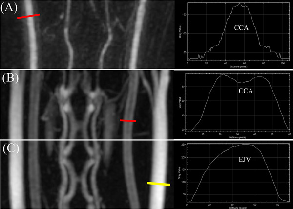 Figure 1
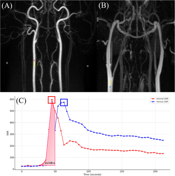 Figure 2
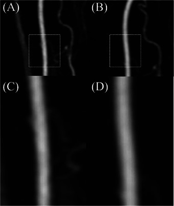 Figure 3
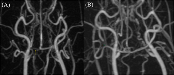 Figure 4
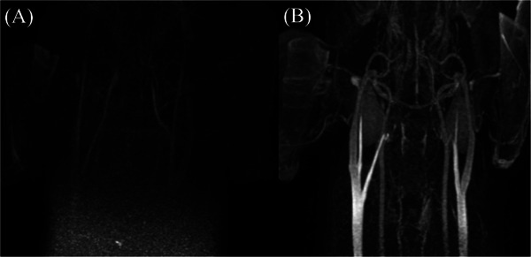 Figure 5
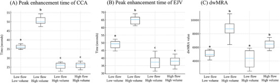 Figure 6
Peer Reviews
No public reviews on file for this paper yet. If you reviewed it on a platform where reviews are public (OpenReview, ICLR, NeurIPS, ICML), you can paste yours below so the community can read it here.
Videos
No videos yet. Explain this paper in a talk, walkthrough, or lecture? Add one.
Taxonomy
TopicsAdvanced MRI Techniques and Applications · Cerebrovascular and Carotid Artery Diseases · MRI in cancer diagnosis