Preliminary comparison of FreeSurfer segmentation algorithms in the Wake Forest community‐based cohort and potential impact on ATN classification
Marc D. Rudolph, Melissa M. Rundle, Kathryn H Alphin, Richard A. Barcus, Timothy M. Hughes, Trey R. Bateman, Kiran K. Solingapuram Sai, Christopher T Whitlow, Suzanne Craft, Da Ma

TL;DR
This study compares two FreeSurfer algorithms for brain MRI segmentation and finds that newer deep learning methods like SynthSeg provide more reliable results, especially in areas affected by aging and disease.
Contribution
The study evaluates the impact of FreeSurfer segmentation algorithms on brain atrophy classification and highlights the benefits of deep learning-based methods like SynthSeg.
Findings
FreeSurfer v7.2 recon-all produced smaller and more variable volume and thickness estimates compared to SynthSeg.
Cortical thickness estimates in key dementia-related regions showed poor to moderate agreement between pipelines.
SynthSeg improved segmentation robustness in poor-quality scans and reduced the need for manual correction.
Abstract
Acquisition and participant‐related artifacts (atrophy, motion) can degrade the quality of acquired images resulting in poor segmentation of tissue compartments. This can bias estimates of brain volume and thickness used quantify age and disease‐related atrophy, a problem particularly salient in clinical populations. In some cases, poor quality scans may be discarded or repeated incurring additional costs. Participants (n = 624; cognitively normal [CU;n = 330]; mild cognitive impairment [MCI;n = 214]; dementia [DEM;n = 75]; otherwise not classified [OTHER;n = 5)] enrolled in the Wake Forest ADRC Clinical Cohort (Table 1). Structural T1‐MRI scans were processed using FreeSurfer (v7.2) recon‐all and FreeSurfer (v7.4) recon‐all‐clinical (SynthSeg) pipelines. Tau‐PET (FTP) images were acquired; global, meta‐temporal, and entorhinal tau‐PET (white+gray matter; SUVr) was quantified. Measures…
Genes, proteins, chemicals, diseases, species, mutations and cell lines named across the full text — each resolved to its canonical identifier and authoritative record.
Click any figure to enlarge with its caption.
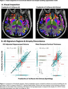 Figure 1
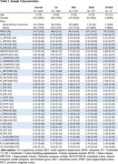 Figure 2
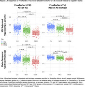 Figure 3
Peer Reviews
No public reviews on file for this paper yet. If you reviewed it on a platform where reviews are public (OpenReview, ICLR, NeurIPS, ICML), you can paste yours below so the community can read it here.
Videos
No videos yet. Explain this paper in a talk, walkthrough, or lecture? Add one.
Taxonomy
TopicsCerebrospinal fluid and hydrocephalus · Dementia and Cognitive Impairment Research · Medical Image Segmentation Techniques