Three‐Dimensional Presentation of Tongue Squamous Cell Carcinoma Histopathology and Magnetic Resonance Imaging—A Novel Image Fusion Method
Anne Koivuholma, Heli J. Sistonen, Katri Aro, Alexandria L. Irace, Antti Mäkitie, Jaana Hagström, Timo Atula

TL;DR
This paper introduces a new 3D imaging method to better visualize and understand oral cancer tumors using MRI and histopathology data.
Contribution
The novel 3D image fusion method combines histopathology and MRI data for soft tissue tumors like OSCC.
Findings
Nine OSCCs were successfully visualized as 3D image fusions.
The 3D model improves understanding of tumor orientation and dimensions.
The method facilitates multidisciplinary discussions and comparisons between MRI and histopathology.
Abstract
Three‐dimensional (3D) modeling has been used in the management of bony head and neck tumors, but not in soft tissue tumors. Currently, histopathological findings of oral squamous cell carcinomas (OSCC) are presented as two‐dimensional images. Previously, we developed a 3D image fusion method that presents tumor histopathology and MRI in 3D form. In this study, we sought to test our method in a series of OSCCs. A 3D table scanner, 3D Slicer software, and 3D modeling software were used to produce 3D image fusions of nine OSCCs. Nine OSCCs are presented as digital, 3D image fusions, showing the resected specimen and the tumor within based on histopathology and MRI. The fused 3D model allows for a better visual understanding of tumor orientation within the resected specimen, provides a tool to compare tumor volume and dimensions between MRI images and histopathology slides, and…
Genes, proteins, chemicals, diseases, species, mutations and cell lines named across the full text — each resolved to its canonical identifier and authoritative record.
Click any figure to enlarge with its caption.
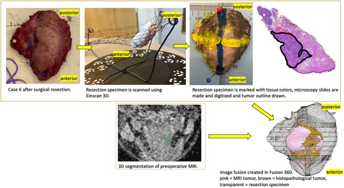 Figure 1
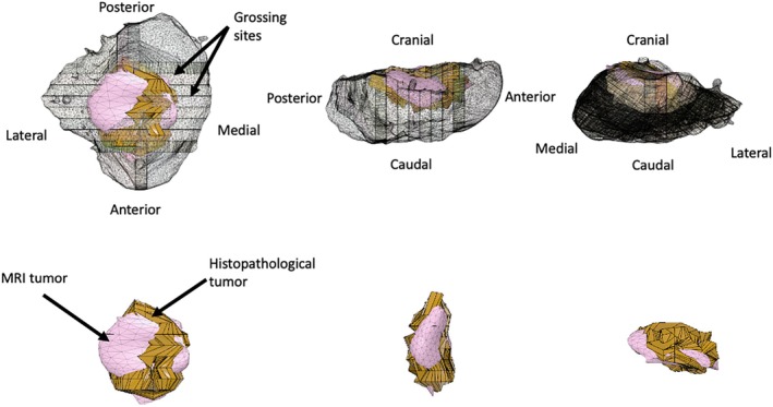 Figure 2
Peer Reviews
No public reviews on file for this paper yet. If you reviewed it on a platform where reviews are public (OpenReview, ICLR, NeurIPS, ICML), you can paste yours below so the community can read it here.
Videos
No videos yet. Explain this paper in a talk, walkthrough, or lecture? Add one.
Taxonomy
TopicsRadiomics and Machine Learning in Medical Imaging · Head and Neck Cancer Studies · Medical Imaging and Analysis