A case of endovascular treatment for pseudoaneurysm of the popliteal artery that developed 3 months after stenting
Takahiro Otsuka, Hideki Wada, Jun Shitara, Hirohisa Endo, Manabu Ogita, Satoru Suwa, Tohru Minamino

TL;DR
This paper describes a successful endovascular treatment of a rare artery issue that developed after a stent placement.
Contribution
The paper presents a novel case demonstrating the effectiveness of stent grafts for post-endovascular pseudoaneurysm.
Findings
A stent graft successfully blocked blood flow to a pseudoaneurysm in a frail patient.
Endovascular treatment was chosen over surgery due to patient frailty and risks.
The case highlights the utility of stent grafts in managing post-procedure pseudoaneurysms.
Abstract
Pseudoaneurysms in lower limb arteries are rare, but can occur after endovascular procedures. We report the case of an 80-year-old man with a chronic limb-threatening ischemia who developed pseudoaneurysm at the site of a stent placed in the left popliteal artery 3 months earlier. Because of the severe pain and risk of aneurysm rupture, intervention was considered necessary. Considering the frailty of the patient, surgical treatment would have likely been difficult and endovascular treatment was therefore planned. A stent graft was placed, blocking blood flow to the pseudoaneurysm. This case demonstrates the effectiveness of stent grafts in managing post-endovascular pseudoaneurysm.
Genes, proteins, chemicals, diseases, species, mutations and cell lines named across the full text — each resolved to its canonical identifier and authoritative record.
Click any figure to enlarge with its caption.
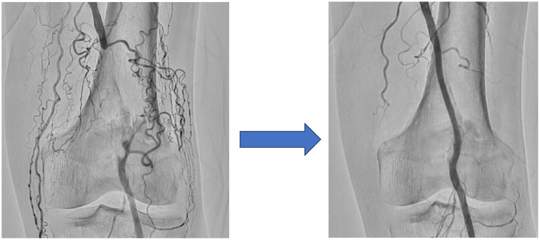 Figure 1
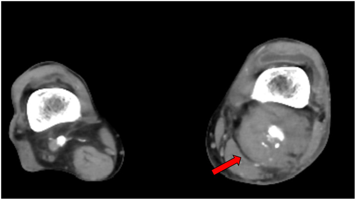 Figure 2
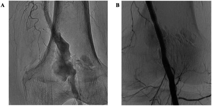 Figure 3
Peer Reviews
No public reviews on file for this paper yet. If you reviewed it on a platform where reviews are public (OpenReview, ICLR, NeurIPS, ICML), you can paste yours below so the community can read it here.
Videos
No videos yet. Explain this paper in a talk, walkthrough, or lecture? Add one.
Taxonomy
TopicsVascular Procedures and Complications · Peripheral Artery Disease Management · Infectious Aortic and Vascular Conditions
Introduction
Lower extremity arterial disease is 1 manifestation of systemic atherosclerosis and is associated with significant morbidity and mortality [1,2]. Although endovascular therapy (EVT) has become a useful treatment option for popliteal artery diseases, the outcomes are not always acceptable [3]. Pseudoaneurysm can arise after a patient experiences complications injuring the vascular system, resulting in blood flow between the tunica media and tunica adventitia [4]. This in turn leads to the development of hematoma and compression of surrounding tissues. Pseudoaneurysms of the popliteal artery are rare and reported causes have involved complications from fractures, surgical interventions, and trauma and idiopathic cases. However, late aneurysm formation after EVT in the popliteal artery is very rare.
We report a case in which pseudoaneurysm developed at the site of a popliteal artery stent placed 3 months earlier. Treatment was provided using a stent graft. We also discuss the rationale for our treatment choice with reference to similar cases in the literature.
Case report
An 80-year-old man with a history of type 2 diabetes mellitus, dyslipidemia, hypertension, and coronary artery bypass grafting visited our hospital due to pain and swelling from the left lower back of the thigh to the back of the knee and an ulcer on the right foot. He had received several EVTs to address chronic limb-threatening ischemia for ulcers and rest pain in both legs. The patient had stenosis with severe calcification from the distal left superficial femoral artery to the popliteal artery, and had undergone a third EVT for the same lesion 3 months earlier. In that treatment, a wire-interwoven stent (Supera peripheral stent, 5.5 × 80 mm; Abbott Vascular Corporation, Chicago, IL, USA) was placed in addition to balloon angioplasty due to repeated restenosis (Fig. 1).Fig. 1. Endovascular treatment performed on the left popliteal artery 3 months before this presentationA previously treated severe calcified lesion in the left popliteal artery was found to be occluding the vessel (left panel). Endovascular treatment was therefore performed and a wire-interwoven stent (Supera peripheral stent, 5.5 × 80 mm) was implanted 3 months prior to this visit (right panel).Fig 1
At the time of this visit, he was admitted to the plastic surgery department for treatment of an ulcer on the right foot. However, symptoms did not improve and he was referred to the department of cardiovascular medicine. The swollen area from the lower left thigh to the dorsal aspect of the knee was pulsatile, so we performed contrast-enhanced computed tomography, which revealed pseudoaneurysm with a maximum dimension of 54 × 55 mm at the popliteal artery (Fig. 2). Angiography performed later also showed aneurysm formation around the stent placed 3 months before (Fig. 3A). Because of the severe pain and risk of aneurysm rupture, intervention was considered necessary. Surgical resection of the aneurysm was an option, but considering the frailty of the patient, EVT using a stent graft was planned.Fig. 2. Popliteal artery imaged by contrast-enhanced computed tomography. Contrast-enhanced computed tomography reveals formation of a pseudoaneurysm with a maximum dimension of 54 × 55 mm in the left popliteal artery (red arrow).Fig 2. Fig. 3Angiographic findings. A. Angiographic findings of the popliteal artery pseudoaneurysm; B. Final angiography after stent graft implantation(A) Selective angiography shows aneurysm formation around the stent placed 3 months earlier. (B) After placement of the stent graft (VIABHN, 6.0 × 100 mm), blood flow to the pseudoaneurysm was confirmed to have ceased.Fig 3
After puncturing the left common femoral artery, a guiding sheath was inserted and a guide wire was advanced to the distal rather than the lesion. We first examined the aneurysm lesion using an intravascular ultrasound device. A stent graft (VIABHN, 6.0 × 100 mm; Gore, Flagstaff, AZ, USA) was then implanted to cover the entire aneurysm lesion and post-dilatation was performed using a 6.0 × 40 mm balloon. Angiography and intravascular ultrasound confirmed excellent apposition of the stent graft and total exclusion of the pseudoaneurysms (Fig. 3B). After EVT, the patient reported rapid resolution of pain and swelling of the left leg gradually decreased.
Discussion
Popliteal artery pseudoaneurysms are rare clinical conditions mainly linked to damage to the arterial wall, and are seen most often among young men. The literature has mentioned that the incidence of popliteal artery pseudoaneurysm ranges from 0.03% to 0.17%, representing around 4% of all popliteal artery aneurysms [5]. The pathology has also been reported to occur following local trauma [6], bony deformation [5], or iatrogenic injuries [7]. Here, we presented the case of an elderly man n with a large popliteal artery pseudoaneurysm after multiple EVTs. Some reports have shown that fracture of a self-expandable stent in the femoropopliteal artery caused pseudoaneurysm [8,9]. Stent fracture was not observed in the present case, but balloon dilatation or stenting might have damaged the vessel because the lesion was severely calcified.
Several options are available for the management of popliteal pseudoaneurysm, including open surgery, EVT or observation. Open surgical repair is considered the first choice for a large popliteal artery. EVTs using a covered stent or coiling may not provide reliable treatment for large aneurysms, and surgical treatment is preferred when possible. However, surgical repair requires the patient to be in a condition capable of withstanding general anesthesia, which may be difficult in frail patients. Endovascular repair has been shown to be safe and efficient for treating popliteal pseudoaneurysm, resulting in reduced morbidity and shorter duration of hospitalization. Rief et al. [7] reported a similar case involving a frail, bedridden, 90-year-old woman treated with a stent graft due to the difficulty of performing general anesthesia or surgical intervention. This approach was selected in that case as a minimally invasive and effective treatment for the pseudoaneurysm, taking into account the condition of the patient. On the other hand, the long-term patency issues and the risk of occlusion by thrombus in treatments with covered stents should also be considered. In addition, complete suppression of endoleak might be difficult when the pseudoaneurysm is large. In the present case, we were fortunate to be able to eliminate any endoleak by adding post-dilation after stent graft implantation.
Conclusion
We achieved successful management of a post-endovascular pseudoaneurysm of the popliteal artery using a stent graft. Stent grafting proved to be an effective and less invasive treatment option, providing symptom relief and reducing aneurysm-related complications.
Patient consent
The patient provided written informed consent for the publication of this case report, including all accompanying images and clinical information.
The reference list from the paper itself. Each links out to its DOI / PubMed record.
- 1Nishikawa R.Shiomi H.Morimoto T.Yamamoto K.Sakamoto H.Tada T.Effects of peripheral artery disease on long-term outcomes after percutaneous coronary intervention versus coronary artery bypass grafting in patients with severe coronary artery disease J Cardiol 84420242792863813514710.1016/j.jjcc.2023.12.004 · doi ↗ · pubmed ↗
- 2Komai H.Ogura M.Sakashita H.Miyama N Yamamoto N Takai K The real-world data of lipid-lowering treatment in patients with peripheral artery disease and its association with severity of disease J Cardiol 841202436403781648110.1016/j.jjcc.2023.10.002 · doi ↗ · pubmed ↗
- 3Yoon Y.H.Lee J.H.Hwang W.M.Park HW Roh JH Lee SJ Treatment extent of femoropopliteal disease and clinical outcomes following endovascular therapy Euro Intervention 20182024 e 1154 e 11623927951610.4244/EIJ-D-24-00037 PMC 11384224 · doi ↗ · pubmed ↗
- 4Stolt M.Braun-Dullaeus R.Herold J.Do not underestimate the femoral pseudoaneurysm Vasa 47320181771852943961110.1024/0301-1526/a 000691 · doi ↗ · pubmed ↗
- 5Ammori M.B.Evans A.R.Mc Lain AD.Popliteal artery pseudoaneurysm after total knee arthroplasty J Arthroplasty 3192016200420072702170110.1016/j.arth.2016.02.041 · doi ↗ · pubmed ↗
- 6Raherinantenaina F.Rajaonanahary T.M.Rakoto Ratsimba H.N.Management of popliteal artery pseudoaneurysms as a result of limb trauma and orthopedic surgery or associated with osteochondromas Ann Cardiol Angeiol (Paris)65420162652742723686610.1016/j.ancard.2016.04.023 · doi ↗ · pubmed ↗
- 7Rief M.Rief A.Bornemann-Cimenti H.Rief P.Idiopathic pseudoaneurysm of the popliteal artery with endovascular treatment: a case report Radiol Case Rep 1892023333633403750213410.1016/j.radcr.2023.06.062PMC 10368536 · doi ↗ · pubmed ↗
- 8Aten K.Gerlt D.Rosol Z.Banerjee S.Femoropopliteal pseudoaneurysms as a complication of self-expanding nitinol stent fracture: a brief report and review Am J Cardiol 223202415173878222610.1016/j.amjcard.2024.05.026 · doi ↗ · pubmed ↗