Topical Carboxytherapy Modulates the Skin Microbiome Following CO2 Laser Resurfacing: A Pilot Study
Barbara Hernandez‐Rovira, Emma Villamaria, Julia Oh, Bengisu Ozarslan, Saranya Wyles

Abstract
Genes, proteins, chemicals, diseases, species, mutations and cell lines named across the full text — each resolved to its canonical identifier and authoritative record.
Click any figure to enlarge with its caption.
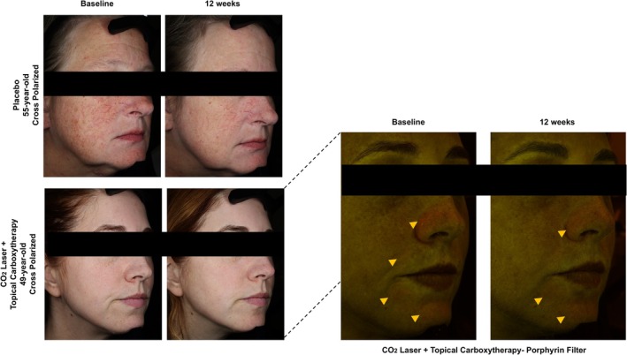 FIGURE 1
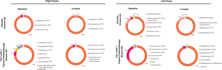 FIGURE 2
- —Lumisque Skincare, LLC
Peer Reviews
No public reviews on file for this paper yet. If you reviewed it on a platform where reviews are public (OpenReview, ICLR, NeurIPS, ICML), you can paste yours below so the community can read it here.
Videos
No videos yet. Explain this paper in a talk, walkthrough, or lecture? Add one.
Taxonomy
TopicsDermatologic Treatments and Research · Wound Healing and Treatments · Skin Protection and Aging
To the Editor,
Skin microbiome contributes to cutaneous homeostasis and barrier defense by regulating keratinocyte proliferation, angiogenesis, and immune responses [1, 2]. Carbon dioxide (CO_2_) laser resurfacing, widely used for photoaging and scar revision, induces controlled dermal injury and transient skin barrier disruption to trigger the wound healing cascade. Topical carboxytherapy, which delivers CO_2_ transdermally, is proposed to enhance tissue regeneration by improving oxygenation and microvascular perfusion [3]; however, its mechanistic impact on skin physiology and microbial communities remains poorly defined. Given the role of skin microbiota in wound healing and inflammation, understanding how these procedures affect microbial dynamics is clinically relevant. This pilot study aimed to characterize the facial skin microbiome following full‐face CO_2_ fractional resurfacing and post‐procedural treatment with topical carboxytherapy (CO_2_ Lift, Lumisque, Weston, FL).
Two participants were enrolled and assigned to one of two post‐laser care groups: the placebo group (n = 1) received standard care with a petroleum‐based ointment, while the intervention group (n = 1) was treated with a gel‐formulated carboxytherapy. VISIA‐CR (Canfield Scientific Inc., Parsippany, NJ) imaging was performed at baseline and 12 weeks post‐procedure (Figure 1). Microbiome samples were collected from the left versus right malar cheek at baseline and week 4 using standardized swabbing. Microbial profiling was performed via 16S rRNA gene sequencing with genus‐level taxonomic assignment; sequencing depth and diversity metrics were evaluated descriptively, without statistical testing, due to the limited sample size. Genus‐level taxonomic composition is shown in Figure 2. Given that only one participant was included per group, this exploratory pilot study was not powered for statistical comparison between groups, and all findings are presented as hypothesis‐generating observations without inference of group differences or treatment effects.
Facial skin imaging before and after fractional CO2 laser resurfacing and topical carboxytherapy. VISIA‐CR imaging captured at baseline and 12 weeks post‐treatment, demonstrating cutaneous changes in participants receiving either placebo or topical carboxytherapy following fractional CO2 laser resurfacing. Top row: Cross‐polarized images from the placebo group. Bottom row: Cross‐polarized and porphyrin‐filter images from the topical carboxytherapy treatment arm, showing follicular porphyrin signal in the nasolabial folds, mental fold, and chin region.
Facial skin microbiome composition before and after fractional CO2 laser resurfacing and topical carboxytherapy. Genus‐level relative abundance based on observed amplicon sequence variants (ASVs) from the right and left cheek at baseline and 4 weeks post‐treatment. The skin microbiome was consistently dominated by Cutibacterium spp. and Corynebacterium spp. across both pre‐ and post‐treatment timepoints. Topical carboxytherapy after laser resurfacing was associated with a greater post‐treatment increase in Cutibacterium spp. abundance. Top row: Placebo group. Bottom row: Topical carboxytherapy treatment arm.
Cutibacterium spp. was the dominant genus across all subjects, with bilateral detection at both time points. Relative abundance increased from 79% at baseline to 92% at week 4. Corynebacterium spp. was the second most abundant genus (11% at baseline, 3% at week 4), followed by low‐abundance taxa including Streptococcus spp. and Staphylococcus spp. Alpha diversity patterns varied between participants at baseline and over time; both groups exhibited a slight decline. These findings are reported descriptively only. Beta diversity analysis suggested overall compositional stability within individuals between baseline and follow‐up, though no formal clustering or between‐group comparisons can be inferred.
An observed increase in the relative abundance of Cutibacterium spp., presumed to be C. acnes, following CO_2_ laser resurfacing may suggest a potential role in maintaining microbial balance and supporting tissue repair. As the primary commensal of sebaceous skin, C. acnes is well‐adapted to lipid‐rich environments and contributes to skin homeostasis through immunomodulation, lipid metabolism, and inhibition of opportunistic pathogens [4]. Reduced C. acnes diversity has been associated with aging and frailty, while its antioxidant activity and ability to suppress biofilm formation support a protective role in wound healing [1, 5]. These mechanistic interpretations are intended to contextualize the observed microbiome patterns rather than establish causality.
Although limited by a small sample size, these findings indicate that neither CO_2_ laser resurfacing nor adjunctive topical carboxytherapy disrupts the composition or diversity of the facial skin microbiome. Given the bidirectional interaction between skin microbiota and wound regeneration, future studies should include larger cohorts and metagenomic sequencing to further define the impact of adjunctive treatments on microbial dynamics. Nonetheless, this pilot study suggests post‐procedural microbiological stability and highlights the feasibility of incorporating microbiome endpoints in studies aimed at optimizing dermatologic interventions.
Author Contributions
Barbara Hernandez‐Rovira: Writing‐original draft (equal); formal analysis (lead); visualization. Emma Villamaria: Writing‐original draft (equal); formal analysis (supporting). Julia Oh: Writing‐review and editing (equal); validation. Bengisu Ozarslan: Writing‐review and editing (equal); formal analysis (supporting). Saranya Wyles: Conceptualization; methodology; supervision; writing‐review and editing (equal).
Funding
This study was supported by Lumisque Skincare, LLC. The funder was not involved in study and manuscript preparation.
Disclosure
AI was not used in the composition of this manuscript.
Ethics Statement
This study was approved by the Mayo Clinic Institutional Review Board (IRB 23‐005182).
Consent
Written informed consent was obtained from all participants included in this study.
Conflicts of Interest
The authors declare no conflicts of interest.
The reference list from the paper itself. Each links out to its DOI / PubMed record.
- 1P. J. Larson , W. Zhou , A. Santiago , et al., “Associations of the Skin, Oral and Gut Microbiome With Aging, Frailty and Infection Risk Reservoirs in Older Adults,” Nature Aging 2, no. 10 (2022): 941–955.36398033 10.1038/s 43587-022-00287-9PMC 9667708 · doi ↗ · pubmed ↗
- 2M. Zielińska , A. Pawłowska , A. Orzeł , et al., “Wound Microbiota and Its Impact on Wound Healing,” International Journal of Molecular Sciences 24, no. 24 (2023): 17318.38139146 10.3390/ijms 242417318 PMC 10743523 · doi ↗ · pubmed ↗
- 3P. Susini , D. di Seclì , S. Bacchini , et al., “Carboxytherapy in Dermatologic Surgery and Aesthetic Medicine: Current Knowledge and Future Perspectives‐A Systematic Review,” Dermatologic Surgery 51, no. 7 (2025): 673–678.40062597 10.1097/DSS.0000000000004608 · doi ↗ · pubmed ↗
- 4M. Rozas , A. de Hart Ruijter , M. J. Fabrega , et al., “From Dysbiosis to Healthy Skin: Major Contributions of Cutibacterium Acnes to Skin Homeostasis,” Microorganisms 9, no. 3 (2021): 628.33803499 10.3390/microorganisms 9030628 PMC 8003110 · doi ↗ · pubmed ↗
- 5J. Oh , J. Robison , and G. A. Kuchel , “Frailty‐Associated Dysbiosis of Human Microbiotas in Older Adults in Nursing Homes,” Nature Aging 2, no. 10 (2022): 876–877.36909933 10.1038/s 43587-022-00289-7PMC 9997047 · doi ↗ · pubmed ↗