Liposome‐Based Potential Vaccines Platforms that Are Noncytotoxic
Saida Mebarek, Killian Jacob, Carmela Ilaria Pierro, Davide Romanini, Michele Fiore

TL;DR
This study introduces a safe, non-toxic liposome-based vaccine platform for cancer immunotherapy using engineered neoglycolipids.
Contribution
A modular, noncytotoxic liposomal vaccine platform using bio-orthogonal chemistry for antigen delivery is proposed.
Findings
Neoglycolipids bearing the Tn antigen were synthesized without detectable cytotoxicity.
Stable liposomes were formed using palmitic acid and 1-palmitoyl-2-oleoyl-sn-glycero-3-phosphocholine via freeze–thaw/extrusion.
The platform offers a tunable and safe method for antigen delivery in cancer immunotherapy.
Abstract
Synthetic vaccines represent a promising avenue in cancer immunotherapy by promoting targeted immune responses. Liposomal technologies have further advanced synthetic vaccinology by enabling the efficient delivery of tumor‐associated carbohydrate antigens. Despite this progress, the toxicity and reproducibility of such platforms remain underexplored. In this preliminary study, we synthesized a series of neoglycolipids bearing the Thomsen–Nouveau (Tn) antigen using bio‐orthogonal thiol–ene click chemistry. Here we present the results obtained using a set of neoglycolipids that were evaluated for their ability to self‐assemble into liposomal vesicles and for in vitro cytotoxicity. The resulting neoglycolipids exhibited no detectable cytotoxicity and formed stable liposomal structures when formulated with palmitic acid and 1‐palmitoyl‐2‐oleoyl‐sn‐glycero‐3‐phosphocholine via a…
Genes, proteins, chemicals, diseases, species, mutations and cell lines named across the full text — each resolved to its canonical identifier and authoritative record.
Click any figure to enlarge with its caption.
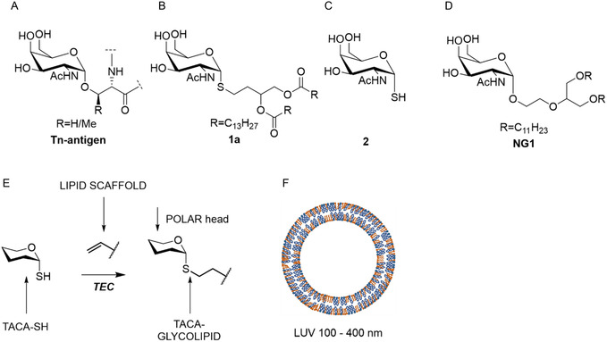 FIGURE 1
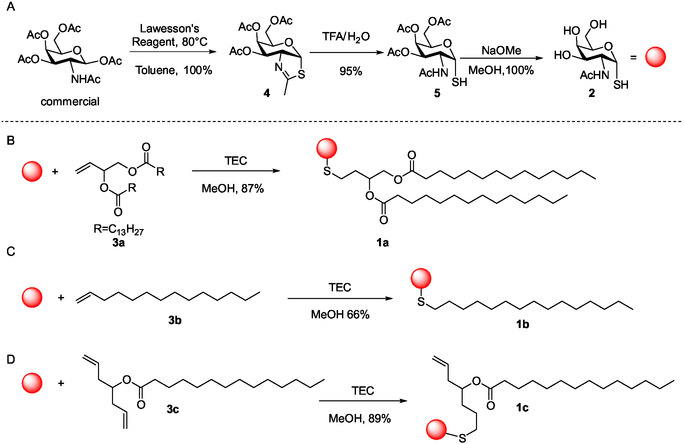 SCHEME 1
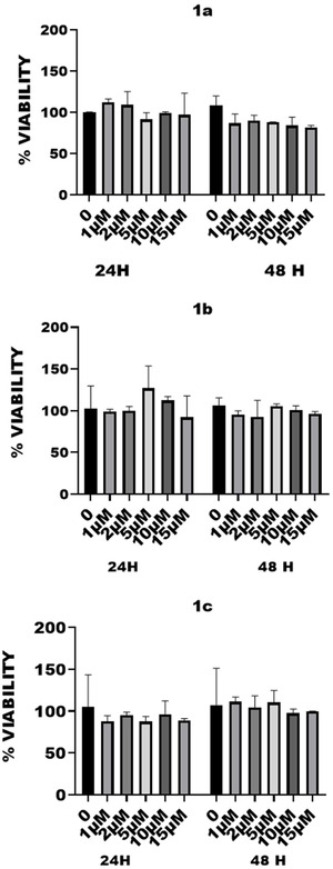 FIGURE 2
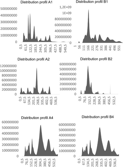 FIGURE 3
| Entry | Lipid scaffold | Product | Y% | liposomes (0.01 mM) | Cytotoxicity | |
|---|---|---|---|---|---|---|
| 1 |
|
|
| 87 | yes | no |
| 2 |
|
|
| 66 | yes | no |
| 3 |
|
|
| 70 | no | no |
| 4 |
| [ | no | no |
| Entry |
|
| Particle number (mL) | |
|---|---|---|---|---|
| 1 | A4/ | 320 | 9.2310 ± 4.189 | |
| 2 | A2/ | 240 | 1.0111 ± 1.1210 | |
| 3 | A1/ | 118 | 2.9310 ± 3.399 | |
| 4 | B4/ | 350 | 9.2310 ± 4.189 | |
| 5 | B2/ | 172 | 1.0111 ± 1.1210 | |
| 6 | B1/ | 130 | 3.3110 ± 2.479 | |
Peer Reviews
No public reviews on file for this paper yet. If you reviewed it on a platform where reviews are public (OpenReview, ICLR, NeurIPS, ICML), you can paste yours below so the community can read it here.
Videos
No videos yet. Explain this paper in a talk, walkthrough, or lecture? Add one.
Taxonomy
TopicsRNA Interference and Gene Delivery · Click Chemistry and Applications · Immunotherapy and Immune Responses
Introduction
1
Dendrimers [1], calixarenes [2], fullerenes [3], cyclodextrins [4], peptides [5, 6, 7, 8, 9], and immunogenic cyclopeptides [10, 11, 12, 13, 14, 15, 16, 17, 18] have been used in the last decades as platforms for anticancer drug delivery or self‐adjuvating vaccine cancer themself. Although encouraging, the outcomes achieved have not extended beyond the initial disclosure of preliminary biological data with some exceptions [6, 19]. The synthesis of the aforementioned glycoclusters typically involves considerable synthetic complexity, multiple iterative steps, often requiring repeated chromatographic purifications (e.g. high‐performance liquid chromatography), which can lead to low overall yields. In some cases, purification and reproducibility present significant bottlenecks [10, 18]. The application of chemoselective and bio‐orthogonal ligation strategies has been employed to partially address these limitations, owing to their operational simplicity and resistance to enzymatic degradation during conjugation processes [20]. Furthermore, the number of exposed antigens remains limited to the structure of the glycocluster [21] itself with the undesired clustering side effect [22]. Recent results showed that glycolipids assembled via thiol–ene click (TEC) chemistry (bearing two GlcNAc‐SH sugar moieties on the same scaffold) exhibited a binding affinity to wheat germ agglutinin 3000‐fold higher than that of the corresponding monosaccharide alone [23]. Recent advances in glycolipid synthesis have demonstrated significant potential for the fabrication of supramolecular assemblies that mimic the morphology and stability of membrane vesicles, ranging from micrometric (giant unilamellar vesicles, GUV) [24, 25, 26] to nanometric scales as large unilamellar vesicles (LUV) [27]. In the context of vaccine design, LUV offer substantial advantages over classical glycoarchitectures such as glycoclusters or multivalent glycosylated scaffolds [1, 2, 3, 4, 5, 6, 7, 8, 9, 10, 11, 12, 13, 14, 15, 16, 17, 18]. A key benefit lies in the synthetic accessibility and scalability of glycolipids, which can be obtained in substantial quantities—typically hundreds of milligrams—via conventional synthetic routes and purified efficiently using standard silica gel chromatography, thereby reducing both time and cost. Due to their amphiphilic nature and structural similarity to phospholipids, glycolipids spontaneously integrate into lipid bilayers and drive vesicle formation [28]. Why use engineered LUV? The resulting LUVs provide an exceptionally high capacity for glycan/epitopes display. To the best of our knowledge, in a few cases glycoclusters can typically accommodate up to 64 glycosyl moieties; in contrast, LUVs offer a dramatically larger presentation platform. For example, a liposome composed solely of 1‐palmitoyl‐2‐oleoyl‐sn‐glycero‐3‐phosphocholine (POPC) with a diameter of 100 nm has an estimated surface area of ~125,000 nm^2^ and can display up to ~190,000 entities per surface, according to measurements by Kučerka et al. [29] for POPC. This enables a vastly enhanced multivalent presentation of carbohydrate antigens. To the best of our knowledge, this represents one of the highest epitopes densities (in the nanometric scale) achievable in a modular and biocompatible format, positioning LUV as a powerful platform for next‐generation glycovaccine development. A notable example was recently reported by the research group of Chen and Zhang, who synthesized a “modified” cholesterol derivative that generates a Tn‐glycolipid antigen (Chol‐GalNAc/CpG), capable of simultaneously eliciting both humoral and cellular immune responses [19]. The vesicle size was characterized by transmission electron microscopy and found to be approximately 200 µm, which, in our assessment, may be excessively large for applications in human therapy. Importantly, the study did not address cytotoxicity assessments nor the absorption, distribution, metabolism, and excretion profile—critical parameters for evaluating the pharmacokinetics and safety of vaccine candidates. From a chemical standpoint, the carbonate linkage employed is known to lack enzymatic stability, potentially compromising in vivo durability. Furthermore, the use of TEC and simpler phospholipid‐based scaffolds compared to cholesterol ensures greater versatility in the synthesis of glycolipids. Additionally, employing Nanometer‐scale vesicles facilitates enhanced bioavailability by allowing LUVs to circulate through the bloodstream without posing a risk of vascular occlusion. In addition, in our long‐term studies, these constructs will demonstrate the ability to facilitate interactions with CD4^+^ T cells, leading to B‐cell activation and proliferation (unpublished). Moreover, engineered LUVs have the potential to increase the number of epitopes simultaneously recognized by B‐cell receptors and CD4^+^ T cells, thereby enhancing the overall efficacy of the vaccine. Notably, oncological vaccines based on this platform can elicit robust antitumor immunity by engaging B cells, CD4^+^ helper T cells, and CD8^+^ cytotoxic T cells [30, 31].
Results and Discussion
2
As part of our rationale, we sought to investigate an often‐overlooked property of glyco‐clusters: their potential cytotoxicity. We decided to produce LUV‐bearing synthetic glycolipids using TEC, a methodology extensively optimized and routinely applied in our laboratory [12, 32, 33, 34]. These LUVs are called synthetic outer membrane vesicles (sOMV) [35] for their similarity with the outer membrane vesicles (OMVs) produced by Gram negative bacteria and extensively used in cancer vaccinology [36]. Mixtures of naturally occurring phospholipids and glycolipids bearing tumor‐associated carbohydrate antigens can be assembled into vesicles using well‐established methodologies. The resulting GUVs can be purified by simple vacuum filtration [37] and, upon application of standardized protocols, these GUVs can be downsized via extrusion to yield LUVs of defined diameters (Figure 1F) with a monodispersed population.
(A) The structure of the Tn Antigen; (B) the first glycolipid bearing a modified Tn antigen (1a) obtained by TEC [35]; (C) the structure of 2; (D) structure of NG1 [38]; (E) generic scheme for TEC applied to the production of thio‐glycolipids; (F) engineered LUVs; based on the surface occupied by the polar head of a molecule of POPC (68 Å2 = 0.68 nm2), each liposome of 100 nm diameter has a surface of ~125,000 nm2 constituted of ~190,000 molecules of POPC, the internal surface is not considered in this calculation.
To obtain biologically active glycolipids, we have selected the Thiol‐GalNAc as an analog of the Thomsen–Nouveau antigen (Tn). The Tn antigen is defined as a single N‐acetylgalactosamine (GalNAc) α‐linked to the hydroxyl group of a serine or a threonine residue (GalNAc‐α1‐O‐Ser/Thr; Figure 1A) and is a hallmark of aberrantly glycosylated mucins, frequently associated with malignant transformation and tumor progression [39]. For example the neoglycolipid 1a is formed by coupling 2 to the lipophilic scaffold 3a (Scheme 1B) through TEC. This stable molecule exhibited amphipathic properties as shown previously [35]. However, its toxicity remained unknown as well as its ability to form liposomes in the nanometric scale. We decided to carry out a new study preparing a small library of neoglycolipids, each formed by coupling different lipidic moieties (3a‐3c) to 2 via TEC but optimizing reaction conditions [23, 40, 41] and analyzing cytotoxicity and assessing their application in liposome preparation, both crucial properties before considering their use in nanomedicine as future vaccine components [42]. The synthesis of 2 (Scheme 1A) was carried out following the procedure described by Knapp and Myers [43] with a few modifications (under Ar atmosphere) that increased the overall yields up to 90%. This synthesis allows the preparation of solely the α anomer via acidic treatment of the thiazolidine intermediary 4, followed by deacetylation in "Zemplén" conditions of 5 (Scheme 1A) [44]. Commercially available 3b was used to prepare 1b (Scheme 1C) and the product was purified by SiO_2_ flash chromatography using the isocratic eluent composed of CHCl_3_:MeOH:H_2_O (65:25:0.4 v/v/v, Eluent A) in 66% of yield. Compound 1c was instead obtained from 3c (Scheme 1D). Before irradiation, all the mixtures were saturated with argon at 1 atm for 30 min. Reactions were carried out in 5 mL test tubes under stirring and were irradiated with blue light (365 nm) at room temperature for 15 min and additional 15 min in the case of 1c. TLC monitoring (DCM:MeOH 9:1, v/v) showed the complete consumption of the starting material 3a, 3b, or 3c. The presence of disulfide was not observed. In the case of 3c we have observed that only one thiol formed a stable thioether bond whereas one of the terminal akenes was unreacted. Two other sets of reactions have been performed: (i) 1c bearing one thioether was submitted to a second reaction with 6 equivalents of 2 and (ii) a second set of synthesis of 1d was performed adding to 3c up to 12 equivalents of 2. In both cases a dramatical formation of the disulfide of 2 occurred. If formed, 1d was probably present in traces and the yields of 1c lowered to 25% per reaction. Thus, 1c was reprepared to establish the yields that were around 50% of conversion using 3 or 6 eq of 2 per double bond in 3c (see Scheme S1, Supporting Information).
(A) Schematic preparation of mimic Tn‐SH antigen 2 [43]. (B–D) Synthesis of 1a [35] and 1b‐1c (this research), see Scheme S1 (Supporting Information) for further details.
All glycolipids exhibited no detectable toxicity after 24 and 48 h at concentrations up to 15 µM (Figure 2). We tested a range of compound concentrations on cells to assess toxicity. We went up to 15 µM because of limited compound solubility, which can cause precipitation at higher concentrations, and because of DMSO toxicity. DMSO becomes cytotoxic above approximately 0.5%–1% (v/v), depending on the cell line, which can confound the assay. Viability tests were conducted on murine vascular smooth muscle cell line as in previously reported researches [45, 46]. The absence of cytotoxicity (Table 1) has been a priority for us, a factor that is poorly reported in similar research [10, 19], while only the absence of immunogenicity of the scaffold is considered (i.e. ref. [47]). The absence of cytotoxicity was also detected for a sister molecule (NG1, Table 1, Figure S1, Supporting Information) presenting the GalNAc residue in the position sn‐2 of glycerol moiety (Figure 1D). These compounds were used to prepare LUV with nominal diameter of 400, 200, and 100 nm (Table 2). The lipid composition was as follows: 49% of either 1a, 1b, 1c, or NG1, 50% of POPC, and 1% of PA. The final concentration of each LUV was 0.01 mM. Only mixtures containing 1a and 1b hydrated enough to prepare LUV, whereas the lack of hydration for 1c and NG1 in mixtures can be explained for a too poor lipophilic part for the former, and the presence of the “polar head” in the sn‐2 position for the latter (Scheme 1D and Figure 1D, respectively), that altered the conical shape required for the formation of a membrane bilayer or disrupted the membrane bilayer upon hydration [48]. The method used is a standardized one involving freeze–thaw cycling, followed by extrusion processes. The rapid freezing in liquid nitrogen (−78°C) and thawing at temperatures above the lipid phase transition (typically ~38°C) induces transient fusion and rupture events, facilitating internal reorganization of the vesicles. Following thermal cycling, the suspension is subjected to extrusion through polycarbonate membranes with defined pore sizes. This step disrupts residual multilamellar vesicles and selectively retains liposomes matching the filter diameter, yielding a population of LUVs with uniform size and improved reproducibility. A predominant and homogeneous size distribution was observed for all concentrations (A1‐A4 and B1‐BA) and liposome types (Table 2, 400–100 nm). Mixtures containing compound 1a were labeled as series A, and those containing 1b as series B. Homogeneous populations were present in each samples prepared using 100 nm (A1/B1), 200 nm (A2/B2), and 400 nm (A4/B4) pore sizes, as reported in Table 2. However, the size distribution does not align with the nominal membrane sizes but corresponds instead to the measured sizes—for example, A4 with a size distribution of 320 nm is comparable to B4 with 350 nm (see also Figure 3). The liposome concentrations (particles per mL) are also consistent across samples.
Results of the viability test on molecules 1a–1c et NG1 after 24 and 48 h of incubation and statistical analysis of the viability assays. Results were represented as mean ± standard error of the mean (SEM) and were expressed in percent as fold change compared to untreated control. For each analysis, three independent experiments were performed. To establish significance of our results, data were analyzed by the use of two‐sided Mann–Whitney U test. The level of significance was set at p < 0.05. Graphs and calculations were done using Prism and Instat 3, respectively (GraphPad software, California, USA). Statistical tests show that the results are not significant. The compounds have no significant effect on cell viability compared to the untreated control.
Size distribution of the population of s‐OMV (LUV) of nominal diameter 100 (A1), 200 (A2), and 400 (A4) nm containing 1a, and size distribution of the population of s‐OMV (LUV) of nominal diameter 100 (B1), 200 (B2), and 400 (B4) nm containing 1b.
Conclusion
3
A set of glycolipids (1a–1c) was chemically and biologically evaluated. None of the compounds exhibited cytotoxicity. Among them, only 1a and 1b were used to generate nanosized sOMVs, with effective diameters ranging from 118 to 350 nm, which are either slightly above or below the nominal pore size of the membranes used for their extrusion from GUVs. The molar composition of each glycolipid in the vesicles was implemented up to 49%, in conformity with previously collected data from 2019 and 2023 [23, 35]. Synthetic procedures evidenced that phospholipid‐shaped glycolipids (like 1a) prevalently formed using thiol‐ene reaction and produced vesicles upon hydration. Based on these findings, we are confident that liposome‐based vaccine platforms can be enhanced, particularly when antigens are integrated into the membrane bilayer. The data from this study, and those of others [10, 18, 19], support the feasibility of developing nanovaccines in the form of s‐OMVs. These promising results encourage us to further pursue the development of liposomes‐based vaccines incorporating CD4^+^ and CD8^+^ peptide antigens, adjuvants like PA, and potentially combinations of natural or fully synthetic phospholipid analogs [48]. In the next future, expected results are to trigger in a “one set” construct both humoral and cellular response, strengthening the immune system against the insurgence ofmalignancies.
Supporting Information
Additional supporting information can be found online in the Supporting Information section. Supporting Scheme S1: Summary of the reactions carried out for the preparation of 1d. Supporting Fig. S1: Results of the viability test on molecule NG1 after 24 h of incubation. To establish significance of our results, data were analyzed by the use of two‐sided Mann‐Whitney U test. The level of significance was set at p < 0.05. Graphs and calculations were done using Prism and Instat 3, respectively (GraphPad software, California, USA). Statistical tests show that the results are not significant. The compounds have no significant effect on cell viability compared to the untreated control. Supporting Fig. S2: ^1^H NMR in DMSO‐d6 of compound 1b (300 MHz). Supporting Fig. S3: ^13^C NMR in DMSO‐d6 of compound 1b (75 MHz). Supporting Fig. S4: MS spectra of compound 1b. Supporting Fig. S5: ^1^H NMR in DMSO‐d6 of compound 1c (300 MHz). Supporting Fig. S6: ^13^C NMR in DMSO‐d6 of compound 1c (75MHz). Supporting Fig. S7: HRMS spectra of compound 1c. Supporting Fig. S8 – 10: ^1^H, g‐COSY, ^13^C and HSQC of compound 1c in MeOD.
Conflicts of Interest
The authors declare no conflicts of interest.
Supporting information
Supplementary Material
The reference list from the paper itself. Each links out to its DOI / PubMed record.
- 1S. E. Sherman , Q. Xiao , and V. Percec , “Mimicking Complex Biological Membranes and Their Programmable Glycan Ligands with Dendrimersomes and Glycodendrimersomes,” Chemical Reviews 117 (2017): 6538–6631, 10.1021/acs.chemrev.7b 00097.28417638 · doi ↗ · pubmed ↗
- 2E. C. Wrobel , L. S. de Lara , Â. de Fátima , and O. N. Oliveira , “Nanoarchitectonics and Simulation on the Molecular‐Level Interactions between, p‐, Sulfonic Acid Calix[4]arene and Langmuir Monolayers Representing Healthy and Cancerous Cell Membranes,” Langmuir 40 (2024): 27010–27027.39663612 10.1021/acs.langmuir.4c 03948 PMC 11673576 · doi ↗ · pubmed ↗
- 3X. Zhang , H. Cong , B. Yu , and Q. Chen , “Recent Advances of Water‐Soluble Fullerene Derivatives in Biomedical Applications,” Mini‐Reviews in Organic Chemistry 16 (2018): 92–99.
- 4N. Bognanni , M. Viale , A. Distefano , et al., “Cyclodextrin Polymers as Delivery Systems for Targeted Anti‐Cancer Chemotherapy,” Molecules 26 (2021): 6046.34641590 10.3390/molecules 26196046 PMC 8512365 · doi ↗ · pubmed ↗
- 5S. D. Kuduk , J. B. Schwarz , X. T. Chen , et al., “Synthetic and Immunological Studies on Clustered Modes of Mucin‐Related Tn and TF O‐Linked Antigens: The Preparation of a Glycopeptide‐Based Vaccine for Clinical Trials against Prostate Cancer,” Journal of the American Chemical Society 120 (1998): 12474–12485.
- 6S. J. Danishefsky and J. R. Allen , “From the Laboratory to the Clinic: A Retrospective on Fully Synthetic Carbohydrate‐Based Anticancer Vaccines,” Angewandte Chemie International Edition 39 (2000): 836–863.10760879 10.1002/(sici)1521-3773(20000303)39:5<836::aid-anie 836>3.0.co;2-i · doi ↗ · pubmed ↗
- 7S. J. Keding , A. Endo , and S. J. Danishefsky , “Synthesis of Non‐Natural Glycosylamino Acids Containing Tumor‐Associated Carbohydrate Antigens,” Tetrahedron 59 (2003): 7023–7031.
- 8H. Herzner and H. Kunz , “Spacer‐Separated Sialyl Lewis X Cyclopeptide Conjugates as Potential E‐Selectin Ligands,” Carbohydrate Research 342 (2007): 541–557.17027941 10.1016/j.carres.2006.09.012 · doi ↗ · pubmed ↗