Abnormalities of resting‐state eyes‐closed electroencephalographic rhythms are not affected by white matter lesions in patients with mild cognitive impairment due and not due to Alzheimer's disease
Roberta Lizio, Claudio Del Percio, Susanna Lopez, Mina De Bartolo, Matteo Carpi, Antonio Pio Afragola, Giuseppe Noce, Raffaele Ferri, Bahar Güntekin, Görsev Yener, Claudio Babiloni

TL;DR
This study finds that brain wave patterns in patients with mild cognitive impairment are not worsened by white matter damage, regardless of whether the impairment is due to Alzheimer's disease.
Contribution
The study shows that white matter lesions do not affect resting-state alpha brain rhythms in mild cognitive impairment patients.
Findings
Posterior alpha brain activity was significantly reduced in MCI groups compared to healthy controls.
Increased white matter lesions were linked to worse executive function in non-Alzheimer's MCI patients.
Neurophysiological mechanisms regulating brain oscillations appear unaffected by white matter damage in MCI.
Abstract
Patients with mild cognitive impairment (MCI) typically show abnormal high delta (<4 Hz) and low alpha (8–12 Hz) rhythms measured from resting‐state eyes‐closed electroencephalographic (rsEEG) source activities as well as white matter lesions (WMLs) measured from magnetic resonance imaging (MRI). Here we tested the hypothesis that rsEEG rhythms may not deteriorate with the increase of WLMs in patients with MCI due and not due to Alzheimer's disease (ADMCI and noADMCI). An international database provided demographic, clinical, and rsEEG datasets for cognitively unimpaired older (Healthy; N = 30), ADMCI (N = 64), and noADMCI (N = 36) participants. The rsEEG rhythms spanned individual delta, theta, and alpha frequency bands. The eLORETA freeware estimated cortical rsEEG sources. The international database also provided MRI datasets for the ADMCI and noADMCI participants. T2 and Fluid…
Genes, proteins, chemicals, diseases, species, mutations and cell lines named across the full text — each resolved to its canonical identifier and authoritative record.
Click any figure to enlarge with its caption.
 Figure 1
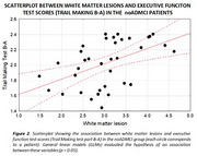 Figure 2
Peer Reviews
No public reviews on file for this paper yet. If you reviewed it on a platform where reviews are public (OpenReview, ICLR, NeurIPS, ICML), you can paste yours below so the community can read it here.
Videos
No videos yet. Explain this paper in a talk, walkthrough, or lecture? Add one.
Taxonomy
TopicsFunctional Brain Connectivity Studies · Vestibular and auditory disorders · Advanced MRI Techniques and Applications