Distinct Sensitivity of MRI Versus [18F]FDG‐PET To Detect Cerebral Changes Across the Alzheimer's Continuum in Down Syndrome: A Multimodal Imaging Study
José Enrique Arriola‐Infante, Alejandra O. Morcillo‐Nieto, Maria Franquesa‐Mullerat, Sara E Zsadanyi, Lídia Vaqué‐Alcázar, Mateus Rozalem Aranha, José Allende Parra, Zili Zhao, Javier Arranz, Íñigo Rodríguez‐Baz, Lucía Maure‐Blesa, Laura Videla, Isabel Barroeta, Laura Del Hoyo

TL;DR
This study compares MRI and PET scans in detecting early brain changes in Alzheimer's disease among people with Down syndrome.
Contribution
The study reveals that MRI is more sensitive than PET in detecting cerebral changes in Alzheimer's stages in Down syndrome.
Findings
MRI shows greater sensitivity than [18F]FDG-PET in detecting brain volume changes in Alzheimer's stages in Down syndrome.
Amyloid pathology in asymptomatic Down syndrome is linked to frontal lobe atrophy without significant hypometabolism.
Symptomatic stages show widespread atrophy and hypometabolism, with MRI detecting more pronounced changes.
Abstract
To date, limited evidence exists on the different sensitivity of MRI versus [18F]FDG‐PET to detect early cerebral changes along the Alzheimer's disease (AD) continuum in Down syndrome (DS). Therefore, we aimed to characterize volume and metabolism alterations in adults with DS and compare their performance in detecting AD clinical stages. Cross‐sectional study, including 92 adults with DS from the Down‐Alzheimer Barcelona Neuroimaging Initiative (34 asymptomatic without amyloid pathology [aDS A−], 20 asymptomatic with amyloid pathology [aDS A+], 12 with prodromal AD [pDS], and 26 with AD dementia [dDS]; Table 1), who underwent 3T‐MRI and [18F]FDG‐PET. Amyloid status in the aDS group was determined using amyloid‐PET (>20 centiloids, n = 29) or CSF Aβ42/Aβ40 ratio (<0.062, Lumipulse, n = 25). Brain volume and metabolism values were extracted using the Hammers atlas, normalizing MRI…
Genes, proteins, chemicals, diseases, species, mutations and cell lines named across the full text — each resolved to its canonical identifier and authoritative record.
Click any figure to enlarge with its caption.
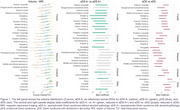 Figure 1
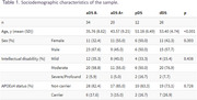 Figure 2
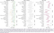 Figure 3
Peer Reviews
No public reviews on file for this paper yet. If you reviewed it on a platform where reviews are public (OpenReview, ICLR, NeurIPS, ICML), you can paste yours below so the community can read it here.
Videos
No videos yet. Explain this paper in a talk, walkthrough, or lecture? Add one.
Taxonomy
TopicsDown syndrome and intellectual disability research · Digital Image Processing Techniques · Cell Image Analysis Techniques