Evaluation discrepancy between [18F]MK‐6240 and [18F]AV‐1451 tau‐PET using tau‐PET overlap index
Seokbeen Lim, Hoon‐Ki Min, Jessica L. Brunn, David N. Soleimani‐Meigooni, Hwamee Oh, Juan Fortea, Belen Pascual, Brian A. Gordon, Pedro Rosa‐Neto, Suzanne L. Baker, Firoza Z Lussier, Guilherme Povala, Tharick A Pascoal, Val J Lowe

TL;DR
This study compares two tau-PET imaging methods, [18F]MK-6240 and [18F]AV-1451, using the overlap index to detect differences in tau accumulation, especially in younger and cognitively unimpaired individuals.
Contribution
The study reveals variability in overlap index results between [18F]MK-6240 and [18F]AV-1451, highlighting off-target binding effects in younger and cognitively unimpaired participants.
Findings
OI values showed broad distribution between the two tracers, especially in cognitively unimpaired and younger individuals.
OI-detected voxels in [18F]MK-6240 were often in the meninges, a site of off-target binding not seen in [18F]AV-1451.
META-ROI SUVr values between the tracers followed an exponential trend, unlike the OI distribution.
Abstract
The overlap index (OI), previously introduced by Lee et al. (2022), has proven to be a reliable method for detecting tau accumulation via tau‐PET imaging with flortaucipir ([18F]AV‐1451). This technique identifies voxel‐wise increases in standardized uptake value ratio (SUVr) across serial scans. However, the relationship between tau‐PET measurements obtained with [18F]MK‐6240 and [18F]AV‐1451 using the OI remains unclear. This study aims to investigate the relationship between [18F]MK‐6240 and [18F]AV‐1451 tau‐PET using the tau‐PET OI. The study included 27 participants from the HEAD project, all of whom underwent two serial tau‐PET scans (each [18F]MK‐6240 and [18F]AV‐1451) along with 3T T1‐weighted MRI, acquired within an average interval of 18 months. SUVr maps for each tau‐PET tracer were normalized to the cerebellar crus grey matter, and MR images were co‐registered to the MCALT…
Genes, proteins, chemicals, diseases, species, mutations and cell lines named across the full text — each resolved to its canonical identifier and authoritative record.
Click any figure to enlarge with its caption.
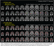 Figure 1
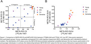 Figure 2
Peer Reviews
No public reviews on file for this paper yet. If you reviewed it on a platform where reviews are public (OpenReview, ICLR, NeurIPS, ICML), you can paste yours below so the community can read it here.
Videos
No videos yet. Explain this paper in a talk, walkthrough, or lecture? Add one.
Taxonomy
TopicsAdvanced MRI Techniques and Applications · Medical Imaging Techniques and Applications · Lanthanide and Transition Metal Complexes