Automated Brain Tissue Segmentation on CT guided by MRI: Advancing AI‐based Neuroimaging for Dementia
Vidya Somashekarappa, Meera Srikrishna, Silke Kern, Joyce R Chong, Eric Westman, Christopher Chen, Ingmar Skoog, Jakob Seidlitz, Michael Schöll

TL;DR
This paper introduces a new AI method to segment brain tissues in CT scans using MRI data, improving dementia diagnosis in resource-limited settings.
Contribution
The study introduces MedNeXt, a novel 3D segmentation model that outperforms nnU-Net in multi-orientation and multi-modal brain tissue segmentation on CT.
Findings
MedNeXt achieved higher Dice Similarity Coefficients (DSCs) and volumetric similarity scores compared to nnU-Net across different orientations.
MedNeXt showed better generalizability in dementia cohorts with higher DSC and volumetric similarity scores.
nnU-Net is more resource-efficient, suitable for limited-resource settings, while MedNeXt excels in accuracy and scalability.
Abstract
Brain tissue segmentation is vital in Alzheimer's and dementia research for creating detailed neuroanatomical maps, diagnosing early‐stage neurodegeneration, and guiding interventions. Although MRI remains the standard approach for its superior soft‐tissue contrast, CT is a more accessible imaging modality in acute and resource‐constrained settings. This study utilized paired CT‐MRI datasets from the Gothenburg H70 Birth Cohort (N = 733) and the Memory Clinic Cohort of the National University Hospital, Singapore (NUS Dementia Cohort, N = 210) to train and evaluate advanced segmentation models—nnUNet (2D & 3D models for 300‐1000 epochs) and MedNeXt (3D‐ Small, Base, Medium and Large models for 3x3x3 & 5x5x5 kernels). MRI‐derived labels were employed to guide CT segmentation, allowing accurate delineation of brain tissue segmentation (Gray Matter: GM, White Matter: WM and Cerebrospinal…
Genes, proteins, chemicals, diseases, species, mutations and cell lines named across the full text — each resolved to its canonical identifier and authoritative record.
Click any figure to enlarge with its caption.
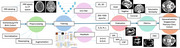 Figure 1
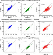 Figure 2
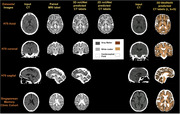 Figure 3
Peer Reviews
No public reviews on file for this paper yet. If you reviewed it on a platform where reviews are public (OpenReview, ICLR, NeurIPS, ICML), you can paste yours below so the community can read it here.
Videos
No videos yet. Explain this paper in a talk, walkthrough, or lecture? Add one.
Taxonomy
TopicsMedical Image Segmentation Techniques · Brain Tumor Detection and Classification · Advanced Neural Network Applications