Imaging‐based biological staging associates with brain atrophy in preclinical Alzheimer's disease
Lorenzo Fontura Brasil Barcellos, João Pedro Ferrari‐Souza, Isabela Just de Jesus Vanni, Marco Antônio Albini Valer, Andrei Bieger, Douglas Teixeira Leffa, Firoza Z Lussier, Wagner S. Brum, Cristiano Aguzzoli, Anderson Corin, Marco Antônio De Bastiani, Giovanna Carello‐Collar

TL;DR
This study shows that brain atrophy rates increase with Alzheimer's disease stages based on imaging markers, suggesting that tracking brain volume changes could help detect early disease progression.
Contribution
The study demonstrates that imaging-based biological staging correlates with cortical atrophy rates in preclinical Alzheimer's disease.
Findings
A+TNEO+ individuals showed significantly higher cortical atrophy rates compared to A+T− and A+TMTL+ groups.
Serial volumetric measurements in temporoparietal regions may serve as sensitive biomarkers for early Alzheimer's progression.
Cortical atrophy rates accelerate progressively across biological AD stages.
Abstract
Alzheimer's disease (AD) diagnostic and staging criteria rely on imaging biomarkers as robust tools for in vivo disease assessment. Among these, cortical brain atrophy is recognized as a key hallmark of disease progression. However, rates of cortical atrophy in preclinical stages remain insufficiently explored within current staging frameworks. In a group of amyloid β‐positive (Aβ+) asymptomatic individuals, we investigated the association of the imaging‐based biological AD staging framework with longitudinal brain atrophy patterns. We included 162 Aβ+ participants from the A4 Study placebo group with available magnetic resonance imaging (MRI) and positron emission tomography (PET) for amyloid‐β (Aβ) plaques ([18F]Florbetapir) and tau ([18F]Flortaucipir) at baseline, along with a follow‐up MRI at least 2 years after baseline. Tau positivity in the medial temporal lobe (TMTL+) and in…
Genes, proteins, chemicals, diseases, species, mutations and cell lines named across the full text — each resolved to its canonical identifier and authoritative record.
Click any figure to enlarge with its caption.
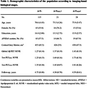 Figure 1
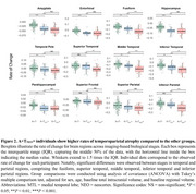 Figure 2
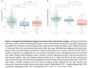 Figure 3
Peer Reviews
No public reviews on file for this paper yet. If you reviewed it on a platform where reviews are public (OpenReview, ICLR, NeurIPS, ICML), you can paste yours below so the community can read it here.
Videos
No videos yet. Explain this paper in a talk, walkthrough, or lecture? Add one.
Taxonomy
TopicsDementia and Cognitive Impairment Research · Alzheimer's disease research and treatments · Functional Brain Connectivity Studies