Detecting Hippocampal Subfield Iron in Alzheimer's Disease using Ultra‐high Resolution in vivo 7T MRI
Reese A. Dunne, Hossein Moein Taghavi, Phillip DiGiacomo, Julian Maclaren, Meghan Bell, Mackenzie L. Carlson, Elizabeth C. Mormino, Victor W. Henderson, Pascal Spincemaille, Hangwei Zhuang, Yi Wang, Brian S. Rutt, Marios Georgiadis, Michael Zeineh

TL;DR
This study uses ultra-high resolution 7T MRI to detect increased iron in the hippocampus of Alzheimer's patients, linking it to memory decline and offering a potential new imaging biomarker.
Contribution
The study introduces a noninvasive method using 7T MRI to visualize hippocampal iron in vivo, supporting its use as a biomarker for Alzheimer's disease.
Findings
R2* values showed a significant increase in hippocampal subfields in AD and MCI compared to healthy controls.
A significant negative correlation was found between memory scores and QSM-χ+ in the CA1 subfield.
7T MRI detected in vivo hippocampal iron deposition consistent with post-mortem findings.
Abstract
Hippocampal iron, as measured with imaging, biofluids, and histology, has been associated with Alzheimer's disease (AD), its progression, and potentially neuroinflammatory disease mechanisms. With its high sensitivity to tissue magnetic susceptibility, 7T MRI offers the potential to detect abnormal iron deposition within the hippocampus of AD and mild cognitive impairment (MCI) brains in vivo, especially when combined with dedicated methods such as quantitative susceptibility mapping (QSM). We aim to utilize ultra‐high resolution 7T MRI and explore conventional and novel source‐separated QSM to quantify hippocampal iron deposition in AD, providing insights into the involvement of brain iron in disease progression. We conducted 7T MRI on 19 ADRC human volunteers, including 8 healthy controls (HC), 6 individuals with MCI, and 5 with AD. MR images were acquired using a GE MR950 scanner…
Genes, proteins, chemicals, diseases, species, mutations and cell lines named across the full text — each resolved to its canonical identifier and authoritative record.
Click any figure to enlarge with its caption.
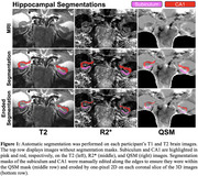 Figure 1
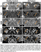 Figure 2
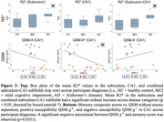 Figure 3
Peer Reviews
No public reviews on file for this paper yet. If you reviewed it on a platform where reviews are public (OpenReview, ICLR, NeurIPS, ICML), you can paste yours below so the community can read it here.
Videos
No videos yet. Explain this paper in a talk, walkthrough, or lecture? Add one.
Taxonomy
TopicsAdvanced MRI Techniques and Applications · Functional Brain Connectivity Studies · Alzheimer's disease research and treatments