Amygdala microstructural changes in subjective cognitive decline as measured by MRI diffusion kurtosis, neurite orientation dispersion and density, and magnetization transfer imaging
Ryn Flaherty, Yu Veronica Sui, Arjun V. Masurkar, Zakia Youss, Steven Baete, Thomas Wisniewski, Henry Rusinek, Mariana Lazar

TL;DR
This study uses MRI techniques to explore microstructural changes in the amygdala of people with subjective cognitive decline, revealing altered tissue organization and potential early signs of brain pathology.
Contribution
The study links diffusion kurtosis imaging with NODDI and MTI metrics to better understand microstructural changes in the amygdala associated with subjective cognitive decline.
Findings
SCD participants showed lower KFA and higher MK and RK in the right amygdala.
MK and RK correlated strongly with NODDI metrics, suggesting changes in neurite organization and density.
No significant correlations were found between DKI metrics and MTR, indicating distinct microstructural features.
Abstract
Our prior work applying diffusion kurtosis imaging (DKI) to subjective cognitive decline (SCD) showed significantly decreased kurtosis fractional anisotropy (KFA) and significantly increased mean kurtosis (MK) for SCD in bilateral amygdala, an early site for tau tangle pathology1. However, the microstructural alterations driving these differences are unclear. Here, we assess associations of MK and KFA with neurite orientation dispersion and density imaging (NODDI) and magnetization transfer imaging (MTI) metrics, which provide a more specific characterization of tissue microstructure. 175 cognitively normal participants from Cam‐Can2 (75 SCD) ages 55‐88 were included in the analysis. Participants were defined as SCD if they endorsed problems with their memory and in the control group otherwise. Diffusion images were processed to obtain MK, Radial Kurtosis (RK), Axial Kurtosis (AK) and…
Genes, proteins, chemicals, diseases, species, mutations and cell lines named across the full text — each resolved to its canonical identifier and authoritative record.
Click any figure to enlarge with its caption.
 Figure 1
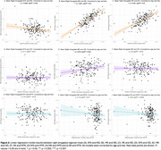 Figure 2
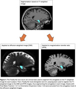 Figure 3
Peer Reviews
No public reviews on file for this paper yet. If you reviewed it on a platform where reviews are public (OpenReview, ICLR, NeurIPS, ICML), you can paste yours below so the community can read it here.
Videos
No videos yet. Explain this paper in a talk, walkthrough, or lecture? Add one.
Taxonomy
TopicsAdvanced Neuroimaging Techniques and Applications · MRI in cancer diagnosis · Functional Brain Connectivity Studies